Genetic identity of thermosensory relay neurons in the lateral parabrachial nucleus - PubMed (original) (raw)
Genetic identity of thermosensory relay neurons in the lateral parabrachial nucleus
Joel C Geerling et al. Am J Physiol Regul Integr Comp Physiol. 2016.
Abstract
The parabrachial nucleus is important for thermoregulation because it relays skin temperature information from the spinal cord to the hypothalamus. Prior work in rats localized thermosensory relay neurons to its lateral subdivision (LPB), but the genetic and neurochemical identity of these neurons remains unknown. To determine the identity of LPB thermosensory neurons, we exposed mice to a warm (36°C) or cool (4°C) ambient temperature. Each condition activated neurons in distinct LPB subregions that receive input from the spinal cord. Most c-Fos+ neurons in these LPB subregions expressed the transcription factor marker FoxP2. Consistent with prior evidence that LPB thermosensory relay neurons are glutamatergic, all FoxP2+ neurons in these subregions colocalized with green fluorescent protein (GFP) in reporter mice for Vglut2, but not for Vgat. Prodynorphin (Pdyn)-expressing neurons were identified using a GFP reporter mouse and formed a caudal subset of LPB FoxP2+ neurons, primarily in the dorsal lateral subnucleus (PBdL). Warm exposure activated many FoxP2+ neurons within PBdL. Half of the c-Fos+ neurons in PBdL were Pdyn+, and most of these project into the preoptic area. Cool exposure activated a separate FoxP2+ cluster of neurons in the far-rostral LPB, which we named the rostral-to-external lateral subnucleus (PBreL). These findings improve our understanding of LPB organization and reveal that Pdyn-IRES-Cre mice provide genetic access to warm-activated, FoxP2+ glutamatergic neurons in PBdL, many of which project to the hypothalamus.
Figures
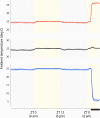
Fig. 1.
Ambient temperature trends inside individual cages from each group (n = 4 each). Thin lines denote temperatures from individual cages in each group: warm-exposed, control, and cool-exposed; thick lines represent group averages. At baseline, ambient cage temperatures remain stable near 24°C during the dark period and approach 25°C during the light period, possibly due to heat generation by fluorescent lamps along the chamber walls. Warm-exposed cage temperatures rapidly increase to 36°C for the last 4 h of the experiment. In the control group, ambient temperatures remain between 24 and 25°C. Cool-exposed cage temperatures fall quickly to 5–6°C for the final 4 h of the experiment. ZT is zeitgeber time (ZT 0 is the time when lights were turned on). The thick black bar beneath the _x_-axis indicates the 4-h period of ambient temperature change before perfusion.
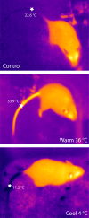
Fig. 2.
Thermal images show mice immediately after removal from a warm (36°C) or cool (4°C) ambient temperature. Images were taken at the same ambient temperature (22°C). The mid-tail temperature, a proxy for thermoregulatory vasodilation (41), is indicated by white star. Tail temperature is near room temperature in control mice, high after warm exposure, and very low after cool exposure.
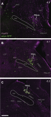
Fig. 3.
Distributions of FoxP2+ and dynorphin neurons in the lateral parabrachial nucleus (LPB). Sections span the caudal midbrain (A and B) through the rostral pons (C). Magenta nuclear labeling marks neurons that express FoxP2. Green somatic labeling (_Pdyn_-GFP) marks cells with Prodynorphin gene expression. These neurons constitute a small, caudal subset of the larger population of FoxP2+ neurons in the LPB. Scale bar: 200 μm. Abbreviations: Cb, cerebellum; Cun, cuneiform nucleus; KF, Kolliker-Fuse nucleus; LDT, laterodorsal tegmental nucleus; Me5, mesencephalic nucleus of the trigeminal nerve; NLL, nucleus of the lateral lemniscus; PB, parabrachial nucleus; PBcL, central lateral subnucleus; PBdL, dorsal lateral subnucleus; PBeL, external lateral subnucleus; PBlc, lateral crescent subnucleus; PBm, medial subnucleus; PBreL, rostral-to-external lateral subnucleus; scp, superior cerebellar peduncle.
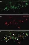
Fig. 4.
Green fluorescent protein (GFP)-immunoreactivity in Pdyn-IRES-Cre reporter mice (_Pdyn_-GFP, green) colocalizes with Pdyn mRNA in the lateral parabrachial nucleus (LPB) (_Pdyn_-FISH, in red). These examples are in the caudal dorsal-lateral parabrachial nucleus (approximate bregma −5.4 mm). Arrowheads indicate neurons with prominent colocalization. Scale bar is 50 μm.
Fig. 5.
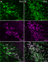
FoxP2+ neurons in most LPB subnuclei are glutamatergic. A: GFP cytoplasmic labeling (green) colocalizes with FoxP2 nuclear immunoreactivity (magenta) in the caudal LPB in a _Vglut2_-GFP reporter mouse [dorsal-lateral (dL) PB; bregma −5.3 mm]. B: GFP and FoxP2 colocalization is also apparent in the rostral to external lateral (PBreL; bregma −4.9 mm). Scale bars: 50 μm.
Fig. 6.
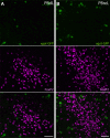
FoxP2+ neurons in PBdL and PBreL are not GABAergic. A: in a _Vgat_-GFP reporter mouse, GFP does not colocalize with FoxP2 immunoreactivity in PBdL (bregma −5.3 mm). B: likewise, FoxP2 and GFP do not colocalize in any neurons of PBreL (bregma −5.0 mm). Scale bars: 50 μm.
Fig. 7.
LPB neurons express c-Fos in distinct patterns after exposure to cool or warm ambient temperature. c-Fos immunofluorescence (red) and _Pdyn_-GFP fluorescence (green) are shown at three rostrocaudal levels of the LPB. A: control mice (24°C) have minimal c-Fos throughout the LPB. B: cool-exposed mice (4°C) express c-Fos in the far-rostral LPB, clustering in PBreL. Cool-activated, Fos+ neurons extend caudally into a rostral, lateral portion of the PBcL, as shown in _B_′, but are absent at caudal levels. C: Warm-exposed mice (36°C) express c-Fos primarily at caudal levels of the LPB. c-Fos labeling in warm-exposed mice colocalizes with and overlaps the dense cluster of _Pdyn_-GFP+ neurons in PBdL. Approximate bregma level (in mm) is indicated in each panel. Scale bar: 200 μm.
Fig. 8.
Cool-exposed (4°C) mice express c-Fos in many FoxP2+ neurons in the far-rostral LPB, primarily in the PBreL. Roughly half of the FoxP2+ neurons in PBreL are cool-activated (Fos+). This rostral region of the LPB contains relatively few _Pdyn_-GFP+ neurons, but some were c-Fos+ (arrowheads). Approximate bregma level is −4.9 mm. Scale bar: 50 μm.
Fig. 9.
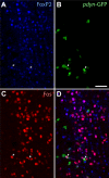
Warm-exposed (36°C) mice express c-Fos in FoxP2+, _Pdyn_-GFP+ neurons in PBdL. Triple-labeled, warm-activated neurons are indicated by white arrows. Most c-Fos+ neurons lacking _Pdyn_-GFP still express FoxP2 (magenta nuclei). Bregma level is approximately −5.4 mm. Scale bar: 50 μm.
Fig. 10.
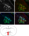
Warm-activated dynorphin neurons in PBdL project to the preoptic area. A: after cholera toxin subunit B (CTb) injection into the medial preoptic area (MPO), many PBdL neurons are retrogradely labeled (white). B: 36°C warm exposure produces abundant c-Fos (red) in _Pdyn_-GFP+ and intermingled neurons in PBdL. C: CTb (blue) labels many _Pdyn_-GFP+ neurons projecting to the preoptic area. D: many PBdL neurons are triple-labeled with CTb, _Pdyn_-GFP, and c-Fos, indicating that they are dynorphinergic, warm-activated, and project to the preoptic area. Arrows indicate triple-labeled neurons. E: CTb injection site involved both the MPO and the median preoptic nucleus (MnPO). Abbreviations: ac, anterior commissure; oc, optic chiasm.
Fig. 11.
Neurons relaying warm and cool thermosensory information exist in largely separate clusters along the rostral to caudal axis of the LPB. At left, in a sagittal brain stem drawing (1.2–1.3 mm lateral to bregma), Pdyn neurons are shown as green dots; aqua dots represent cholinergic nuclei, as anatomic landmarks. The expanded inset at right shows the PB region. We observed two patterns of c-Fos expression: cool-activated neurons cluster in the far-rostral LPB and are predominantly FoxP2+ and nondynorphinergic. Warm-activated neurons are clustered caudally and include many FoxP2+ dynorphin neurons. Abbreviations: Cb, cerebellum; IC, inferior colliculus; M, medulla; SNc, substantia nigra, pars compacta; PB, parabrachial nucleus; PBdl, dorsolateral parabrachial nucleus; PBeL, external lateral PB; PBreL, rostral-to-external PB; PPT, pedunculopontine tegmental nucleus; SC, superior colliculus; scp, superior cerebellar peduncle; SO, superior olivary nuclear complex; V, trigeminal motor nucleus (cholinergic motor neurons in dark blue); VII, facial motor nucleus; VIIn, facial nerve fascicles (postgenu).
Similar articles
- Efferent projections of Vglut2, Foxp2, and Pdyn parabrachial neurons in mice.
Huang D, Grady FS, Peltekian L, Geerling JC. Huang D, et al. J Comp Neurol. 2021 Mar;529(4):657-693. doi: 10.1002/cne.24975. Epub 2020 Sep 21. J Comp Neurol. 2021. PMID: 32621762 Free PMC article. - Two Ascending Thermosensory Pathways from the Lateral Parabrachial Nucleus That Mediate Behavioral and Autonomous Thermoregulation.
Yahiro T, Kataoka N, Nakamura K. Yahiro T, et al. J Neurosci. 2023 Jul 12;43(28):5221-5240. doi: 10.1523/JNEUROSCI.0643-23.2023. Epub 2023 Jun 20. J Neurosci. 2023. PMID: 37339876 Free PMC article. - Parabrachial opioidergic projections to preoptic hypothalamus mediate behavioral and physiological thermal defenses.
Norris AJ, Shaker JR, Cone AL, Ndiokho IB, Bruchas MR. Norris AJ, et al. Elife. 2021 Mar 5;10:e60779. doi: 10.7554/eLife.60779. Elife. 2021. PMID: 33667158 Free PMC article. - Central circuitries for body temperature regulation and fever.
Nakamura K. Nakamura K. Am J Physiol Regul Integr Comp Physiol. 2011 Nov;301(5):R1207-28. doi: 10.1152/ajpregu.00109.2011. Epub 2011 Sep 7. Am J Physiol Regul Integr Comp Physiol. 2011. PMID: 21900642 Review. - Prostaglandin E2 production in the brainstem parabrachial nucleus facilitates the febrile response.
Blomqvist A. Blomqvist A. Temperature (Austin). 2024 Sep 24;11(4):309-317. doi: 10.1080/23328940.2024.2401674. eCollection 2024. Temperature (Austin). 2024. PMID: 39583895 Free PMC article. Review.
Cited by
- Median preoptic area neurons are required for the cooling and febrile activations of brown adipose tissue thermogenesis in rat.
da Conceição EPS, Morrison SF, Cano G, Chiavetta P, Tupone D. da Conceição EPS, et al. Sci Rep. 2020 Oct 22;10(1):18072. doi: 10.1038/s41598-020-74272-w. Sci Rep. 2020. PMID: 33093475 Free PMC article. - Parabrachial Complex: A Hub for Pain and Aversion.
Chiang MC, Bowen A, Schier LA, Tupone D, Uddin O, Heinricher MM. Chiang MC, et al. J Neurosci. 2019 Oct 16;39(42):8225-8230. doi: 10.1523/JNEUROSCI.1162-19.2019. J Neurosci. 2019. PMID: 31619491 Free PMC article. Review. - A parabrachial-hypothalamic parallel circuit governs cold defense in mice.
Yang WZ, Xie H, Du X, Zhou Q, Xiao Y, Zhao Z, Jia X, Xu J, Zhang W, Cai S, Li Z, Fu X, Hua R, Cai J, Chang S, Sun J, Sun H, Xu Q, Ni X, Tu H, Zheng R, Xu X, Wang H, Fu Y, Wang L, Li X, Yang H, Yao Q, Yu T, Shen Q, Shen WL. Yang WZ, et al. Nat Commun. 2023 Aug 15;14(1):4924. doi: 10.1038/s41467-023-40504-6. Nat Commun. 2023. PMID: 37582782 Free PMC article. - Thermosensory thalamus: parallel processing across model organisms.
Leva TM, Whitmire CJ. Leva TM, et al. Front Neurosci. 2023 Oct 13;17:1210949. doi: 10.3389/fnins.2023.1210949. eCollection 2023. Front Neurosci. 2023. PMID: 37901427 Free PMC article. Review. - Galaninergic and hypercapnia-activated neuronal projections to the ventral respiratory column.
Dereli AS, Oh AYS, McMullan S, Kumar NN. Dereli AS, et al. Brain Struct Funct. 2024 Jun;229(5):1121-1142. doi: 10.1007/s00429-024-02782-8. Epub 2024 Apr 5. Brain Struct Funct. 2024. PMID: 38578351 Free PMC article.
References
- Bernard JF, Besson JM. The spino(trigemino)pontoamygdaloid pathway: electrophysiological evidence for an involvement in pain processes. J Neurophysiol 63: 473–490, 1990. - PubMed
- Bernard JF, Dallel R, Raboisson P, Villanueva L, Le Bars D. Organization of the efferent projections from the spinal cervical enlargement to the parabrachial area and periaqueductal gray: a PHA-L study in the rat. J Comp Neurol 353: 480–505, 1995. - PubMed
- Bester H, Chapman V, Besson JM, Bernard JF. Physiological properties of the lamina I spinoparabrachial neurons in the rat. J Neurophysiol 83: 2239–2259, 2000. - PubMed
- Blair ML, Mickelsen D. Activation of lateral parabrachial nucleus neurons restores blood pressure and sympathetic vasomotor drive after hypotensive hemorrhage. Am J Physiol Regul Integr Comp Physiol 291: R742–R750, 2006. - PubMed
- Bratincsák A, Palkovits M. Activation of brain areas in rat following warm and cold ambient exposure. Neuroscience 127: 385–397, 2004. - PubMed
Publication types
MeSH terms
Substances
Grants and funding
- R01 DK089044/DK/NIDDK NIH HHS/United States
- R01 DK096010/DK/NIDDK NIH HHS/United States
- T32 AG000222/AG/NIA NIH HHS/United States
- T32 HL007901/HL/NHLBI NIH HHS/United States
- NIH-T32 AG000222/AG/NIA NIH HHS/United States
- R25 NS070682/NS/NINDS NIH HHS/United States
- P01 HL095491/HL/NHLBI NIH HHS/United States
- P30 DK057521/DK/NIDDK NIH HHS/United States
- R01 DK075632/DK/NIDDK NIH HHS/United States
- R01 DE022912/DE/NIDCR NIH HHS/United States
LinkOut - more resources
Full Text Sources
Other Literature Sources
Medical
Molecular Biology Databases
