Inhibition of Lysyl Oxidases Improves Drug Diffusion and Increases Efficacy of Cytotoxic Treatment in 3D Tumor Models - PubMed (original) (raw)
Inhibition of Lysyl Oxidases Improves Drug Diffusion and Increases Efficacy of Cytotoxic Treatment in 3D Tumor Models
Friedrich Schütze et al. Sci Rep. 2015.
Abstract
Tumors are characterized by a rigid, highly cross-linked extracellular matrix (ECM), which impedes homogeneous drug distribution and potentially protects malignant cells from exposure to therapeutics. Lysyl oxidases are major contributors to tissue stiffness and the elevated expression of these enzymes observed in most cancers might influence drug distribution and efficacy. We examined the effect of lysyl oxidases on drug distribution and efficacy in 3D in vitro assay systems. In our experiments elevated lysyl oxidase activity was responsible for reduced drug diffusion under hypoxic conditions and consequently impaired cytotoxicity of various chemotherapeutics. This effect was only observed in 3D settings but not in 2D-cell culture, confirming that lysyl oxidases affect drug efficacy by modification of the ECM and do not confer a direct desensitizing effect. Both drug diffusion and efficacy were strongly enhanced by inhibition of lysyl oxidases. The results from the in vitro experiments correlated with tumor drug distribution in vivo, and predicted response to therapeutics in murine tumor models. Our results demonstrate that lysyl oxidase activity modulates the physical barrier function of ECM for small molecule drugs influencing their therapeutic efficacy. Targeting this process has the potential to significantly enhance therapeutic efficacy in the treatment of malignant diseases.
Figures
Figure 1. Tumor Spheroid Assay for Determining Drug Diffusion Rates.
(A) Micrograph of multicellular tumor spheroid generated from 4T1 breast carcinoma cells. Scale Bar: 250 μm. (B) Experimental setup. (C) Confocal image of tumor spheroid generated from GFP-expressing 4T1 tumor cells after exposure to doxorubicin (red channel). Scale bar: 100 μm.
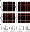
Figure 2. Lysyl Oxidase Activity reduces Drug Diffusion Rate in Multicellular Tumor Spheroids.
(A) Time course of doxorubicin diffusion in tumor spheroids generated from murine 4T1 breast carcinoma, MT6 fibrosarcoma, EMT6 breast carcinoma and Lewis Lung Carcinoma cells after cultivation at 20% or 2% oxygen. (B) Absolute diffusion rates into tumor spheroids. Images acquired on a laser scanning confocal microscope (n = 12), Scale bars: 100 μm, Error bars: ±SEM.
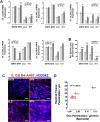
Figure 3. Lysyl Oxidase Expression is Increased Under Reduced Oxygen Supply.
(A) mRNA expression of the five lysyl oxidase family members in murine tumor cells cultivated at 20% or 2% oxygen levels (n = 3). (B) Lysyl oxidase activity in the supernatant of murine tumor cells cultivated at 20% or 2% oxygen levels (n = 4). (C) Extravasation of Hoechst 33342 (blue) from vessels (Isolectin GS-B4, red) in implanted tumors. (D) Correlation between Hoechst 33342 penetration in tumors and DOX diffusion in tumor spheroids generated from different murine cell lines. Error bars: ±SEM. Scale bars: 100 μm. *indicate statistical significance versus respective controls, *P < 0.05, **P < 0.01, ***P < 0.001 and mRNA levels changed at least 2-fold.
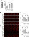
Figure 4. Recombinant Overexpression of Lysyl Oxidases Reduces Drug Diffusion in Tumor Spheroids.
(A) Relative lysyl oxidase activity in the supernatant of transfected and selected 4T1 cells (n = 4). (B) Time course of doxorubicin diffusion in tumor spheroids generated from 4T1 cells overexpressing hLOX versus control transfected cells. (C) Absolute diffusion rates into tumor spheroids generated from 4T1 cells overexpressing hLOX. (D) Time course of doxorubicin diffusion in tumor spheroids generated from 4T1 cells overexpressing hLOXL2 versus control transfected cells. (E) Absolute diffusion rates into tumor spheroids generated from 4T1 cells overexpressing hLOX. Tumor spheroids cultivated at 20% oxygen (n = 12). All images acquired on a laser scanning confocal microscope, Scale bars: 100 μm, Error bars: ±SEM.
Figure 5. Cell Toxicity of Cytotoxic Drugs in 2D and 3D Culture.
(A) Experimental setup of drug exposure times and cultivation conditions. (B) EC50 values for PTX and DOX after exposure of cells cultivated in 2D and exposure to the drugs for 72 h. (C) EC50 values for PTX and DOX after exposure of cells cultivated in 2D and exposure to the drugs for 30 min. (D) EC50 values for PTX and DOX after exposure of cells cultivated in 3D embedded in collagen I and exposure to the drugs for 30 min. (E) EC50 values for 4T1 cells overexpressing hLOX or hLOXL2 cultivated in 2D and 3D culture and exposure to DOX for 30 min. Error bars: ±SEM, n = 4.
Figure 6. Results from 3D Cytotoxicity Assays Predict Tumor Resistance to Cytotoxic Drugs.
(A) Dependency of drug efficacy on cultivation and exposure conditions. Effect of (B) DOX and (C) PTX treatment on MT6 tumors. Fully established tumors were treated at the indicated days with DOX (5 mg/kg BW) or PTX (20 mg/kg BW). Displayed are tumor volume measured at indicated days by caliper and mass of dissected tumors. Effect of (D) DOX and (E) PTX treatment on 4T1 tumors. Error bars: ±SEM. *indicates statistical significance versus control, *P < 0.05, **P < 0.01.
Similar articles
- VEGF-ablation therapy reduces drug delivery and therapeutic response in ECM-dense tumors.
Röhrig F, Vorlová S, Hoffmann H, Wartenberg M, Escorcia FE, Keller S, Tenspolde M, Weigand I, Gätzner S, Manova K, Penack O, Scheinberg DA, Rosenwald A, Ergün S, Granot Z, Henke E. Röhrig F, et al. Oncogene. 2017 Jan 5;36(1):1-12. doi: 10.1038/onc.2016.182. Epub 2016 Jun 6. Oncogene. 2017. PMID: 27270432 Free PMC article. - Pan-lysyl oxidase inhibition disrupts fibroinflammatory tumor stroma, rendering cholangiocarcinoma susceptible to chemotherapy.
Burchard PR, Ruffolo LI, Ullman NA, Dale BS, Dave YA, Hilty BK, Ye J, Georger M, Jewell R, Miller C, De Las Casas L, Jarolimek W, Perryman L, Byrne MM, Loria A, Marin C, Chávez Villa M, Yeh JJ, Belt BA, Linehan DC, Hernandez-Alejandro R. Burchard PR, et al. Hepatol Commun. 2024 Aug 5;8(8):e0502. doi: 10.1097/HC9.0000000000000502. eCollection 2024 Aug 1. Hepatol Commun. 2024. PMID: 39101793 Free PMC article. - Targeting the lysyl oxidases in tumour desmoplasia.
Chitty JL, Setargew YFI, Cox TR. Chitty JL, et al. Biochem Soc Trans. 2019 Dec 20;47(6):1661-1678. doi: 10.1042/BST20190098. Biochem Soc Trans. 2019. PMID: 31754702 Review. - Lysyl Oxidase Isoforms and Potential Therapeutic Opportunities for Fibrosis and Cancer.
Trackman PC. Trackman PC. Expert Opin Ther Targets. 2016 Aug;20(8):935-45. doi: 10.1517/14728222.2016.1151003. Epub 2016 Mar 3. Expert Opin Ther Targets. 2016. PMID: 26848785 Free PMC article. Review. - CEP-7055: a novel, orally active pan inhibitor of vascular endothelial growth factor receptor tyrosine kinases with potent antiangiogenic activity and antitumor efficacy in preclinical models.
Ruggeri B, Singh J, Gingrich D, Angeles T, Albom M, Yang S, Chang H, Robinson C, Hunter K, Dobrzanski P, Jones-Bolin S, Pritchard S, Aimone L, Klein-Szanto A, Herbert JM, Bono F, Schaeffer P, Casellas P, Bourie B, Pili R, Isaacs J, Ator M, Hudkins R, Vaught J, Mallamo J, Dionne C. Ruggeri B, et al. Cancer Res. 2003 Sep 15;63(18):5978-91. Cancer Res. 2003. PMID: 14522925
Cited by
- Bystander Effects of Hypoxia-Activated Prodrugs: Agent-Based Modeling Using Three Dimensional Cell Cultures.
Hong CR, Bogle G, Wang J, Patel K, Pruijn FB, Wilson WR, Hicks KO. Hong CR, et al. Front Pharmacol. 2018 Sep 18;9:1013. doi: 10.3389/fphar.2018.01013. eCollection 2018. Front Pharmacol. 2018. PMID: 30279659 Free PMC article. - Overcoming physical stromal barriers to cancer immunotherapy.
Chung SW, Xie Y, Suk JS. Chung SW, et al. Drug Deliv Transl Res. 2021 Dec;11(6):2430-2447. doi: 10.1007/s13346-021-01036-y. Epub 2021 Aug 5. Drug Deliv Transl Res. 2021. PMID: 34351575 Free PMC article. Review. - Lysyl Oxidase: Its Diversity in Health and Diseases.
Kumari S, Panda TK, Pradhan T. Kumari S, et al. Indian J Clin Biochem. 2017 Jun;32(2):134-141. doi: 10.1007/s12291-016-0576-7. Epub 2016 May 11. Indian J Clin Biochem. 2017. PMID: 28428687 Free PMC article. Review. - The Dynamic Interaction between Extracellular Matrix Remodeling and Breast Tumor Progression.
Martinez J, Smith PC. Martinez J, et al. Cells. 2021 Apr 29;10(5):1046. doi: 10.3390/cells10051046. Cells. 2021. PMID: 33946660 Free PMC article. Review. - The extracellular matrix of hematopoietic stem cell niches.
Lee-Thedieck C, Schertl P, Klein G. Lee-Thedieck C, et al. Adv Drug Deliv Rev. 2022 Feb;181:114069. doi: 10.1016/j.addr.2021.114069. Epub 2021 Nov 25. Adv Drug Deliv Rev. 2022. PMID: 34838648 Free PMC article. Review.
References
- Kratz F. In Drug Delivery in Oncology − Challenges and Perspectives in Drug Delivery in Oncology – from Research Concepts to Cancer Therapy Vol. 1 (eds Kratz F., Steinhagen H. & Senter P. ) LIX–LXXXV (VCM, 2011).
- Gangloff A. et al. Estimation of paclitaxel biodistribution and uptake in human-derived xenografts in vivo with (18)F-fluoropaclitaxel. J Nucl Med 46, 1866–1871 (2005). - PubMed
- Phillips P. G. et al. Increased tumor uptake of chemotherapeutics and improved chemoresponse by novel non-anticoagulant low molecular weight heparin. Anticancer Res 31, 411–419 (2011). - PubMed
Publication types
MeSH terms
Substances
LinkOut - more resources
Full Text Sources
Other Literature Sources


