Daily Carnosine and Anserine Supplementation Alters Verbal Episodic Memory and Resting State Network Connectivity in Healthy Elderly Adults - PubMed (original) (raw)
Daily Carnosine and Anserine Supplementation Alters Verbal Episodic Memory and Resting State Network Connectivity in Healthy Elderly Adults
Jaroslav Rokicki et al. Front Aging Neurosci. 2015.
Abstract
Carnosine and anserine are strong antioxidants, previously demonstrated to reduce cognitive decline in animal studies. We aimed to investigate their cognitive and neurophysiological effects, using functional MRI, on humans. Thirty-one healthy participants (age 40-78, 10 male/21 female) were recruited to a double-blind placebo-controlled study. Participants were assigned to twice-daily doses of imidazole dipeptide formula (n = 14), containing 500 mg (carnosine/anserine, ratio 1/3) or an identical placebo (n = 17). Functional MRI and neuropsychological assessments were carried out at baseline and after 3 months of supplementation. We analyzed resting state functional connectivity with the FSL fMRI analysis package. There were no differences in neuropsychological scores between the groups at baseline. After 3 months of supplementation, the carnosine/anserine group had better verbal episodic memory performance and decreased connectivity in the default mode network, the posterior cingulate cortex and the right fronto parietal network, as compared with the placebo group. Furthermore, there was a correlation between the extents of cognitive and neuroimaging changes. These results suggest that daily carnosine/anserine supplementation can impact cognitive function and that network connectivity changes are associated with its effects.
Keywords: aging; anserine; carnosine; connectivity; default mode network; elderly; memory; resting-state fMRI.
Figures
Figure 1
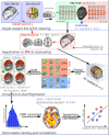
Analysis workflow. FC, functional connectivity.
Figure 2
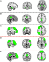
Five masks used in further dual-regression analysis. The figure shows the three most informative orthogonal slices for each mask. Green colored mask is scaled to fit and superimposed onto the 1-mm standard MNI152 standard space template image. Masks are: (A) left and right hippocampus (Montreal Neurological institute (MNI) coordinates x = 3, y = −35, z = 28), (B) posterior cingulate cortex (x = −28, y = −24, z = −14), (C) right fronto parietal network (x = 32, y = 7, z = 35), (D) left fronto parietal network (x = −47, y = −44, z = −4), (E) default mode network (x = −4, y = −53, z = −35). First two masks (A,B) are binary masks, while last three (C–E) are components produced by the group ICA decomposition of rsfMRI data and converted to Z statistic images via normalized mixture-model fit, and thresholded at arbitrary threshold Z = 3 for visualization purposes.
Figure 3
Results for the default mode network (DMN) mask (two dimensional image on top, with mask shaded in green, centered at x = −4, y = −53, z = −35). Warm colors present the cluster of decreased connectivity in carnosine/anserine subjects (multiple comparison FWE corrected, p < 0.05). Left (L) and right (R) hemispheres are presented separately.
Figure 4
Results for the posterior cingulate cortex (PCC) mask (two dimensional image on top, with mask shaded in green, centered at x = −28, y = −24, z = −14). Warm colors present the cluster of decreased connectivity in carnosine/anserine subjects (multiple comparison FWE corrected, p < 0.05). Bluish colors present the areas in which carnosine/anserine subjects have decreased connectivity as compared to placebo with additional Bonferroni correction (p < 0.005). Left (L) and right (R) hemispheres are presented separately.
Figure 5
Results for the right fronto-parietal network (RFPN) mask (two dimensional image on top, with mask shaded in green, centered at x = 32, y = 7, z = 35). Warm colors present the cluster of decreased connectivity in carnosine/anserine subjects (multiple comparison FWE corrected, p < 0.05). Bluish colors present the areas in which carnosine/anserine subjects have decreased connectivity as compared to placebo with additional Bonferroni correction (p < 0.005). Left (L) and right (R) hemispheres are presented separately.
Figure 6
Dependency between change in WMS-LM2 score and change in connectivity between DMN and Left hippocamus/WM voxel (MNI coordinates: x = −18, y = −34, z = −8; Spearman's r = 0.446, p = 0.043). Carnosine/anserine (car/ans) subjects are presented with red crosses and placebo subjects with blue circles.
Figure 7
Dependency between age and change in WMS-LM2 score (Spearman's r = 0.493, p = 0.023). Carnosine/anserine (car/ans) subjects are presented with red crosses and placebo subjects with blue circles.
Similar articles
- Influence of Imidazole-Dipeptides on Cognitive Status and Preservation in Elders: A Narrative Review.
Masuoka N, Lei C, Li H, Hisatsune T. Masuoka N, et al. Nutrients. 2021 Jan 27;13(2):397. doi: 10.3390/nu13020397. Nutrients. 2021. PMID: 33513893 Free PMC article. Review. - Effect of Anserine/Carnosine Supplementation on Verbal Episodic Memory in Elderly People.
Hisatsune T, Kaneko J, Kurashige H, Cao Y, Satsu H, Totsuka M, Katakura Y, Imabayashi E, Matsuda H. Hisatsune T, et al. J Alzheimers Dis. 2016;50(1):149-59. doi: 10.3233/JAD-150767. J Alzheimers Dis. 2016. PMID: 26682691 Free PMC article. Clinical Trial. - Anserine/Carnosine Supplementation Suppresses the Expression of the Inflammatory Chemokine CCL24 in Peripheral Blood Mononuclear Cells from Elderly People.
Katakura Y, Totsuka M, Imabayashi E, Matsuda H, Hisatsune T. Katakura Y, et al. Nutrients. 2017 Oct 31;9(11):1199. doi: 10.3390/nu9111199. Nutrients. 2017. PMID: 29088099 Free PMC article. Clinical Trial. - Anserine and carnosine supplementation in the elderly: Effects on cognitive functioning and physical capacity.
Szcześniak D, Budzeń S, Kopeć W, Rymaszewska J. Szcześniak D, et al. Arch Gerontol Geriatr. 2014 Sep-Oct;59(2):485-90. doi: 10.1016/j.archger.2014.04.008. Epub 2014 May 2. Arch Gerontol Geriatr. 2014. PMID: 24880197 - Effects of Anserine/Carnosine Supplementation on Mild Cognitive Impairment with APOE4.
Masuoka N, Yoshimine C, Hori M, Tanaka M, Asada T, Abe K, Hisatsune T. Masuoka N, et al. Nutrients. 2019 Jul 17;11(7):1626. doi: 10.3390/nu11071626. Nutrients. 2019. PMID: 31319510 Free PMC article. Clinical Trial.
Cited by
- A genome-wide approach for identification and characterisation of metabolite-inducible systems.
Hanko EKR, Paiva AC, Jonczyk M, Abbott M, Minton NP, Malys N. Hanko EKR, et al. Nat Commun. 2020 Mar 5;11(1):1213. doi: 10.1038/s41467-020-14941-6. Nat Commun. 2020. PMID: 32139676 Free PMC article. - Determination of Imidazole Dipeptides and Related Amino Acids in Natural Seafoods by Liquid Chromatography-Tandem Mass Spectrometry Using a Pre-Column Derivatization Reagent.
Onozato M, Horinouchi M, Yoshiba Y, Sakamoto T, Sugasawa H, Fukushima T. Onozato M, et al. Foods. 2024 Jun 20;13(12):1951. doi: 10.3390/foods13121951. Foods. 2024. PMID: 38928892 Free PMC article. - Enhancing cognitive function in older adults: dietary approaches and implications.
Polis B, Samson AO. Polis B, et al. Front Nutr. 2024 Jan 31;11:1286725. doi: 10.3389/fnut.2024.1286725. eCollection 2024. Front Nutr. 2024. PMID: 38356861 Free PMC article. Review. - Improving Cognition with Nutraceuticals Targeting TGF-β1 Signaling.
Grasso M, Caruso G, Godos J, Bonaccorso A, Carbone C, Castellano S, Currenti W, Grosso G, Musumeci T, Caraci F. Grasso M, et al. Antioxidants (Basel). 2021 Jul 5;10(7):1075. doi: 10.3390/antiox10071075. Antioxidants (Basel). 2021. PMID: 34356309 Free PMC article. Review. - Influence of Imidazole-Dipeptides on Cognitive Status and Preservation in Elders: A Narrative Review.
Masuoka N, Lei C, Li H, Hisatsune T. Masuoka N, et al. Nutrients. 2021 Jan 27;13(2):397. doi: 10.3390/nu13020397. Nutrients. 2021. PMID: 33513893 Free PMC article. Review.
References
- Abbas K., Shenk T. E., Poole V. N., Breedlove E. L., Leverenz L. J., Nauman E. A., et al. . (2014). Alteration of default mode network in high school football athletes due to repetitive subconcussive mild traumatic brain injury: a resting-state functional magnetic resonance imaging study. Brain Connect. 5, 91–101. 10.1089/brain.2014.0279 - DOI - PubMed
- Beck A. T., Steer R., Brown G. K. (1996). Manual for Beck Depression Inventory-II. San Antonio, TX: Psychological Corporation.
LinkOut - more resources
Full Text Sources
Other Literature Sources


