Microscopy Image Browser: A Platform for Segmentation and Analysis of Multidimensional Datasets - PubMed (original) (raw)
Microscopy Image Browser: A Platform for Segmentation and Analysis of Multidimensional Datasets
Ilya Belevich et al. PLoS Biol. 2016.
Abstract
Understanding the structure-function relationship of cells and organelles in their natural context requires multidimensional imaging. As techniques for multimodal 3-D imaging have become more accessible, effective processing, visualization, and analysis of large datasets are posing a bottleneck for the workflow. Here, we present a new software package for high-performance segmentation and image processing of multidimensional datasets that improves and facilitates the full utilization and quantitative analysis of acquired data, which is freely available from a dedicated website. The open-source environment enables modification and insertion of new plug-ins to customize the program for specific needs. We provide practical examples of program features used for processing, segmentation and analysis of light and electron microscopy datasets, and detailed tutorials to enable users to rapidly and thoroughly learn how to use the program.
Conflict of interest statement
The authors have declared that no competing interests exist.
Figures
Fig 1. MIB recognizes a large array of imaging formats and offers many essential image processing tools.
(A) Most common image formats can be imported to (left column) and exported from (right column) MIB. Most commonly used image processing tools are listed in the middle column (see MIB website for the full list). (B) Image segmentation workflow usually comprises a combination of several approaches. Datasets can be segmented iteratively using various manual and semiautomatic tools combined with quantification filtering of results in order to generate a model. Data in MIB are organized in four layers: Image (raw data), Selection (active layer for segmentation), Mask (an optional supporting layer for temporal storage of the segmentation results for evaluation and filtering), and Model (containing the final segmentation).
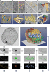
Fig 2. The selection of the best suitable tool for segmentation depends on specimen and object of interest.
Five examples are given: in (A–D), the top row of each image shows a preprocessed slice and a segmentation overlay, and the bottom row shows the final 3-D visualization. Segmentation was done with MIB, and the 3-D rendering using different freeware (A–D) and commercial (E) software packages. (A) Trypanosoma brucei was chemically fixed and imaged with electron tomography (ET). Each Golgi cisternae (four shades of blue) was manually segmented using the brush tool, while ER and ER-derived vesicles (yellow) were segmented using a combination of the brush tool and shape interpolation. The resulting 3-D model was rendered directly in MIB. (B) A Huh-7 cell was high-pressure frozen and freeze-substituted, and a portion of the cell was subjected to ET [11]. ER (yellow) was segmented using the brush tool with shape interpolation and microtubules (magenta) using the line tracker tool. The resulting model was exported in the IMOD-compatible format and rendered in IMOD [6]. (C) A Huh-7 cell transiently expressing ssHRP-KDEL was cytochemically stained (dark precipitate) and imaged with a serial block-face scanning electron microscope (SB-EM) [11]. The ER network (yellow) was segmented semiautomatically using global black-and-white thresholding and further polished using quantification filtering [11]. The model was exported in the nearly raw raster data (NRRD) format and rendered in 3D Slicer [8]. (D) Mouse cochlea was perilymphatically fixed, and the sensory epithelium of the medial part of the cochlear duct was imaged with SB-EM [13]. Different cell types of the organ of Corti (inner hairs in green, outer hair cells in yellow, external rod in vermilion, internal rod in sapphire blue, and phalangeal part of the Deiters’ cells in greyish blue) were segmented using local thresholding combined with shape interpolation and model rendered using 3D Slicer. (E) Stepwise segmentation workflow is needed to generate a 3-D model of a complex structure. A metaphase Huh-7 cell transiently expressing ssHRP-KDEL was cytochemically stained (dark precipitate in ER lumen) and imaged with SB-EM [10]. The modelling workflow for ER consists of 13 steps (S1 Table). For the visualization, the model was exported in the AmiraMesh format and rendered in Amira. Scale bars: A, B 500 nm; C, D, E 5 μm.
Fig 3. Semiautomatic image segmentation in MIB can dramatically decrease the time spent on modelling.
(A) The Random Forest classifier was used to segment ER from wide-field time-lapse LM videos of Huh-7 cells. Labels were assigned (central image) to mark ER (Hsp47-GFP marker seen in green) and background (red), which were then used to train the classifier and segment ER throughout the time-lapse video (right image). (B) The semiautomatic watershed segmentation was used for segmentation of nucleus in the sieve element of A. thaliana root imaged with SB-EM [14]. Assigning of just two labels (green for nucleus and vermilion for background) was sufficient to segment the complete nucleus in 3-D (light blue, image on the right). (C) The separation of the fused objects using the watershed segmentation. The human U251MG astrocytoma cells were loaded with oleic acid producing a large amount of lipid droplets (LDs) (left image) that tend to form clusters. LDs were segmented using marker-controlled watershed; however, because of close proximity, most of the LDs appear merged (the second image). The object separation mode of the watershed tool was used to separate individual LDs for quantitative analysis (third and fourth images). The 3-D models were rendered with 3D Slicer [8]. Scale bars: 2 μm.
Fig 4. Quantification and visualization of results are the final steps of the imaging workflow.
Generated models may be quantified to extract different parameters of the segmented objects and visualized using a number of programs. As an example, the volumes (μm3) and numbers of segmented mitochondria were calculated (the plot in red). The quantifications results can either be plotted directly in MIB or exported to MATLAB or Microsoft Excel. The manual measurements of angles, distances, caliper, and radius complement automatic quantification. The visualization of the mitochondria model (in green) is demonstrated using six alternative programs (the lower row).
Fig 5. MIB has a user-friendly graphical user interface and is freely available from the website.
(A) A screenshot of the MIB user interface. The program menu, toolbar, and panels are highlighted. A brief description of each element is provided. (B) A dedicated website includes direct links for software download and covers various topics and aspects of MIB functionality. Image credit: Ilya Belevich, on behalf of MIB.
Similar articles
- Bi-channel image registration and deep-learning segmentation (BIRDS) for efficient, versatile 3D mapping of mouse brain.
Wang X, Zeng W, Yang X, Zhang Y, Fang C, Zeng S, Han Y, Fei P. Wang X, et al. Elife. 2021 Jan 18;10:e63455. doi: 10.7554/eLife.63455. Elife. 2021. PMID: 33459255 Free PMC article. - Modular segmentation, spatial analysis and visualization of volume electron microscopy datasets.
Müller A, Schmidt D, Albrecht JP, Rieckert L, Otto M, Galicia Garcia LE, Fabig G, Solimena M, Weigert M. Müller A, et al. Nat Protoc. 2024 May;19(5):1436-1466. doi: 10.1038/s41596-024-00957-5. Epub 2024 Feb 29. Nat Protoc. 2024. PMID: 38424188 Review. - LOBSTER: an environment to design bioimage analysis workflows for large and complex fluorescence microscopy data.
Tosi S, Bardia L, Filgueira MJ, Calon A, Colombelli J. Tosi S, et al. Bioinformatics. 2020 Apr 15;36(8):2634-2635. doi: 10.1093/bioinformatics/btz945. Bioinformatics. 2020. PMID: 31860062 - The ImageJ ecosystem: Open-source software for image visualization, processing, and analysis.
Schroeder AB, Dobson ETA, Rueden CT, Tomancak P, Jug F, Eliceiri KW. Schroeder AB, et al. Protein Sci. 2021 Jan;30(1):234-249. doi: 10.1002/pro.3993. Epub 2020 Nov 20. Protein Sci. 2021. PMID: 33166005 Free PMC article. - The ImageJ ecosystem: An open platform for biomedical image analysis.
Schindelin J, Rueden CT, Hiner MC, Eliceiri KW. Schindelin J, et al. Mol Reprod Dev. 2015 Jul-Aug;82(7-8):518-29. doi: 10.1002/mrd.22489. Epub 2015 Jul 7. Mol Reprod Dev. 2015. PMID: 26153368 Free PMC article. Review.
Cited by
- Presynaptic Mitochondria Volume and Abundance Increase during Development of a High-Fidelity Synapse.
Thomas CI, Keine C, Okayama S, Satterfield R, Musgrove M, Guerrero-Given D, Kamasawa N, Young SM Jr. Thomas CI, et al. J Neurosci. 2019 Oct 9;39(41):7994-8012. doi: 10.1523/JNEUROSCI.0363-19.2019. Epub 2019 Aug 27. J Neurosci. 2019. PMID: 31455662 Free PMC article. - Weighted average ensemble-based semantic segmentation in biological electron microscopy images.
Shaga Devan K, Kestler HA, Read C, Walther P. Shaga Devan K, et al. Histochem Cell Biol. 2022 Nov;158(5):447-462. doi: 10.1007/s00418-022-02148-3. Epub 2022 Aug 20. Histochem Cell Biol. 2022. PMID: 35988009 Free PMC article. - Morphology of Mitochondria in Syncytial Annelid Female Germ-Line Cyst Visualized by Serial Block-Face SEM.
Urbisz AZ, Student S, Śliwińska MA, Małota K. Urbisz AZ, et al. Int J Cell Biol. 2020 Jan 7;2020:7483467. doi: 10.1155/2020/7483467. eCollection 2020. Int J Cell Biol. 2020. PMID: 32395131 Free PMC article. - Automated 3D Axonal Morphometry of White Matter.
Abdollahzadeh A, Belevich I, Jokitalo E, Tohka J, Sierra A. Abdollahzadeh A, et al. Sci Rep. 2019 Apr 15;9(1):6084. doi: 10.1038/s41598-019-42648-2. Sci Rep. 2019. PMID: 30988411 Free PMC article. - Progression of herpesvirus infection remodels mitochondrial organization and metabolism.
Leclerc S, Gupta A, Ruokolainen V, Chen JH, Kunnas K, Ekman AA, Niskanen H, Belevich I, Vihinen H, Turkki P, Perez-Berna AJ, Kapishnikov S, Mäntylä E, Harkiolaki M, Dufour E, Hytönen V, Pereiro E, McEnroe T, Fahy K, Kaikkonen MU, Jokitalo E, Larabell CA, Weinhardt V, Mattola S, Aho V, Vihinen-Ranta M. Leclerc S, et al. PLoS Pathog. 2024 Apr 15;20(4):e1011829. doi: 10.1371/journal.ppat.1011829. eCollection 2024 Apr. PLoS Pathog. 2024. PMID: 38620036 Free PMC article.
References
Publication types
MeSH terms
LinkOut - more resources
Full Text Sources
Other Literature Sources
Molecular Biology Databases
Research Materials


