Structural basis for the endoribonuclease activity of the type III-A CRISPR-associated protein Csm6 - PubMed (original) (raw)
Structural basis for the endoribonuclease activity of the type III-A CRISPR-associated protein Csm6
Ole Niewoehner et al. RNA. 2016 Mar.
Abstract
Prokaryotic CRISPR-Cas systems provide an RNA-guided mechanism for genome defense against mobile genetic elements such as viruses and plasmids. In type III-A CRISPR-Cas systems, the RNA-guided multisubunit Csm effector complex targets both single-stranded RNAs and double-stranded DNAs. In addition to the Csm complex, efficient anti-plasmid immunity mediated by type III-A systems also requires the CRISPR-associated protein Csm6. Here we report the crystal structure of Csm6 from Thermus thermophilus and show that the protein is a ssRNA-specific endoribonuclease. The structure reveals a dimeric architecture generated by interactions involving the N-terminal CARF and C-terminal HEPN domains. HEPN domain dimerization leads to the formation of a composite ribonuclease active site. Consistently, mutations of invariant active site residues impair catalytic activity in vitro. We further show that the ribonuclease activity of Csm6 is conserved across orthologs, suggesting that it plays an important functional role in CRISPR-Cas systems. The dimer interface of the CARF domains features a conserved electropositive pocket that may function as a ligand-binding site for allosteric control of ribonuclease activity. Altogether, our work suggests that Csm6 proteins provide an auxiliary RNA-targeting interference mechanism in type III-A CRISPR-Cas systems that operates in conjunction with the RNA- and DNA-targeting endonuclease activities of the Csm effector complex.
Keywords: CRISPR; Cas protein; Csm6; HEPN domain; crRNA; ribonuclease.
© 2016 Niewoehner and Jinek; Published by Cold Spring Harbor Laboratory Press for the RNA Society.
Figures
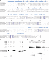
FIGURE 1.
Thermus thermophilus Csm6 (TtCsm6) is a single-strand-specific endoribonuclease. (A) Multiple sequence alignment of Csm6 and Csx1 proteins from T. thermophilus (TtCsm6, GI:55978335), S. epidermidis (SeCsm6, GI:488416649), S. mutans (SmCsm6, GI:24379650), S. thermophilus (StCsm6, GI:585230687), and P. furiosus Csx1 (PfCsx1, GI:33359545), performed using ClustalOmega (McWilliam et al. 2013). HEPN domain active site residues are marked with asterisks. Secondary structure elements of TtCsm6 are indicated above the sequences. Disordered amino acid residues are indicated with dashed lines. (B) Nuclease activity assays performed with single- (ss) and double-stranded (ds) RNA and DNA oligonucleotide substrates. The 24-nt substrates have identical sequences and carry a Cy5 fluorophore group covalently attached to the 3′-terminus. Double-stranded substrates were prepared by annealing an unlabeled complementary strand to the respective single-stranded substrate. TtCsm6 (400 nM final concentration) was incubated with substrates (200 nM) at 37°C for 1 h. Cleavage products were analyzed by electrophoresis on a 15% denaturing polyacrylamide gel and detected using a fluorescence gel scanner. (C) Nuclease activity assays performed using TtCsm6 and identical ssRNA oligonucleotide substrates (24 nt) labeled with Cy5 at the 5′ or 3′ ends. The assay was performed as in B. A control digest with RNase T1 was used to generate RNA fragments of defined size, as indicated.
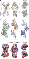
FIGURE 2.
TtCsm6 is a helical homodimer containing N-terminal CARF and C-terminal HEPN domains. (A) Overall architecture of the TtCsm6 dimer shown in four orientations. (B) Comparison of the three-dimensional structures of COG1517 family proteins: P. furiosus Csx1 (PfCsx1, PDB ID 4EOG), S. mutans Csm6 (SmCsm6, PDB ID 4RGP), and S. solfataricus Csa3 (SsCsa3, PDB ID 2WTE). Structural superpositions were performed using DALI (Hasegawa and Holm 2009). (C) Surface representation of the TtCsm6 dimer colored according to electrostatic surface potential. The molecule is displayed in the same orientations as in A.
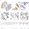
FIGURE 3.
The C-terminal HEPN domains of TtCsm6 form a composite endoribonuclease active site. (A) Overall view of the HEPN domain interface in the TtCsm6 dimer. The inset shows a close-up view of the HEPN domain active site. Conserved active site residues are shown in stick format. The Ni2+ ion present in the TtCsm6 crystal structure is shown as a green sphere. (B) Comparison of the HEPN ribonuclease active sites of yeast Ire1 (left), TtCsm6 (middle), and SmCsm6 (right). Active site residues are shown in stick format. The structures were aligned by least-squares superposition of the active site Asn, Arg, and His residues and are shown in identical orientations. (C) Nuclease activity assays of active site mutants of TtCsm6. The assays were performed using a 24-nt ssRNA substrate labeled with Cy5 at the 3′-end. Reactions were resolved on a 16% denaturing polyacrylamide gel and analyzed on a fluorescence gel scanner. For conciseness, the panel depicts a cropped region of the denaturing gel; a full-size image of the gel is shown in Supplemental Figure 7. (D) Ribonuclease activities of Csm6 orthologs. Recombinant T. thermophilus, S. epidermidis (SeCsm6), and P. horikoshii (PhCsm6) Csm6 proteins were incubated with a 24-nt Cy5-labeled ssRNA substrate at 37°C. Reactions were sampled at indicated time points and analyzed as for Figure 1C. For conciseness, the panel depicts a cropped region of the denaturing gel; a full-size image of the gel is shown in Supplemental Figure 8.
Similar articles
- The ribonuclease activity of Csm6 is required for anti-plasmid immunity by Type III-A CRISPR-Cas systems.
Foster K, Kalter J, Woodside W, Terns RM, Terns MP. Foster K, et al. RNA Biol. 2019 Apr;16(4):449-460. doi: 10.1080/15476286.2018.1493334. Epub 2018 Aug 1. RNA Biol. 2019. PMID: 29995577 Free PMC article. - Conservation and variability in the structure and function of the Cas5d endoribonuclease in the CRISPR-mediated microbial immune system.
Koo Y, Ka D, Kim EJ, Suh N, Bae E. Koo Y, et al. J Mol Biol. 2013 Oct 23;425(20):3799-810. doi: 10.1016/j.jmb.2013.02.032. Epub 2013 Mar 7. J Mol Biol. 2013. PMID: 23500492 - Activation and self-inactivation mechanisms of the cyclic oligoadenylate-dependent CRISPR ribonuclease Csm6.
Garcia-Doval C, Schwede F, Berk C, Rostøl JT, Niewoehner O, Tejero O, Hall J, Marraffini LA, Jinek M. Garcia-Doval C, et al. Nat Commun. 2020 Mar 27;11(1):1596. doi: 10.1038/s41467-020-15334-5. Nat Commun. 2020. PMID: 32221291 Free PMC article. - Cutting it close: CRISPR-associated endoribonuclease structure and function.
Hochstrasser ML, Doudna JA. Hochstrasser ML, et al. Trends Biochem Sci. 2015 Jan;40(1):58-66. doi: 10.1016/j.tibs.2014.10.007. Epub 2014 Nov 18. Trends Biochem Sci. 2015. PMID: 25468820 Review. - The Cmr complex: an RNA-guided endoribonuclease.
Bailey S. Bailey S. Biochem Soc Trans. 2013 Dec;41(6):1464-7. doi: 10.1042/BST20130216. Biochem Soc Trans. 2013. PMID: 24256238 Review.
Cited by
- Structural insight into the Csx1-Crn2 fusion self-limiting ribonuclease of type III CRISPR system.
Zhang D, Du L, Gao H, Yuan C, Lin Z. Zhang D, et al. Nucleic Acids Res. 2024 Aug 12;52(14):8419-8430. doi: 10.1093/nar/gkae569. Nucleic Acids Res. 2024. PMID: 38967023 Free PMC article. - Two HEPN domains dictate CRISPR RNA maturation and target cleavage in Cas13d.
Zhang B, Ye Y, Ye W, Perčulija V, Jiang H, Chen Y, Li Y, Chen J, Lin J, Wang S, Chen Q, Han YS, Ouyang S. Zhang B, et al. Nat Commun. 2019 Jun 11;10(1):2544. doi: 10.1038/s41467-019-10507-3. Nat Commun. 2019. PMID: 31186424 Free PMC article. - CRISPR-cas systems based molecular diagnostic tool for infectious diseases and emerging 2019 novel coronavirus (COVID-19) pneumonia.
Xiang X, Qian K, Zhang Z, Lin F, Xie Y, Liu Y, Yang Z. Xiang X, et al. J Drug Target. 2020 Aug-Sep;28(7-8):727-731. doi: 10.1080/1061186X.2020.1769637. Epub 2020 May 26. J Drug Target. 2020. PMID: 32401064 Free PMC article. Review. - Bioinformatic analysis of type III CRISPR systems reveals key properties and new effector families.
Hoikkala V, Graham S, White MF. Hoikkala V, et al. Nucleic Acids Res. 2024 Jul 8;52(12):7129-7141. doi: 10.1093/nar/gkae462. Nucleic Acids Res. 2024. PMID: 38808661 Free PMC article. - Controlling and enhancing CRISPR systems.
Shivram H, Cress BF, Knott GJ, Doudna JA. Shivram H, et al. Nat Chem Biol. 2021 Jan;17(1):10-19. doi: 10.1038/s41589-020-00700-7. Epub 2020 Dec 16. Nat Chem Biol. 2021. PMID: 33328654 Free PMC article. Review.
References
- Barrangou R, Fremaux C, Deveau H, Richards M, Boyaval P, Moineau S, Romero DA, Horvath P. 2007. CRISPR provides acquired resistance against viruses in prokaryotes. Science 315: 1709–1712. - PubMed
Publication types
MeSH terms
Substances
LinkOut - more resources
Full Text Sources
Other Literature Sources
Research Materials


