Ultrasonography of the Kidney: A Pictorial Review - PubMed (original) (raw)
Review
Ultrasonography of the Kidney: A Pictorial Review
Kristoffer Lindskov Hansen et al. Diagnostics (Basel). 2015.
Abstract
Ultrasonography of the kidneys is essential in the diagnosis and management of kidney-related diseases. The kidneys are easily examined, and most pathological changes in the kidneys are distinguishable with ultrasound. In this pictorial review, the most common findings in renal ultrasound are highlighted.
Keywords: pictorial review; practical guide; renal ultrasound.
Figures
Figure 1
Normal adult kidney. Measurement of kidney length on the US image is illustrated by ‘+’ and a dashed line. * Column of Bertin; ** pyramid; *** cortex; **** sinus.
Figure 2
Normal pediatric kidney. * Column of Bertin; ** pyramid; *** cortex; **** sinus.
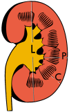
Figure 3
Measures of the kidney. L = length. P = parenchymal thickness. C = cortical thickness.
Figure 4
Doppler ultrasound (US) of a normal adult kidney with the estimation of the systolic velocity (Vs), the diastolic velocity (Vd), acceleration time (AoAT), systolic acceleration (Ao Accel) and resistive index (RI). Red and blue colors in the color box represent flow towards and away from the transducer, respectively. The specrogram below the B-mode image shows flow velocity (m/s) against time (s) obtained within the range gate. The small flash icons on the spectrogram represent initiation of the flow measurement.
Figure 5
Simple cyst with posterior enhancement in an adult kidney. Measurement of kidney length on the US image is illustrated by ‘+’ and a dashed line.
Figure 6
Complex cyst with thickened walls and membranes in the lower pole of an adult kidney. Measurements of kidney length and the complex cyst on the US image are illustrated by ‘+’ and dashed lines.
Figure 7
Advanced polycystic kidney disease with multiple cysts.
Figure 8
Cortical solid mass, which later was shown to be renal cell carcinoma. Measurement of the solid mass on the US image is illustrated by ‘+’ and a dashed line.
Figure 9
Renal cell carcinoma with both cystic and solid components located in the cortex. Measurement of tumor on the US image is illustrated by ‘+’ and a dashed line.
Figure 10
Solid tumor in the renal sinus seen as a hypoechoic mass, later found to be lymphoma. The ‘1’ and ‘2’ on the US image are reference points used for CT fusion (not shown).
Figure 11
Angiomyolipoma seen as a hyperechoic mass in the upper pole of an adult kidney.
Figure 12
Patient with tuberous sclerosis and multiple angiomyolipomas in the kidney. Measurement of kidney length on the US image is illustrated by ‘+’ and a dashed line.
Figure 13
Hydronephrosis due to ureteropelvic junction obstruction in a pediatric patient.
Figure 14
Bilateral dilatation of the ureters due to vesicoureteric reflux in a pediatric patient.
Figure 15
End-stage hydronephrosis with cortical thinning. Measurement of pelvic dilatation on the US image is illustrated by ‘+’ and a dashed line.
Figure 16
Hydronephrosis with dilated anechoic pelvis and calyces, along with cortical atrophy. The width of a calyx is measured on the US image in the longitudinal scan plane, and illustrated by ‘+’ and a dashed line.
Figure 17
Same patient as in Figure 16 with measurement of the pelvis dilation in the transverse scan plane illustrated on the US image with ‘+’ and a dashed line.
Figure 18
Renal stone located at the pyeloureteric junction with accompanying hydronephrosis.
Figure 19
Centrally-located stone with posterior shadowing. No hydronephrosis is present. Measurement of kidney length on the US image is illustrated by ‘+’ and a dashed line.
Figure 20
Staghorn calculi filling the entire collecting system and creating pronounced shadowing.
Figure 21
Left hydroureter with ureteric jet. No stone is visible. The red color in the color box represents motion towards the transducer as defined by the color bar.
Figure 22
Chronic renal disease caused by glomerulonephritis with increased echogenicity and reduced cortical thickness. Measurement of kidney length on the US image is illustrated by ‘+’ and a dashed line.
Figure 23
Nephrotic syndrome. Hyperechoic kidney without demarcation of cortex and medulla.
Figure 24
Chronic pyelonephritis with reduced kidney size and focal cortical thinning. Measurement of kidney length on the US image is illustrated by ‘+’ and a dashed line.
Figure 25
End-stage chronic kidney disease with increased echogenicity, homogenous architecture without visible differentiation between parenchyma and renal sinus and reduced kidney size. Measurement of kidney length on the US image is illustrated by ‘+’ and a dashed line.
Figure 26
Acute pyelonephritis with increased cortical echogenicity and blurred delineation of the upper pole.
Figure 27
Postoperative renal failure with increased cortical echogenicity and kidney size. Biopsy showed acute tubular necrosis.
Figure 28
Renal trauma with laceration of the lower pole and subcapsular fluid collection below the kidney.
Figure 29
(A) Percutaneous nephrostomy tube placed through a calyx in the lower pole of a kidney with hydronephrosis. (B) The pigtail catheter is placed in the dilated calyx. The tube in (A) and the pigtail in (B) are marked with white arrows.
Figure 30
Renal cell carcinoma successfully treated with thermal ablation, as no contrast enhancement is seen.
Figure 31
Unspecific cortical lesion on CT is confirmed cystic and benign with contrast-enhanced ultrasound (CEUS) using image fusion.
Figure 32
Strain elastography of a normal kidney. Red depicts soft areas, and blue depicts hard areas relative to the entire elastography image. Note that the medulla is softer than the cortex. A color bar is shown to the left of the image, where ‘S’ and ‘H’ denote soft and hard tissue, respectively.
Similar articles
- Pictorial review: Renal ultrasound.
Gulati M, Cheng J, Loo JT, Skalski M, Malhi H, Duddalwar V. Gulati M, et al. Clin Imaging. 2018 Sep-Oct;51:133-154. doi: 10.1016/j.clinimag.2018.02.012. Epub 2018 Feb 16. Clin Imaging. 2018. PMID: 29477809 Review. - Contrast enhanced ultrasound of kidneys. Pictorial essay.
Prakash A, Tan GJ, Wansaicheong GK. Prakash A, et al. Med Ultrason. 2011 Jun;13(2):150-6. Med Ultrason. 2011. PMID: 21655542 Review. - Pediatric cystic diseases of the kidney.
Ferro F, Vezzali N, Comploj E, Pedron E, Di Serafino M, Esposito F, Pelliccia P, Rossi E, Zeccolini M, Vallone G. Ferro F, et al. J Ultrasound. 2019 Sep;22(3):381-393. doi: 10.1007/s40477-018-0347-9. Epub 2019 Jan 1. J Ultrasound. 2019. PMID: 30600488 Free PMC article. - Thoracic wall ultrasonography - normal and pathological findings. Pictorial essay.
Chira R, Chira A, Mircea PA. Chira R, et al. Med Ultrason. 2011 Sep;13(3):228-33. Med Ultrason. 2011. PMID: 21894294 - Common pitfalls in renal mass evaluation: a practical guide.
Leão LRS, Mussi TC, Yamauchi FI, Baroni RH. Leão LRS, et al. Radiol Bras. 2019 Jul-Aug;52(4):254-261. doi: 10.1590/0100-3984.2018.0007. Radiol Bras. 2019. PMID: 31435088 Free PMC article.
Cited by
- Ultrasonographic measurement of the renal resistive index in the cynomolgus monkey (Macaca fascicularis) under conscious and ketamine-immobilized conditions.
Konno H, Ishizaka T, Chiba K, Mori K. Konno H, et al. Exp Anim. 2020 Jan 29;69(1):119-126. doi: 10.1538/expanim.19-0084. Epub 2019 Oct 22. Exp Anim. 2020. PMID: 31645524 Free PMC article. - Gender and side distribution of urinary calculi using ultrasound imaging.
Alshoabi SA, Alhamodi DS, Gameraddin MB, Babiker MS, Omer AM, Al-Dubai SA. Alshoabi SA, et al. J Family Med Prim Care. 2020 Mar 26;9(3):1614-1616. doi: 10.4103/jfmpc.jfmpc_1153_19. eCollection 2020 Mar. J Family Med Prim Care. 2020. PMID: 32509660 Free PMC article. - Molecular Gold Nanoclusters for Advanced NIR-II Bioimaging and Therapy.
Baghdasaryan A, Dai H. Baghdasaryan A, et al. Chem Rev. 2025 Jun 11;125(11):5195-5227. doi: 10.1021/acs.chemrev.4c00835. Epub 2025 May 28. Chem Rev. 2025. PMID: 40435324 Free PMC article. Review. - Classification of Chronic Kidney Disease in Sonography Using the GLCM and Artificial Neural Network.
Kim DH, Ye SY. Kim DH, et al. Diagnostics (Basel). 2021 May 11;11(5):864. doi: 10.3390/diagnostics11050864. Diagnostics (Basel). 2021. PMID: 34064910 Free PMC article. - Evaluation of Tryptic Podocin Peptide in Urine Sediment Using LC-MS-MRM Method as a Potential Biomarker of Glomerular Injury in Dogs with Clinical Signs of Renal and Cardiac Disorders.
Szczepankiewicz B, Bąchor R, Pasławski R, Siwińska N, Pasławska U, Konieczny A, Szewczuk Z. Szczepankiewicz B, et al. Molecules. 2019 Aug 26;24(17):3088. doi: 10.3390/molecules24173088. Molecules. 2019. PMID: 31454880 Free PMC article.
References
- Wilson D.A. Ultrasonic scanning of the kidneys. Ann. Clin. Lab. Sci. 1981;11:367–376. - PubMed
- Rumack C.M., Wilson S.R., Charboneau J.W. Diagnostic Ultrasound. 3rd ed. Elsevier Health Sciences; St. Louis, MO, USA: 2005.
- Pedersen M.H., Nielsen M.B., Skjoldbye B. Basics of Clinical Ultrasound. UltraPocketBooks; Copenhagen, Denmark: 2006.
Publication types
LinkOut - more resources
Full Text Sources
Other Literature Sources


