Parallel Transformation of Tactile Signals in Central Circuits of Drosophila - PubMed (original) (raw)
Parallel Transformation of Tactile Signals in Central Circuits of Drosophila
John C Tuthill et al. Cell. 2016.
Abstract
To distinguish between complex somatosensory stimuli, central circuits must combine signals from multiple peripheral mechanoreceptor types, as well as mechanoreceptors at different sites in the body. Here, we investigate the first stages of somatosensory integration in Drosophila using in vivo recordings from genetically labeled central neurons in combination with mechanical and optogenetic stimulation of specific mechanoreceptor types. We identify three classes of central neurons that process touch: one compares touch signals on different parts of the same limb, one compares touch signals on right and left limbs, and the third compares touch and proprioceptive signals. Each class encodes distinct features of somatosensory stimuli. The axon of an individual touch receptor neuron can diverge to synapse onto all three classes, meaning that these computations occur in parallel, not hierarchically. Representing a stimulus as a set of parallel comparisons is a fast and efficient way to deliver somatosensory signals to motor circuits.
Copyright © 2016 Elsevier Inc. All rights reserved.
Figures
Figure 1. Genetic tools for targeting mechanoreceptor neurons of the Drosophila leg
(A) Projection of a confocal stack through the prothoracic leg. GFP (green) is expressed in sensory neurons (under the control of ChAT-Gal4). Magenta shows cuticle autofluorescence. (B) Schematic diagrams of each mechanoreceptor type. Associated mechanoreceptor neurons are in green. (C) Projections of confocal stacks showing sensory neurons within each mechanoreceptor type. GFP (green) is expressed under the control of LexA. Magenta shows cuticle autofluorescence. Scale bar in each image is 10 Im. See also, S1 and S2. See Supplemental Experimental Procedures for all genotypes.
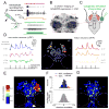
Figure 2. Propagation of touch signals in the fly ventral nerve cord (VNC)
(A) Left: Schematic of bristle recording configuration. Right: Responses of a bristle neuron to mechanical (red) and optogenetic (green) stimuli. Signals are band-pass filtered to facilitate spike identification (see Supplemental Experimental Procedures). All bristle neuron recordings are made from the same bristle on the prothoracic leg, near the femur-tibia joint (Figure S5A). (B) Projection of a coronal stack through a region (~180 μm x 180 μm) of the prothoracic VNC showing resting GCaMP6f fluorescence. GCaMP6f is expressed pan-neuronally and imaged with a two-photon microscope. Outlined in white dashed lines are the neuropil regions (neuromeres); these regions do not contain neuronal cell bodies. (C) Schematic of optogenetic bristle stimulation for calcium imaging experiments. The fly is positioned ventral side up. Light is directed at the femur/tibia joint of a prothoracic leg. The imaged region of the VNC is outlined in red. (D) GCaMP signals recorded during periodic optogenetic stimulation of leg bristles. The left and right panels show color-coded ΔF/F responses of example neurons from a single imaging plane, illustrated in the center panel. Cross-correlation values, computed between each neuron’s ΔF/F signal and the stimulus waveform, are indicated alongside each trace. (E) Map of all 699 neurons in the prothoracic region of a typical VNC, with individual neurons color-coded by their correlation value. In this projection, neurons with higher correlation are displayed on top. Neurons with correlation values below the threshold for statistical significance (0.19) are blue (n.s.). (F) Top: distribution of correlation values between calcium signals (ΔF/F) and the stimulus waveform for all 699 neurons. Bottom: correlation values after shuffling the stimulus waveform; the 95th percentile of this distribution was taken as the threshold for significance. (G) Correlation map of VNC neurons (same as panel E) but excluding motor neurons. The arrow points to the cluster of neurons that we identified for further scrutiny.
Figure 3. Three classes of VNC neurons that receive direct synaptic input from the same femur bristle
(A) Left: morphology of a biocytin-filled neuron which expressed GFP in the indicated genotype. Red arrows indicate cell body position. Right: maximum intensity projection of GFP expression within the prothoracic neuromere of the VNC (black), co-labeled with the axonal arbor of a single femur bristle neuron filled with DiI (red); we always targeted the same femur bristle (Figure S5A). All three central neuron classes overlap with this bristle neuron axon. Scale bars, 10 Im. See also Figures S3–S5. (B) Each row shows a typical in vivo whole-cell current-clamp recording from a central neuron and the simultaneously-recorded signal from a bristle neuron. As before, we targeted the same bristle on the femur, near the femur-tibia joint (Figure S5A). (C) Single spikes in this bristle neuron reliably trigger excitatory postsynaptic potentials (EPSPs) in each class of central neuron. As before, we targeted the same bristle on the posterior femur, near the femur-tibia joint (Figure S5A). Examples of bristle neuron spikes are shown at bottom. The left column shows representative single-trial EPSPs recorded from each corresponding central neuron class. At right are spike-triggered-averages of the postsynaptic voltage, averaged across all paired recordings from the same central neuron class where a connection was detected.
Figure 4. Intersegmental neurons compare touch and proprioceptive inputs
(A) Top: membrane potential responses of intersegmental neurons to optogenetic stimulation of bristle neurons (individual cells in gray, average in green, n = 9). Bottom: responses of a subset of the same neurons to mechanical stimulation of small numbers of bristles (individual cells in gray, average in black, n = 6). (B) Average spike rates and peak voltage changes for the cells shown in (A), mean ± SEM across cells, plotted versus stimulus location. Optogenetic responses are in green, mechanical responses in black. (C) Top: bristle neurons on the distal tibia are stimulated mechanically, and the femoral chordotonal organ is stimulated optogenetically. Bottom: proposed circuit diagram for sensory inputs converging onto intersegmental neurons, with proprioceptive inhibition routed via an interposed inhibitory interneuron. Chordotonal neurons also drive excitation (directly or indirectly), but this is normally masked by inhibition. (D) Inhibitory input driven by chordotonal neurons suppresses excitatory input from leg bristle neurons. The top and middle rows show responses of a typical intersegmental neuron to stimulation of bristle neurons or chordotonal neurons alone. In the bottom row, the two stimuli are delivered together. The optogenetic stimulus precedes the mechanical stimulus, and is more prolonged, in order to increase the effect of inhibition. Antagonists of synaptic inhibition (100 μM picrotoxin and 50 μM CGP54626) block the suppressive effect of chordotonal neuron stimulation, revealing underlying excitation; similar effects were seen in a total of 6 experiments (data not shown). The postsynaptic neuron is not spiking in the presence of antagonists because the neuron has been depolarized to the point where it cannot initiate spikes. (E) Average spike rates and peak voltage changes, ± SEM across cells, for all experiments like that shown in the left column of (D). Inhibition driven by chordotonal neurons significantly suppressed responses to bristle neuron stimulation (n = 9 cells, **, P = 0.03 for depolarization and P=0.03 for spikes, Wilcoxon signed rank test).
Figure 5. Midline local neurons compare proximal and distal touch within a leg
(A) Responses of midline local neurons, as in Figure 4 (n = 10 for optogenetic stimuli, n = 9 for mechanical stimuli). (B) Average spike rates and peak voltage changes for the cells shown in (A), mean ± SEM across cells. (C) Top: to elicit combined distal and proximal touch inputs, bristle neurons on the distal tibia are stimulated mechanically, and bristle neurons on the femur are stimulated optogenetically. Bottom: proposed circuit diagram for sensory inputs converging onto local neurons, with proximal inhibition routed via an interposed inhibitory interneuron. (D) Inhibitory input driven by proximal femur bristle neurons suppresses excitatory input from distal tibia bristle neurons. Top and middle: responses of a midline local neuron to stimulation of femur or tibia bristle neurons alone. Bottom: the two stimuli are delivered together. Picrotoxin (10 μM) blocks the suppressive effects of femur bristle stimulation, revealing underlying excitation; similar effects were seen in a total of 4 experiments. (E) Average spike rates and peak voltage changes, ± SEM across cells, for all experiments like those shown in the left column of (D). Inhibition from femur bristles significantly suppressed excitatory signals from tibia bristles (n = 7 cells, **, P = 0.02 for depolarization and P=0.03 for spikes, Wilcoxon signed rank test).
Figure 6. Midline projection neurons compare touch stimuli across the body midline
(A) Trial-averaged responses of 10 midline projection neurons to optogenetic stimulation of bristle neurons. For each neuron, responses were measured for both the ipsilateral (blue) and contralateral (red) prothoracic legs. Ipsilateral is defined as the side containing the reconstructed neuron’s primary arborization (schematic at left). The right column shows trial-averaged spike rates versus stimulus location. (B) Left: schematic of the recording configuration. Bristles on the contralateral and ipsilateral tibia are stimulated optogenetically. Right: proposed circuit diagram for those midline projection neurons that combine ipsilateral excitation with contralateral inhibition. (C) Integration of touch signals across legs. Top and middle: responses of local neurons to independent stimulation of ipsilateral and contralateral bristles. Bottom: the two stimuli are delivered together. Picrotoxin (10 μM) blocks inhibition from contralateral bristles, revealing excitation (Figure S7B). Data for all experiments of this type are shown in Figure S7.
Figure 7. Parallel coding of complex stimuli in simultaneously-recorded central neuron pairs
(A) Paired whole-cell recording from an intersegmental neuron and a midline local neuron. Example traces show the simultaneous responses of the two neurons to mechanical stimulation of a femur bristle. These responses confirm that this particular pair of neurons share input from some of the same bristles (Figure 3). (B) During this epoch of the experiment, the fly makes large movements which cause both neurons to depolarize and fire correlated bursts of spikes (e.g., at the arrow). Fly movement is quantified in arbitrary units (see Supplemental Experimental Procedures). Movement bouts are shaded in gray. Spike times are represented with rasters above the raw voltage traces. (C) During a later epoch of the same experiment, the midline local neuron stops being excited during movement bouts, and is instead inhibited by movement (e.g., at the arrow). This change corresponds to a switch between large movements of multiple legs, to smaller movements of the prothoracic leg. (D) Same as (A) but for a simultaneously-recorded intersegmental and a midline projection neuron. (E) The same pair of neurons as in (D), but now responding to spontaneous movement. Small periodic twitching movements of the fly’s leg (gray shading) evoke reliable responses in the midline projection neuron, but not in the intersegmental neuron. The periodic responses of the intersegmental neuron are interrupted by barrages of inhibitory postsynaptic potentials (e.g., at the arrow). Note the expanded vertical scale of the movement measurements. (F) During a later epoch of the same experiment, experiment, the fly spontaneously switches from twitching to making larger movements of the entire leg. The midline projection neuron is depolarized during large movement bouts, while the intersegmental neuron responds with sequences of inhibition and excitation at movement onset (e.g., at the arrow). The inset in the movement trace (bottom) shows periodic movement on a 10-fold expanded vertical scale.
Similar articles
- Mechanosensory circuits coordinate two opposing motor actions in Drosophila feeding.
Zhou Y, Cao LH, Sui XW, Guo XQ, Luo DG. Zhou Y, et al. Sci Adv. 2019 May 22;5(5):eaaw5141. doi: 10.1126/sciadv.aaw5141. eCollection 2019 May. Sci Adv. 2019. PMID: 31131327 Free PMC article. - Scanned optogenetic control of mammalian somatosensory input to map input-specific behavioral outputs.
Schorscher-Petcu A, Takács F, Browne LE. Schorscher-Petcu A, et al. Elife. 2021 Jul 29;10:e62026. doi: 10.7554/eLife.62026. Elife. 2021. PMID: 34323214 Free PMC article. - Cortical responses to touch reflect subcortical integration of LTMR signals.
Emanuel AJ, Lehnert BP, Panzeri S, Harvey CD, Ginty DD. Emanuel AJ, et al. Nature. 2021 Dec;600(7890):680-685. doi: 10.1038/s41586-021-04094-x. Epub 2021 Nov 17. Nature. 2021. PMID: 34789880 Free PMC article. - Neural Basis of Touch and Proprioception in Primate Cortex.
Delhaye BP, Long KH, Bensmaia SJ. Delhaye BP, et al. Compr Physiol. 2018 Sep 14;8(4):1575-1602. doi: 10.1002/cphy.c170033. Compr Physiol. 2018. PMID: 30215864 Free PMC article. Review. - The biology of skin wetness perception and its implications in manual function and for reproducing complex somatosensory signals in neuroprosthetics.
Filingeri D, Ackerley R. Filingeri D, et al. J Neurophysiol. 2017 Apr 1;117(4):1761-1775. doi: 10.1152/jn.00883.2016. Epub 2017 Jan 25. J Neurophysiol. 2017. PMID: 28123008 Free PMC article. Review.
Cited by
- Mechanosensation and Adaptive Motor Control in Insects.
Tuthill JC, Wilson RI. Tuthill JC, et al. Curr Biol. 2016 Oct 24;26(20):R1022-R1038. doi: 10.1016/j.cub.2016.06.070. Curr Biol. 2016. PMID: 27780045 Free PMC article. Review. - Mechanoreceptor synapses in the brainstem shape the central representation of touch.
Lehnert BP, Santiago C, Huey EL, Emanuel AJ, Renauld S, Africawala N, Alkislar I, Zheng Y, Bai L, Koutsioumpa C, Hong JT, Magee AR, Harvey CD, Ginty DD. Lehnert BP, et al. Cell. 2021 Oct 28;184(22):5608-5621.e18. doi: 10.1016/j.cell.2021.09.023. Epub 2021 Oct 11. Cell. 2021. PMID: 34637701 Free PMC article. - Conserved neural circuit structure across Drosophila larval development revealed by comparative connectomics.
Gerhard S, Andrade I, Fetter RD, Cardona A, Schneider-Mizell CM. Gerhard S, et al. Elife. 2017 Oct 23;6:e29089. doi: 10.7554/eLife.29089. Elife. 2017. PMID: 29058674 Free PMC article. - Dopamine promotes head direction plasticity during orienting movements.
Fisher YE, Marquis M, D'Alessandro I, Wilson RI. Fisher YE, et al. Nature. 2022 Dec;612(7939):316-322. doi: 10.1038/s41586-022-05485-4. Epub 2022 Nov 30. Nature. 2022. PMID: 36450986 Free PMC article. - Somatotopic organization among parallel sensory pathways that promote a grooming sequence in Drosophila.
Eichler K, Hampel S, Alejandro-García A, Calle-Schuler SA, Santana-Cruz A, Kmecova L, Blagburn JM, Hoopfer ED, Seeds AM. Eichler K, et al. bioRxiv [Preprint]. 2023 Dec 15:2023.02.11.528119. doi: 10.1101/2023.02.11.528119. bioRxiv. 2023. PMID: 36798384 Free PMC article. Updated. Preprint.
References
- Ahn AN, Meijer K, Full RJ. In situ muscle power differs without varying in vitro mechanical properties in two insect leg muscles innervated by the same motor neuron. J Exp Biol. 2006;209:3370–3382. - PubMed
- Barlow HB. Possible principles underlying the transformation of sensory messages. In: Rosenblith WA, editor. Sensory Communication. Cambridge, MA: MIT Press; 1961. pp. 217–234.
- Brown AG, Franz DN. Responses of spinocervical tract neurones to natural stimulation of identified cutaneous receptors. Experimental brain research. 1969;7:231–249. - PubMed
- Burrows M. Responses of spiking local interneurones in the locust to proprioceptive signals from the femoral chordotonal organ. J Comp Physiol A. 1988;164:207–217. - PubMed
Publication types
MeSH terms
Grants and funding
- U01-NS090514/NS/NINDS NIH HHS/United States
- F32-NS089259/NS/NINDS NIH HHS/United States
- HHMI/Howard Hughes Medical Institute/United States
- U01 NS090514/NS/NINDS NIH HHS/United States
- F32 NS089259/NS/NINDS NIH HHS/United States
LinkOut - more resources
Full Text Sources
Other Literature Sources
Molecular Biology Databases
Miscellaneous


