Antibody-dependent enhancement of dengue virus infection inhibits RLR-mediated Type-I IFN-independent signalling through upregulation of cellular autophagy - PubMed (original) (raw)
Antibody-dependent enhancement of dengue virus infection inhibits RLR-mediated Type-I IFN-independent signalling through upregulation of cellular autophagy
Xinwei Huang et al. Sci Rep. 2016.
Abstract
Antibody dependent enhancement (ADE) of dengue virus (DENV) infection is identified as the main risk factor of severe Dengue diseases. Through opsonization by subneutralizing or non-neutralizing antibodies, DENV infection suppresses innate cell immunity to facilitate viral replication. However, it is largely unknown whether suppression of type-I IFN is necessary for a successful ADE infection. Here, we report that both DENV and DENV-ADE infection induce an early ISG (NOS2) expression through RLR-MAVS signalling axis independent of the IFNs signaling. Besides, DENV-ADE suppress this early antiviral response through increased autophagy formation rather than induction of IL-10 secretion. The early induced autophagic proteins ATG5-ATG12 participate in suppression of MAVS mediated ISGs induction. Our findings suggest a mechanism for DENV to evade the early antiviral response before IFN signalling activation. Altogether, these results add knowledge about the complexity of ADE infection and contribute further to research on therapeutic strategies.
Figures
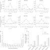
Figure 1. DENV-ADE infection increased virus replication through an intrinsic pathway.
K562 cells were infected with DENV3 at a MOG of 2000, 1000 and 500 complexed with 4-fold dilutions of anti-prM mAb or mock IgG. At 48 h post-infection, cells (a–c) and supernatants (d–f) were collected for total RNA extraction; the DENV genome RNA was quantified (copies/μl) using qRT-PCR. N = 3; error bars show the means ± SEM. (g) Kinetics of DENV3 intracellular RNA accumulation analysed by relative qRT-PCR. K562 cells were infected with DENV3 alone or DENV3-Ab complex at a peak enhancement dilution (1/64) at MOG = 1000 and harvested at the indicated times post-inoculation. An equal amount of total RNA from each sample was subjected to relative quantitation and GAPDH was used as an internal control. N = 2. (h) Binding and internalization results of DENV3 and DENV3-Ab complex on K562 cells by qRT-PCR. N = 3; error bars show the means ± SEM.
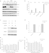
Figure 2. The suppression of NOS2 expression and subsequent NO production in DENV-ADE infected K562 cells.
(a) K562 cells were infected with W/WT DENV or DENV-antibody complex at a MOG of 1000, and at the indicated time points, cellular NO production was quantified by the Griess reaction. N = 3; error bars show the means ± SEM. The results from each group were compared using Student’s t test. **P < 0.01; *P < 0.05. (b,d) The expression of NOS2 at the indicated time points was determined at the gene transcription level by qRT-PCR (b) and at the protein level by western blotting (d). N = 3; error bars show the means ± SEM. (c) K562 cells were pretreated with different concentrations of SMT 1 h before DENV-3 or DENV-ADE infection, and cellular DENV genome RNA was extracted 48 h post-infection and quantified using qRT-PCR, with GAPDH as an internal control. N = 3; error bars show the means ± SEM.
Figure 3. Suppression of the RIG-I and MDA-5 signalling pathways in DENV-ADE infected K562.
K562 cells were infected with DENV alone or DENV-antibody complex or were mock infected. (a–c) The relative quantification of total cellular RNA was performed using qRT-PCR with GAPDH as an internal control. The transcription level of each gene in cells before infection was designated as a basal transcription level and normalized as 1. N = 3; error bars show the means ± SEM. Student’s t test was performed for each group. **P < 0.01; *P < 0.05. (d) Cell lysates post infection were subjected to sodium dodecyl sulfate polyacrylamide gel electrophoresis and stained with specific antibodies against RIG-I, MDA-5, iκ-B, NF-κB p65 and IRF-1 at the indicated time points. N = 3.
Figure 4. DENV/DENV-ADE-induced RLR activation mediates NOS2 expression independent of IFN signalling.
K562 cells was stably transfected with plasmid expressing RIG-I W/WT MDA-5. All the cells were subjected to DENV and DENV-ADE infection. (a) Cell lysates were subjected to sodium dodecyl sulfate polyacrylamide gel electrophoresis and stained with specific antibodies against RIG-I, MDA-5, iκ-B, NF-κB and NOS2 6 h post-inoculation. N = 3. (b) Cells were harvested for total RNA 48 h post-inoculation; the intracellular viral RNA was detected by qRT-PCR, with GAPDH as an internal control. The fold enhancement was expressed as the ratio of DENV/DENV-ADE. N = 3; error bars show the means ± SEM. K562 cells was transfected with siRNA target MAVS or siRNA control. (c) cell lysates were subjected to detection of MAVS and NOS2 4 h post-infection. (d) Total cellular RNA was extracted for intracellular viral RNA quantitation 48 h post-inoculation. (e) K562 cells were co-transfected with pLR-TK plasmid (internal control) and pGL3 -hIFNβ or pGL3-ISRE plasmid (experimental reporter) and then subjected to DENV or DENV-ADE infection. At the indicated time points post-infection, reporter gene activity was measured by a dual-luciferase reporter assay. N = 3; error bars show the means ± SEM. (f) K562 cells were infected with DENV alone, DENV-antibody complex, or mock infected. Cellular RNA was extracted and used for gene expression quantification of IFNγ and IFNλ using qRT-PCR at indicated time points. N = 3; error bars show the means ± SEM.
Figure 5
DENV-ADE infection of K562 is independent of IL-10 function (a) K562 cells were infected with DENV alone or DENV-antibody complex or were mock infected. Cellular RNA was extracted and use for gene expression quantification of IL-10, IL-6 and SOCS3 using qRT-PCR; the supernatants of cell cultures were tested for IL-10 using an ELISA kit. N = 3; error bars show the means ± SEM. (b) Four pairs of sgRNAs were designed to target the human IL-10 loci. Target sequences and PAMs (red) are shown in respective colours, and sites of cleavage by Cas9 are indicated by red triangles. A predicted junction is shown below. (c) Cell populations transfected with paired sgRNAs (1.1 + 2.1, 1.1 + 2.2, 1.2 + 2.1, 1.2 + 2.2) were assayed by PCR (Out-Fwd and Out-Rev primers were used), reflecting a deletion of ~270 bp. (d) Paired sgRNA transfected cells were clonally isolated and expanded for genotyping analysis of deletions using PCR (using the Out-Fwd and Out-Rev primers) and sequencing. Each isolate of K562 with homozygous deletions was subjected to DENV-ADE/DENV infections. (e) Cellular dengue genome RNA was quantified using qRT-PCR with GAPDH as an internal control; the fold enhancement in each K562 isolate was expressed as a ratio of DENV/DENV-ADE. N = 3; error bars show the means ± SEM.
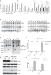
Figure 6. DENV-ADE infection of K562 induces greater autophagy to facilitate viral production.
K562 cells pretreated with different concentrations of rapamycin (a) or 3-MA (b) 1 h before infection with DENV-3 or DENV-ADE. Forty-eight hours post-infection, intracellular DENV RNA was extracted and quantified using qRT-PCR. N = 3; error bars show the means ± SEM. (c) K562 cells were infected with DENV alone, DENV-antibody complex, or mock. At both 0.5 h and 1 h post-inoculation, cellular RNA was extracted and used for gene expression quantification of ATG5 and ATG12 using qRT-PCR. N = 3; error bars show the means ± SEM, the results from each group were compared using Student’s t test. **P < 0.01; *P < 0.05. (**d**) K562 cells were infected with DENV alone, DENV-antibody complex, or mock. Cell lysates were subjected to detection of LC3 I/II, ATG5 and β-actin at the indicated time points. N > 3. (e) K562 cells transfected with paired sgRNA (1.1 + 1.2, 1.1 + 2.3) targeting the ATG5 locus were assayed by PCR (using the ATG5-Out-Fwd and ATG5-Out-Rev primers). Clonal isolates carrying a homozygous deletion in the ATG5 locus was were using PCR and sequencing. (f) Each isolate was subjected to DENV-ADE/DENV infection. Intracellular dengue genome RNA was quantified using the qRT-PCR method with GAPDH as an internal control, and the fold enhancement in each K562 isolate was expressed as a ratio of DENV/DENV-ADE. N = 3; error bars show the means ± SEM, the results from each group were compared using Student’s t test. **P < 0.01; *P < 0.05. K562 cells were stably transfected with plasmid expressing ATG5 or empty plasmid (control). All cell lines were subjected to DENV and DENV-ADE infection. (g) 4 h post-inoculation, cell lysates were subjected to detection of iκ-B, NF-κB, NOS2 and β-actin. N = 3. (h) At 48 h post-inoculation, cells were harvested for total RNA, and the intracellular viral RNA was detected using qRT-PCR with GAPDH as an internal control. N = 3; error bars show the means ± SEM, the results from each group were compared using Student’s t test. **P < 0.01; *P < 0.05.
Similar articles
- Mechanisms of immune evasion induced by a complex of dengue virus and preexisting enhancing antibodies.
Ubol S, Phuklia W, Kalayanarooj S, Modhiran N. Ubol S, et al. J Infect Dis. 2010 Mar 15;201(6):923-35. doi: 10.1086/651018. J Infect Dis. 2010. PMID: 20158392 - Dengue virus replication in infected human keratinocytes leads to activation of antiviral innate immune responses.
Surasombatpattana P, Hamel R, Patramool S, Luplertlop N, Thomas F, Desprès P, Briant L, Yssel H, Missé D. Surasombatpattana P, et al. Infect Genet Evol. 2011 Oct;11(7):1664-73. doi: 10.1016/j.meegid.2011.06.009. Epub 2011 Jun 21. Infect Genet Evol. 2011. PMID: 21722754 - RIG-I-like receptor activation by dengue virus drives follicular T helper cell formation and antibody production.
Sprokholt JK, Kaptein TM, van Hamme JL, Overmars RJ, Gringhuis SI, Geijtenbeek TBH. Sprokholt JK, et al. PLoS Pathog. 2017 Nov 29;13(11):e1006738. doi: 10.1371/journal.ppat.1006738. eCollection 2017 Nov. PLoS Pathog. 2017. PMID: 29186193 Free PMC article. - How Dengue Virus Circumvents Innate Immunity.
Kao YT, Lai MMC, Yu CY. Kao YT, et al. Front Immunol. 2018 Dec 4;9:2860. doi: 10.3389/fimmu.2018.02860. eCollection 2018. Front Immunol. 2018. PMID: 30564245 Free PMC article. Review. - Interplay between dengue virus and Toll-like receptors, RIG-I/MDA5 and microRNAs: Implications for pathogenesis.
Urcuqui-Inchima S, Cabrera J, Haenni AL. Urcuqui-Inchima S, et al. Antiviral Res. 2017 Nov;147:47-57. doi: 10.1016/j.antiviral.2017.09.017. Epub 2017 Sep 28. Antiviral Res. 2017. PMID: 28965915 Review.
Cited by
- Role of autophagy during the replication and pathogenesis of common mosquito-borne flavi- and alphaviruses.
Echavarria-Consuegra L, Smit JM, Reggiori F. Echavarria-Consuegra L, et al. Open Biol. 2019 Mar 29;9(3):190009. doi: 10.1098/rsob.190009. Open Biol. 2019. PMID: 30862253 Free PMC article. Review. - Dengue: A Minireview.
Harapan H, Michie A, Sasmono RT, Imrie A. Harapan H, et al. Viruses. 2020 Jul 30;12(8):829. doi: 10.3390/v12080829. Viruses. 2020. PMID: 32751561 Free PMC article. Review. - Autophagy during viral infection - a double-edged sword.
Choi Y, Bowman JW, Jung JU. Choi Y, et al. Nat Rev Microbiol. 2018 Jun;16(6):341-354. doi: 10.1038/s41579-018-0003-6. Nat Rev Microbiol. 2018. PMID: 29556036 Free PMC article. Review. - Significance of Autophagy in Dengue Virus Infection: A Brief Review.
Acharya B, Gyeltshen S, Chaijaroenkul W, Na-Bangchang K. Acharya B, et al. Am J Trop Med Hyg. 2019 Apr;100(4):783-790. doi: 10.4269/ajtmh.18-0761. Am J Trop Med Hyg. 2019. PMID: 30761986 Free PMC article. Review.
References
- Kliks S. C., Nimmanitya S., Nisalak A. & Burke D. S. Evidence that maternal dengue antibodies are important in the development of dengue hemorrhagic fever in infants. The American journal of tropical medicine and hygiene 38, 411–419 (1988). - PubMed
Publication types
MeSH terms
Substances
LinkOut - more resources
Full Text Sources
Other Literature Sources
Medical
Miscellaneous


