Human mitochondrial DNA replication machinery and disease - PubMed (original) (raw)
Review
Human mitochondrial DNA replication machinery and disease
Matthew J Young et al. Curr Opin Genet Dev. 2016 Jun.
Abstract
The human mitochondrial genome is replicated by DNA polymerase γ in concert with key components of the mitochondrial DNA (mtDNA) replication machinery. Defects in mtDNA replication or nucleotide metabolism cause deletions, point mutations, or depletion of mtDNA. The resulting loss of cellular respiration ultimately induces mitochondrial genetic diseases, including mtDNA depletion syndromes (MDS) such as Alpers or early infantile hepatocerebral syndromes, and mtDNA deletion disorders such as progressive external ophthalmoplegia, ataxia-neuropathy, or mitochondrial neurogastrointestinal encephalomyopathy. Here we review the current literature regarding human mtDNA replication and heritable disorders caused by genetic changes of the POLG, POLG2, Twinkle, RNASEH1, DNA2, and MGME1 genes.
Published by Elsevier Ltd.
Figures
Figure 1
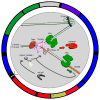
Map of the human mitochondrial genome and the mtDNA replication fork. The outer circle represents the 16,569 bp covalently closed circular double-stranded mtDNA. Counterclockwise from the top of the circle: Grey, control region including the heavy-strand origin of replication (OH) and the displacement-loop (D-loop); Green; 12 and 16 S rRNA; Blue, NADH dehydrogenase (ND) 1 and 2; Red, cytochrome oxidase (COX) I and II; Yellow, ATPase 8 and 6; Red, COX III; Blue, ND 3, 4L, 4, 5, 6; Purple, cytochrome b. The D-loop form of mtDNA is a triple-stranded structure that results from the template-directed termination of H-strand synthesis soon after initiation resulting in mtDNA molecules with nascent H-strand annealed to them [83]. Recent evidence supports that the loading of the Twinkle helicase at the 3′-end of the D-loop is reversible, indicating that this site is critical to regulating the switch between formation of D-loop molecules and initiation of mtDNA replication [84]. Black rectangles represent the 22 tRNA genes. The inset illustrates the replisome at an area near the light-strand origin (OL) of replication located within the WANCY cluster of genes, which encode for tryptophan, alanine, asparagine, cysteine, and tyrosine tRNAs. Black lines represent template mtDNA while green lines represent nascent mtDNA. Main factors highlighted at the replication fork include: 1) the 5′-3′ DNA polymerase pol γ 2) the enzyme topoisomerase (Topo) required for mtDNA unwinding ahead of the replication fork. The phospodiester backbones of both mtDNA strands are enzymatically broken and rejoined allowing relaxation of positive supercoils introduced ahead of the replisome during replication fork elongation, 3) the hexameric replicative Twinkle mtDNA helicase required for ATP-dependent disruption of the hydrogen bonds that hold the two DNA strands together causing mtDNA duplex denaturation (strand separation), 4) mitochondrial RNA polymerase (mtRNAP) required for mitochondrial transcription as well as for RNA primer formation to initiate DNA replication, 5) RNaseH1 required for RNA primer removal [31,70,85], 6) mitochondrial single-stranded DNA (ssDNA) binding protein (mtSSB) required for ssDNA stabilization during mtDNA replication, 7) DNA ligase III (mtLigIII) required for mtDNA break (nick) sealing, 8) mitochondrial transcription factor A (TFAM), 9) mitochondrial genome maintenance 5′-3′ exonuclease 1 (MGME1), 10) flap endonuclease (FEN1), and 11) the helicase/nuclease, DNA2.
Figure 2
DNA polymerase γ ternary structure. The p140 catalytic subunit consist of: 1) an amino terminal domain (NTD, light grey), 2) an exonuclease domain (exo, dark grey), 3) a spacer domain comprised of an intrinsic processivity (IP) subdomain (yellow) plus the accessory-interacting determinant (AID) subdomain (orange), and 4) a DNA polymerase (pol) domain, which folds to resemble a “right-hand” comprised of three subdomains: the thumb (green), fingers (dark blue), and palm (red). The p55 processivity subunit dimer is comprised of the proximal monomer (purple) and the distal protomer, light blue. The DNA primer strand is colored red while the template strand is colored pink. The figure was generated using UCSF Chimera and the published 3.3 Å crystal structure PDB ID 4ZTU; Szymanski et al. [15].
Figure 3
Schematic diagram of POLG, the human DNA polymerase γ catalytic subunit gene, and the linear sequence of the p140 amino acid residues. Amino acid substitutions encoded by POLG disease mutations are listed on the linear map and p140 domains and subdomains are color coded as in Figure 2.
Similar articles
- Defects in mitochondrial DNA replication and human disease.
Copeland WC. Copeland WC. Crit Rev Biochem Mol Biol. 2012 Jan-Feb;47(1):64-74. doi: 10.3109/10409238.2011.632763. Crit Rev Biochem Mol Biol. 2012. PMID: 22176657 Free PMC article. Review. - [Mitochondrial disease and mitochondrial DNA depletion syndromes].
Huang CC, Hsu CH. Huang CC, et al. Acta Neurol Taiwan. 2009 Dec;18(4):287-95. Acta Neurol Taiwan. 2009. PMID: 20329599 Review. Chinese. - Defects of mitochondrial DNA replication.
Copeland WC. Copeland WC. J Child Neurol. 2014 Sep;29(9):1216-24. doi: 10.1177/0883073814537380. Epub 2014 Jun 30. J Child Neurol. 2014. PMID: 24985751 Free PMC article. Review. - Consequences of compromised mitochondrial genome integrity.
Gustafson MA, Sullivan ED, Copeland WC. Gustafson MA, et al. DNA Repair (Amst). 2020 Sep;93:102916. doi: 10.1016/j.dnarep.2020.102916. DNA Repair (Amst). 2020. PMID: 33087282 Free PMC article. Review. - Progressive External Ophthalmoplegia in Polish Patients-From Clinical Evaluation to Genetic Confirmation.
Kierdaszuk B, Kaliszewska M, Rusecka J, Kosińska J, Bartnik E, Tońska K, Kamińska AM, Kostera-Pruszczyk A. Kierdaszuk B, et al. Genes (Basel). 2020 Dec 31;12(1):54. doi: 10.3390/genes12010054. Genes (Basel). 2020. PMID: 33396418 Free PMC article.
Cited by
- Lysosomal dysfunction impairs mitochondrial quality control and is associated with neurodegeneration in TBCK encephaloneuronopathy.
Tintos-Hernández JA, Santana A, Keller KN, Ortiz-González XR. Tintos-Hernández JA, et al. Brain Commun. 2021 Sep 16;3(4):fcab215. doi: 10.1093/braincomms/fcab215. eCollection 2021. Brain Commun. 2021. PMID: 34816123 Free PMC article. - Nuclear genes involved in mitochondrial diseases caused by instability of mitochondrial DNA.
Rusecka J, Kaliszewska M, Bartnik E, Tońska K. Rusecka J, et al. J Appl Genet. 2018 Feb;59(1):43-57. doi: 10.1007/s13353-017-0424-3. Epub 2018 Jan 17. J Appl Genet. 2018. PMID: 29344903 Free PMC article. Review. - Neurotoxicity of cytarabine (Ara-C) in dorsal root ganglion neurons originates from impediment of mtDNA synthesis and compromise of mitochondrial function.
Zhuo M, Gorgun MF, Englander EW. Zhuo M, et al. Free Radic Biol Med. 2018 Jun;121:9-19. doi: 10.1016/j.freeradbiomed.2018.04.570. Epub 2018 Apr 23. Free Radic Biol Med. 2018. PMID: 29698743 Free PMC article. - Mitochondrial DNA heteroplasmy in disease and targeted nuclease-based therapeutic approaches.
Nissanka N, Moraes CT. Nissanka N, et al. EMBO Rep. 2020 Mar 4;21(3):e49612. doi: 10.15252/embr.201949612. Epub 2020 Feb 19. EMBO Rep. 2020. PMID: 32073748 Free PMC article. Review. - PRIMPOL ready, set, reprime!
Tirman S, Cybulla E, Quinet A, Meroni A, Vindigni A. Tirman S, et al. Crit Rev Biochem Mol Biol. 2021 Feb;56(1):17-30. doi: 10.1080/10409238.2020.1841089. Epub 2020 Nov 12. Crit Rev Biochem Mol Biol. 2021. PMID: 33179522 Free PMC article. Review.
References
- Hensen F, Cansiz S, Gerhold JM, Spelbrink JN. To be or not to be a nucleoid protein: a comparison of mass-spectrometry based approaches in the identification of potential mtDNA-nucleoid associated proteins. Biochimie. 2014;100:219–226. - PubMed
- Spelbrink JN. Functional organization of mammalian mitochondrial DNA in nucleoids: history, recent developments, and future challenges. IUBMB Life. 2010;62:19–32. - PubMed
- Wallace DC. Mitochondrial diseases in man and mouse. Science. 1999;283:1482–1488. - PubMed
Publication types
MeSH terms
Substances
LinkOut - more resources
Full Text Sources
Other Literature Sources
Medical
Research Materials
Miscellaneous


