Hematoma Changes During Clot Resolution After Experimental Intracerebral Hemorrhage - PubMed (original) (raw)
Hematoma Changes During Clot Resolution After Experimental Intracerebral Hemorrhage
Shenglong Cao et al. Stroke. 2016 Jun.
Abstract
Background and purpose: Hematoma clearance occurs in the days after intracerebral hemorrhage (ICH) and has not been well studied. In the current study, we examined changes in the hematoma in a piglet ICH model. The effect of deferoxamine on hematoma was also examined.
Methods: The ICH model was induced by an injection of autologous blood into the right frontal lobe of piglets. First, a natural time course of hematoma changes ≤7 days was determined. Second, the effect of deferoxamine on hematoma changes was examined. Hemoglobin and membrane attack complex levels in the hematoma were examined by enzyme-linked immunosorbent assay. Immunohistochemistry and Western blotting were used to examine CD47 (a regulator of erythrophagocytosis), CD163 (a hemoglobin scavenger receptor), and heme oxygenase-1 (a heme degradation enzyme) in the clot.
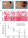
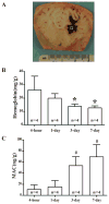
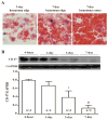
Results: After ICH, there was a reduction in red blood cell diameter within the clot with time. This was accompanied by membrane attack complex accumulation and decreased hemoglobin levels. Erythrophagocytosis occurred in the hematoma, and this was associated with reduced clot CD47 levels. Activated macrophages/microglia were CD163 and hemeoxygenase-1 positive, and these accumulated in the clot with time. Deferoxamine treatment attenuated the process of hematoma resolution by reducing member attack complex formation and inhibiting CD47 loss in the clot.
Conclusions: These results indicate that membrane attack complex and erythrophagocytosis contribute to hematoma clearance after ICH, which can be altered by deferoxamine treatment.
Keywords: cerebral hemorrhage; deferoxamine; hemolysis; membrane attack complex; swine.
© 2016 American Heart Association, Inc.
Conflict of interest statement
Potential Conflicts of Interest: We declare that we have no conflict of interest.
Figures
Figure 1
(A) Coronal sections of perfused piglet brain showing hematomas. (B) Time course of clot changes in hematoma edge and center. Scale bar = 10 μm, scale bar = 2.5 μm in the insets. (C) Diameter of RBC in hematoma edge and center. Values are mean ± SD, *p <0.05, #p <0.01 vs. 4-hour.
Figure 2
(A) Coronal section of an in situ frozen piglet brain after ICH. The square represents the sample area. (B) Time course of hemoglobin content in hematoma. Values are mean ± SD, *p <0.05, #p <0.01 vs. 4-hour. (C) Time course of MAC content in the hematoma. Values are mean ± SD, *p <0.05, #p <0.01 vs. 4-hour.
Figure 3
(A) Erythrophagocytosis (arrows) at day-3 and day-7 in the hematoma and hemosiderin deposition (arrowhead) at day-7. Scale bar = 10 μm. (B) Time course of CD47 levels in the hematoma. Values are mean ± SD, *p <0.05, #p <0.01 vs. 4-hour.
Figure 4
(A) Time course of CD163 immunoreactivity and number of positive cells in the hematoma. Upper scale bar = 50μm, lower scale bar = 10μm. Values are mean ± SD, *p <0.05, #p <0.01 vs. 4-hour. (B) Time course of HO-1 immunoreactivity and protein levels. Upper scale bar = 50 μm, lower scale bar = 10 μm. Values are mean ± SD, *p <0.05, #p <0.01 vs. 4-hour.
Figure 5
(A) Hemoglobin, (B) MAC and (C) CD47 protein levels in the hematoma in vehicle- and DFX-treated groups at 3 days after ICH. Values are mean ± SD, *p <0.05 vs. vehicle treated group.
Figure 6
(A) CD163 immunoreactivity and numbers of positive cells in the hematoma in vehicle- and DFX-treated groups at 3 days after ICH. Upper scale bar = 50 μm, lower scale bar = 10 μm. Values are mean ± SD, *p <0.05. (B) HO-1 immunoreactivity and proteins levels in the hematoma in vehicle- and DFX-treated groups at 3 days after ICH. Upper scale bar = 50 μm, lower scale bar =10 μm. Values are mean ± SD, *p <0.05.
Similar articles
- CD163 Expression in Neurons After Experimental Intracerebral Hemorrhage.
Liu R, Cao S, Hua Y, Keep RF, Huang Y, Xi G. Liu R, et al. Stroke. 2017 May;48(5):1369-1375. doi: 10.1161/STROKEAHA.117.016850. Epub 2017 Mar 30. Stroke. 2017. PMID: 28360115 Free PMC article. - Deferoxamine therapy reduces brain hemin accumulation after intracerebral hemorrhage in piglets.
Hu S, Hua Y, Keep RF, Feng H, Xi G. Hu S, et al. Exp Neurol. 2019 Aug;318:244-250. doi: 10.1016/j.expneurol.2019.05.003. Epub 2019 May 10. Exp Neurol. 2019. PMID: 31078524 Free PMC article. Review. - Role of Erythrocyte CD47 in Intracerebral Hematoma Clearance.
Ni W, Mao S, Xi G, Keep RF, Hua Y. Ni W, et al. Stroke. 2016 Feb;47(2):505-11. doi: 10.1161/STROKEAHA.115.010920. Epub 2016 Jan 5. Stroke. 2016. PMID: 26732568 Free PMC article. - Tin-mesoporphyrin, a potent heme oxygenase inhibitor, for treatment of intracerebral hemorrhage: in vivo and in vitro studies.
Wagner KR, Hua Y, de Courten-Myers GM, Broderick JP, Nishimura RN, Lu SY, Dwyer BE. Wagner KR, et al. Cell Mol Biol (Noisy-le-grand). 2000 May;46(3):597-608. Cell Mol Biol (Noisy-le-grand). 2000. PMID: 10872746 - The Critical Role of Erythrolysis and Microglia/Macrophages in Clot Resolution After Intracerebral Hemorrhage: A Review of the Mechanisms and Potential Therapeutic Targets.
Zheng Y, Tan X, Cao S. Zheng Y, et al. Cell Mol Neurobiol. 2023 Jan;43(1):59-67. doi: 10.1007/s10571-021-01175-3. Epub 2022 Jan 4. Cell Mol Neurobiol. 2023. PMID: 34981286 Review.
Cited by
- Complement Drives Chronic Inflammation and Progressive Hydrocephalus in Murine Neonatal Germinal Matrix Hemorrhage.
Alshareef M, Hatchell D, Vasas T, Mallah K, Shingala A, Cutrone J, Alawieh A, Guo C, Tomlinson S, Eskandari R. Alshareef M, et al. Int J Mol Sci. 2023 Jun 15;24(12):10171. doi: 10.3390/ijms241210171. Int J Mol Sci. 2023. PMID: 37373319 Free PMC article. - The Chemical Basis of Intracerebral Hemorrhage and Cell Toxicity With Contributions From Eryptosis and Ferroptosis.
Derry PJ, Vo ATT, Gnanansekaran A, Mitra J, Liopo AV, Hegde ML, Tsai AL, Tour JM, Kent TA. Derry PJ, et al. Front Cell Neurosci. 2020 Dec 8;14:603043. doi: 10.3389/fncel.2020.603043. eCollection 2020. Front Cell Neurosci. 2020. PMID: 33363457 Free PMC article. - Mechanism of White Matter Injury and Promising Therapeutic Strategies of MSCs After Intracerebral Hemorrhage.
Li J, Xiao L, He D, Luo Y, Sun H. Li J, et al. Front Aging Neurosci. 2021 Apr 13;13:632054. doi: 10.3389/fnagi.2021.632054. eCollection 2021. Front Aging Neurosci. 2021. PMID: 33927608 Free PMC article. Review. - CD47 Blockade Accelerates Blood Clearance and Alleviates Early Brain Injury After Experimental Subarachnoid Hemorrhage.
Xu CR, Li JR, Jiang SW, Wan L, Zhang X, Xia L, Hua XM, Li ST, Chen HJ, Fu XJ, Jing CH. Xu CR, et al. Front Immunol. 2022 Feb 25;13:823999. doi: 10.3389/fimmu.2022.823999. eCollection 2022. Front Immunol. 2022. PMID: 35281006 Free PMC article. - Resolution metabolomes activated by hypoxic environment.
Norris PC, Libreros S, Serhan CN. Norris PC, et al. Sci Adv. 2019 Oct 23;5(10):eaax4895. doi: 10.1126/sciadv.aax4895. eCollection 2019 Oct. Sci Adv. 2019. PMID: 31681846 Free PMC article.
References
- Xi G, Keep RF, Hoff JT. Mechanisms of brain injury after intracerebral hemorrhage. Lancet Neurol. 2006;5:53–63. - PubMed
- Hua Y, Xi G, Keep RF, Hoff JT. Complement activation in the brain after experimental intracerebral hemorrhage. J Neurosurg. 2000;92:1016–1022. - PubMed
- Xi G, Hua Y, Keep RF, Younger JG, Hoff JT. Systemic complement depletion diminishes perihematomal brain edema in rats. Stroke. 2001;32:162–167. - PubMed
- Zhao X, Grotta J, Gonzales N, Aronowski J. Hematoma resolution as a therapeutic target: The role of microglia/macrophages. Stroke. 2009;40:S92–94. - PubMed
- Zhao X, Sun G, Zhang J, Strong R, Song W, Gonzales N, et al. Hematoma resolution as a target for intracerebral hemorrhage treatment: Role for peroxisome proliferator-activated receptor gamma in microglia/macrophages. Ann Neurol. 2007;61:352–362. - PubMed
Publication types
MeSH terms
Substances
Grants and funding
- R21 NS091545/NS/NINDS NIH HHS/United States
- R01 NS096917/NS/NINDS NIH HHS/United States
- R01 NS090925/NS/NINDS NIH HHS/United States
- R01 NS079157/NS/NINDS NIH HHS/United States
- R01 NS073595/NS/NINDS NIH HHS/United States
- R21 NS084049/NS/NINDS NIH HHS/United States
LinkOut - more resources
Full Text Sources
Other Literature Sources
Research Materials


