Irreversible APC(Cdh1) Inactivation Underlies the Point of No Return for Cell-Cycle Entry - PubMed (original) (raw)
Irreversible APC(Cdh1) Inactivation Underlies the Point of No Return for Cell-Cycle Entry
Steven D Cappell et al. Cell. 2016.
Abstract
Proliferating cells must cross a point of no return before they replicate their DNA and divide. This commitment decision plays a fundamental role in cancer and degenerative diseases and has been proposed to be mediated by phosphorylation of retinoblastoma (Rb) protein. Here, we show that inactivation of the anaphase-promoting complex/cyclosome (APC(Cdh1)) has the necessary characteristics to be the point of no return for cell-cycle entry. Our study shows that APC(Cdh1) inactivation is a rapid, bistable switch initiated shortly before the start of DNA replication by cyclin E/Cdk2 and made irreversible by Emi1. Exposure to stress between Rb phosphorylation and APC(Cdh1) inactivation, but not after APC(Cdh1) inactivation, reverted cells to a mitogen-sensitive quiescent state, from which they can later re-enter the cell cycle. Thus, APC(Cdh1) inactivation is the commitment point when cells lose the ability to return to quiescence and decide to progress through the cell cycle.
Copyright © 2016. Published by Elsevier Inc.
Figures
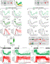
Figure 1.. Rapid and Near-Complete APCCdh1 Inactivation Shortly before S Phase Entry
(A) Schematic diagram of the cell-cycle commitment model. (B) Schematic diagram of the APC-degron reporter (Geminin: aa1–110). (C) Single-cell trace of APC-degron reporter levels in a representative cell released from mitogen starvation. Inset: snapshots of the APC-degron reporter. (D) Single-cell trace of the APC-degron reporter in a representative cycling cell as in (C). (E) MCF10A cells expressing mVenus-APC-degron wild-type and either mChy-APC-degron KEN mutant or mChy-APC-degron KEN/RxxL mutant. Lines are median traces ± SEM. (n = 205 cells, wild-type; n = 800, KEN; n = 600, RxxL). (F) Cells were imaged for ~4 hr then fixed and stained with α-cyclin A2. Cells were binned by the time since mitosis. Data represent median intensity ± SEM of either cyclin A2 or APC-degron reporter. (G) HeLa cells transfected with the APC-degron reporter and mCitrine-Aurora-A K162R (three representative cells shown). (H) APC-degron reporter levels and the derived APC activity for a single cell. Time of mitosis and the G1/S transition are computationally identified. (I) Left: Single-cell traces of APCCdh1 activity computationally aligned to 50% APCCdh1 activity (random selection of 91 out of 431 cells analyzed). Right: Median APCCdh1 activity trace ± SD (n = 861). (J) Scatterplot of BrdU levelsversusthetimesinceAPCCdh1 started to inactivate. Fixed cells were mapped back to live-cell data. Single-cell data were binned and data points are median ± SEM (n = 1100). (K) MCF10A cells were treated with either control siRNA or Cdh1 siRNA. Fixed cells were mapped back to live-cell data. Single-cell data were binned and data points are median ± SEM. n > 30,000 cells per condition. Arrow highlights the shift in timing of EdU incorporation after Cdh1 siRNA. (L) Schematic when APCCdh1 is on and off during the cell cycle. See also Figure S1.
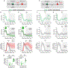
Figure 2.. APCCdh1 Inactivation Is Irreversible and Requires Cyclin E/Cdk2
(A) Schematic of the Cdk2 sensor. Cdk2-mediated phosphorylation controls export of the reporter from the nucleus. NLS, nuclear localization signal; NES, nuclear export signal; S, CDK consensus site on serine. (B) APCCdh1 and Cdk2 activities in a representative single cycling cell. (C) Histogram of the time between the initial rise of Cdk2 activity and APCCdh1 inactivation. n > 1000 cells. (D) Histogram of Cdk2 activity when APCCdh1 inactivates (green). Basal Cdk2 activity after treatment with a Cdk1/2 inhibitor (gray) is added for reference. (E-G) APC and Cdk2 activity aligned in silico to mitosis treated with either control (E), cyclin E1 and E2 (F), or cyclin A2 siRNA(G). Grey line is median trace. n = 54, 26, and 48, respectively. (H) Schematic diagram showing that cyclin E/Cdk2 mediates inactivation of APCCdh1 and that Cdk2 inhibition is expected to prevent APCCdh1 inactivation. (I and J) APC activity traces treated with either DMSO or Cdk1/2 inhibitor. Time of treatment is indicated by the dashed line.(I) Cells treated while in G1 phase (n = 41, DMSO; n = 45, Cdk1/2i).(J) Cells treated while in S phase (n = 47, DMSO; n = 45, Cdk1/2i). Black line is median trace. See also Figure S2.
Figure 3.. Emi1 Is Required for Rapid and Irreversible APCCdh1 Inactivation
(A-D) Single-cell APC activity traces aligned to the time that APCCdh1 starts to turn off in cells treated with either control (A), Usp37 (B), or Emi1 siRNA (C) or cells overexpressing (oe) wild-type Emi1 (D). Dashed line added for reference between panels. (E) Median APC activity trace ± SEM from cells in (A-D). n = 276, 201, 214, 152, respectively. (F) Schematic of experimental design for (G-I). (G-I) Single-cell APC activity traces treated with either control (G) or Emi1 siRNA (H and I). Cells were exposed to either DMSO (H) or Cdk1/2 inhibitor (G and I) at the indicated time. Data from all cells is in Figure S3J. (J) Bar graph of the percent of cells that reactivate APCCdh1 upon Cdk1/2 inhibitor addition. n≈ 250 cells per condition. See also Figure S3.
Figure 4.. A Large Time Gap Separates the Restriction Point and pRb-E2F Induction from APCCdh1 Inactivation
(A) Probability of entering the cell cycle (i.e., EdU incorporation after 24 hr) as a function of the length of mitogen or MEKi pulse after release from starvation. Dashed line indicates when 50% of cells enter the cell cycle. Error bars are SD from four experiments, n > 3000 cells per data point. (B) APC activity in cells released from mitogen-starvation (n = 114). (C) Cumulative distribution function (CDF) of the time cells inactivated APCCdh1 after release from mitogen-starvation (red; n = 9,068). Scatterplot of the percent of cells with phospho-Rb (S807/S811) as a function of time since mitogen stimulation (n > 15,000 cells per data point). Green line is sigmoidal best-fit line. Dashed lines indicate when half max is reached. (D) Histograms of pRb807/811 levels taken at 1 hr intervals after release from mitogen starvation (n > 15,000 cells per histogram). (E) Single-cell mRNA FISH of E2F1 and cycE1 after release from mitogen starvation. Cells were separated into pRb negative and positive based on bimodal histogram as in (D). Error bars are SEM from four experiments, n > 1000 cells per condition. (F) Schematic diagram of the timing of cell-cycle events in cells released from mitogen starvation. (G) Scatterplot of the percent of cells with pRb807/811 at each bin relative to mitosis (green; n « 400 cells per data point). CDF of the time cells inactivated APCCdh1 relative to mitosis (red; n = 1042). (H) Histograms of pRb807/811 levels divided into bins since mitosis. (n ≈ 400 cells per histogram). (I) Single-cell traces of APC activity in asynchronous cells, aligned to mitosis (n = 140). (J) Schematic of the timing of cell-cycle events in cycling cells. Following mitosis, cells either maintain Rb hyper-phosphorylation and stay in the cell cycle or they lose Rb phosphorylation and enter a transient G0-like state. (K) Schematic showing when cells are sensitive to mitogens relative to Rb phosphorylation and APCCdh1 inactivation. (L) Median traces ± SD of APC activity in cells treated with the indicated drug in G1 phase. n = 113, control; n = 107, aphidicolin; n = 116, thymidine; n = 84, hydroxyurea. See also Figure S4.
Figure 5.. Stress Signaling Can Induce Cell-Cycle Exit to Mitogen-Regulated Quiescence after pRb-E2F Induction but Only until APCCdh1 Inactivation
(A) Experimental setup for stress administration after the restriction point but before APCCdh1 inactivation. (B) Schematic showing cells that have increasing Cdk2 activity (Cdk2lnc) or basal Cdk2 activity (Cdk2low) 2 hr after mitosis. Horizontal dashed line indicates the threshold level of Cdk2 activity used to categorize a cell as either Cdk2inc or Cdk2low. Only Cdk2inc cells were considered in Figures 5C–5H. (C and D) Cdk2 and APCCdh1 activities in cells exposed to NCS after Cdk2 activity started to rise. Light gray band represents time when cells were exposed to stress. Top: two individual cells are shown. Middle: median Cdk2 activity ± SD. Bottom: median APCCdh1 activity. Traces are colored if Cdk2 inactivated after initially turning on or are gray if Cdk2 stayed active and APCCdh1 inactivated. Inset: percentage of cells that inactivated Cdk2 activity despite initially turning on. (C) DMSO or (D) 40ng/mL Neocarzinostatin (NCS) (n = 270, DMSO; n = 554, NCS; see Figure S5E). (E) Scatterplot of pRb807/811 levels versus Cdk2 activity 8 hr after the initial rise in Cdk2 activity. (F) Experimental setup for stress administered after APCCdh1 inactivation. (G and H) Cdk2 and APCCdh1 activities exposed to stress after APCCdh1 inactivation. Top: two cells are highlighted. Middle: median Cdk2 activity ± SD. Bottom: median APCCdh1 activity. (G) DMSO or (H) 40ng/mL Neocarzinostatin (NCS). Traces are colored if Cdk2 inactivated despite initially turning on or are gray if Cdk2 stayed active and APCCdh1 inactivated. Note no traces were colored (n = 587, DMSO; n = 1284). (I) Cdk2 and APCCdh1 activity traces of cells treated with NCS after crossing the restriction point. These cells inactivated Cdk2, only to re-activate Cd-k2 and reenter the cell cycle several hours later (n = 35). (J) Cdk2 and APCCdh1 activity traces treated with 200 ng/mL NCS for 12 min, then washed with mitogen-free media, and monitored for an additional 32 hr. (n = 33). (K) Cdk2 and APC traces treated with 200 ng/mL NCS for 12 min, then washed with mitogen-free media, and monitored for an additional 21 hr. After 21 hr, full- growth media was added and cells were monitored for an additional 10 hr. Cells re-entered the cell cycle after stimulation with mitogens (n = 61). (L) Cells stressed in G1 enter a quiescent state while cells stressed in S or G2 pause before continuing through the cell cycle. See also Figure S5.
Figure 6.. Multiple Types of Stresses Can Cause Cell-Cycle Exit until, but Not after APCCdh1 Inactivation
(A) Experimental setup for stress administration after the restriction point but before APCCdh1 inactivation. (B and C) Representative examples and median traces ± SD of Cdk2 and APC activities in cells exposed to stress after Cdk2 activity started to rise. Light gray band represents time when cells were exposed to stress. Top: two cells are highlighted. Middle: median Cdk2 activity. Bottom: median APC activity. Traces are colored ifCdk2 inactivated despite initially turning on or are gray ifCdk2 stayed active and APCCdh1 inactivated. Inset: percentage of cells that inactivated Cdk2 activity despite initially turning on. (B) 200 μM Hydrogen peroxide (H2O2) or (C) 100 mM NaCl (salt) (n = 821, H2O2; n = 223, salt). See Figure S6C. (D) Scatterplot of pRb807/811 levels versus Cdk2 activity 8 hr after the initial rise in Cdk2 activity (n = 1152, H2O2; n = 194, salt). (E) Experimental setup for stress administered after APCCdh1 inactivation. (F and G) Median traces ± SD of Cdk2 and APC activity exposed to stress afterAPCCdh1 inactivation. Top: two cells are highlighted. Middle: median Cdk2 activity. Bottom: median APC activity. (F) 200 μM Hydrogen peroxide (H2O2) or(G) 100mM NaCl (salt).Traces are colored ifCdk2 inactivated despite initially turning on or are gray if Cdk2 stayed active and APCCdh1 inactivated. Note no traces were colored. See also Figure S6.
Figure 7.. APCCdh1 Inactivation Is a Rapid, Irreversible, and Bistable Switch that Commits Cells to Progress through the Cell Cycle
(A) Cdk2 and APCCdh1 activities in cells exposed to NCS and treated with either control (top) or Emil siRNA (bottom). Cells treated with Emil siRNA re-activate APCCdh1 and inactivate Cdk2. Control siRNA treated cells re-activate APCCdh1 much later during mitosis (n = 28, si Control; n = 23, siEmil). (B) Windows of mitogen and stress sensitivity during cell-cycle entry and exit. Timing of cell-cycle entry and exit shows that mitogens and stress regulate entry during different time windows. Approximate cell-cycle phase durations for MCF10Aare highlighted to place APCCdh1 inactivation and cell-cycle commitment into the overall cell-cycle context. (C) Cell-cycle entry and exit is characterized by three regulatory states: quiescence, a window of reversibility, and a proliferative state, defined by changes in APCCdh1 and E2F activities. The scheme shows the relationship of these states with the cell-cycle phases. See also Figure S7.
Similar articles
- EMI1 switches from being a substrate to an inhibitor of APC/CCDH1 to start the cell cycle.
Cappell SD, Mark KG, Garbett D, Pack LR, Rape M, Meyer T. Cappell SD, et al. Nature. 2018 Jun;558(7709):313-317. doi: 10.1038/s41586-018-0199-7. Epub 2018 Jun 6. Nature. 2018. PMID: 29875408 Free PMC article. - Cdh1 degradation is mediated by APC/C-Cdh1 and SCF-Cdc4 in budding yeast.
Nagai M, Shibata A, Ushimaru T. Nagai M, et al. Biochem Biophys Res Commun. 2018 Dec 2;506(4):932-938. doi: 10.1016/j.bbrc.2018.10.179. Epub 2018 Nov 2. Biochem Biophys Res Commun. 2018. PMID: 30396569 - An APC/C-Cdh1 Biosensor Reveals the Dynamics of Cdh1 Inactivation at the G1/S Transition.
Ondracka A, Robbins JA, Cross FR. Ondracka A, et al. PLoS One. 2016 Jul 13;11(7):e0159166. doi: 10.1371/journal.pone.0159166. eCollection 2016. PLoS One. 2016. PMID: 27410035 Free PMC article. - APC/C and retinoblastoma interaction: cross-talk of retinoblastoma protein with the ubiquitin proteasome pathway.
Ramanujan A, Tiwari S. Ramanujan A, et al. Biosci Rep. 2016 Sep 16;36(5):e00377. doi: 10.1042/BSR20160152. Print 2016 Oct. Biosci Rep. 2016. PMID: 27402801 Free PMC article. Review. - Cdh1-APC/C, cyclin B-Cdc2, and Alzheimer's disease pathology.
Aulia S, Tang BL. Aulia S, et al. Biochem Biophys Res Commun. 2006 Jan 6;339(1):1-6. doi: 10.1016/j.bbrc.2005.10.059. Epub 2005 Oct 21. Biochem Biophys Res Commun. 2006. PMID: 16253208 Review.
Cited by
- The G1-S transition is promoted by Rb degradation via the E3 ligase UBR5.
Zhang S, Valenzuela LF, Zatulovskiy E, Mangiante L, Curtis C, Skotheim JM. Zhang S, et al. Sci Adv. 2024 Oct 25;10(43):eadq6858. doi: 10.1126/sciadv.adq6858. Epub 2024 Oct 23. Sci Adv. 2024. PMID: 39441926 Free PMC article. - The oscillation of mitotic kinase governs cell cycle latches in mammalian cells.
Dragoi CM, Kaur E, Barr AR, Tyson JJ, Novák B. Dragoi CM, et al. J Cell Sci. 2024 Feb 1;137(3):jcs261364. doi: 10.1242/jcs.261364. Epub 2024 Feb 13. J Cell Sci. 2024. PMID: 38206091 Free PMC article. - Postmitotic G1 phase survivin drives mitogen-independent cell division of B lymphocytes.
Singh A, Spitzer MH, Joy JP, Kaileh M, Qiu X, Nolan GP, Sen R. Singh A, et al. Proc Natl Acad Sci U S A. 2022 May 3;119(18):e2115567119. doi: 10.1073/pnas.2115567119. Epub 2022 Apr 27. Proc Natl Acad Sci U S A. 2022. PMID: 35476510 Free PMC article. - The RepID-CRL4 ubiquitin ligase complex regulates metaphase to anaphase transition via BUB3 degradation.
Jang SM, Nathans JF, Fu H, Redon CE, Jenkins LM, Thakur BL, Pongor LS, Baris AM, Gross JM, OʹNeill MJ, Indig FE, Cappell SD, Aladjem MI. Jang SM, et al. Nat Commun. 2020 Jan 7;11(1):24. doi: 10.1038/s41467-019-13808-9. Nat Commun. 2020. PMID: 31911655 Free PMC article. - Mechanisms of Cell Cycle Arrest and Apoptosis in Glioblastoma.
Gousias K, Theocharous T, Simon M. Gousias K, et al. Biomedicines. 2022 Feb 28;10(3):564. doi: 10.3390/biomedicines10030564. Biomedicines. 2022. PMID: 35327366 Free PMC article. Review.
References
- Aleem E, Kiyokawa H, and Kaldis P (2005). Cdc2-cyclin E complexes regulate the G1/S phase transition. Nat. Cell Biol. 7, 831–836. - PubMed
- Borel F, Lacroix FB, and Margolis RL (2002). Prolonged arrest of mammalian cellsattheG1/S boundary results in permanent S phase stasis. J. Cell Sci. 115, 2829–2838. - PubMed
- Davis PK, Ho A, and Dowdy SF (2001). Biological methods for cell-cycle synchronization of mammalian cells. BioTechniques 30, 1322–1326, 1328, 1330–1331. - PubMed
Publication types
MeSH terms
Substances
Grants and funding
- R01 GM118377/GM/NIGMS NIH HHS/United States
- P50 GM107615/GM/NIGMS NIH HHS/United States
- S10 OD018073/OD/NIH HHS/United States
- R01 GM030179/GM/NIGMS NIH HHS/United States
- R37 GM030179/GM/NIGMS NIH HHS/United States
LinkOut - more resources
Full Text Sources
Other Literature Sources
Miscellaneous


