Convergence of Afrotherian and Laurasiatherian Ungulate-Like Mammals: First Morphological Evidence from the Paleocene of Morocco - PubMed (original) (raw)
Convergence of Afrotherian and Laurasiatherian Ungulate-Like Mammals: First Morphological Evidence from the Paleocene of Morocco
Emmanuel Gheerbrant et al. PLoS One. 2016.
Abstract
Molecular-based analyses showed that extant "ungulate" mammals are polyphyletic and belong to the two main clades Afrotheria (Paenungulata) and Laurasiatheria (Euungulata: Cetartiodactyla-Perissodactyla). However, paleontological and neontological studies hitherto failed to demonstrate the morphological convergence of African and Laurasian "ungulate" orders. They support an "Altungulata" group including the Laurasian order Perissodactyla and the African superorder Paenungulata and characterized especially by quadritubercular and bilophodont molars adapted for a folivorous diet. We report new critical fossils of one of the few known African condylarth-like mammal, the enigmatic Abdounodus from the middle Paleocene of Morocco. They show that Abdounodus and Ocepeia display key intermediate morphologies refuting the homology of the fourth main cusp of upper molars in Paenungulata and Perissodactyla: Paenungulates unexpectedly have a metaconule-derived pseudohypocone, instead of a cingular hypocone. Comparative and functional dental anatomy of Abdounodus demonstrates indeed the convergence of the quadritubercular and bilophodont pattern in "ungulates". Consistently with our reconstruction of the structural evolution of paenungulate bilophodonty, the phylogenetic analysis relates Abdounodus and Ocepeia to Paenungulata as stem taxa of the more inclusive new clade Paenungulatomorpha which is distinct from the Perissodactyla and Anthracobunidae. Abdounodus and Ocepeia help to identify the first convincing synapomorphy within the Afrotheria-i.e., the pseudohypocone-that demonstrates the morphological convergence of African and Laurasian ungulate-like placentals, in agreement with molecular phylogeny. Abdounodus and Ocepeia are the only known representatives of the early African ungulate radiation predating the divergence of extant paenungulate orders. Paenungulatomorpha evolved in Africa since the early Tertiary independently from laurasiatherian euungulates and "condylarths" such as apheliscids. The rapid early Tertiary radiation of the Afrotheria and Paenungulatomorpha, as illustrated by the Paleocene Moroccan mammals, is concurrent with that of the Laurasiatheria in a general, explosive mammal evolution in both the South and North Tethyan continents following the K/Pg event.
Conflict of interest statement
Competing Interests: The authors have declared that no competing interests exist.
Figures
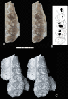
Fig 1. Abdounodus hamdii, Selandian (Phosphate level IIa) of the Ouled Abdoun Basin, Morocco.
MHNM.KHG.154 (collections of the Natural History Museum of Marrakech), left maxillary of preserving P3-4, M1-3; (A) strereophotographic pair of P3-4, M1-3 in occlusal view; (B) occlusal sketch; (C) strereophotographic pair of 3D models reconstructed from CT scans in occlusal view. The 3D models of MHNM.KHG.154 were reconstructed by F. Goussard (MNHN) from the CT scans using the computer programs Materialise Mimics Innovation Suite 18.0 Research Edition (x64), and Maxon Cinema 4D R15. Scale bar = 10 mm.
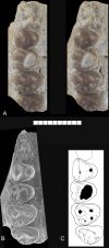
Fig 2. Abdounodus hamdii, Selandian (Phosphate level IIa) of the Ouled Abdoun Basin, Morocco.
MHNM.KHG.154 (collections of the Natural History Museum of Marrakech), right maxillary of Abdounodus hamdii preserving P3-4, M1-3; (A) strereophotographic pair of P3-4, M1-3 in occlusal view; B, s.e.m. photograph of occlusal view; (C) occlusal sketch. Scale bar = 10 mm.
Fig 3. Abdounodus hamdii, Selandian (Phosphate level IIa) of the Ouled Abdoun Basin, Morocco.
MNHN.F PM92, left dentary bearing roots of M3 and M2, crown of M1, P4, P3 (damaged) and root of P2 or P1 or C1. (A) stereophotographic pair in occlusal view and 3D model of the isolated teeth in occlusal view reconstructed from the CT scans; (B) Labial view; (C) Transparent 3D model in labial view reconstructed from the CT scans and showing the roots of the teeth; (D) 3D model of the isolated teeth in labial view, reconstructed from CT scans. (E-F) Same in lingual view. Scale bar = 10 mm.
Fig 4. Abdounodus hamdii, Selandian (Phosphate level IIa) of the Ouled Abdoun Basin, Morocco.
Transverse horizontal CT scan section of the partial lower jaw PM67 preserving M2 and M3 and showing the vertical root furrow and infilling crest of bone (arrows). A: anterior; P: posterior. Scale bar = 1 mm.
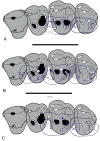
Fig 5. Occlusal sketch of the molars of Abdounodus hamdii reconstructed with the maxillary dentition of specimen MHNM.KHG.154.
(A) occlusal sketch of MHNM.KHG.154 with M2-3 of specimen PM68; (B) cclusal sketch of MHNM.KHG.154 with M1, M2, M3 of specimen OCP DEK/GE 310; (C) occlusal sketch of MHNM.KHG.154 with M2-3 of specimen PM67. The occlusion of opposed molars is reconstructed here in sub-centric position.
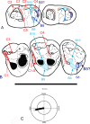
Fig 6. Wear facets of upper (MHNM.KHG.154) and lower molars of Abdounodus hamdii, and mastication compass following Koenigswald et al.
[30]. (A) Right lower molars M2 and M3; (B) left upper molars M1, M2, M3. C1-C10: Crompton [31] wear facets of tribosphenic molars (in red); B1-10, Butler [32] wear facets of lophodont molars (in blue). (C) Mastication compass of Abdounodus hamdii, indicating the inclination and direction of the lower jaw motion during power stroke of mastication. Note that the lower jaw motion is mostly labio-lingual (transverse) and horizontal, and is restricted to phase I of mastication.
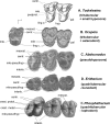
Fig 7. Comparison of the upper molar pattern (occlusal view) of early paenungulatomorphans (Afrotheria) with respect to the tribosphenic-tritubercular pattern of Todralestes, with indication of significant homologous structures.
(A) Todralestes variabilis (Eutheria, Pantolesta?), M2-1; (B) Ocepeia grandis (left), M3, and O. daouiensis (right), M2-1 (Paenungulatomorpha); (C) Abdounodus hamdii (Paenungulatomorpha), M3-1; (D) Eritherium azzouzorum (Paenungulata, Proboscidea), M3-1; (E) Phosphatherium escuilliei (Paenungulata, Proboscidea), M3-1. Not to scale. All teeth figured as right teeth. Abbreviations: centr: centrocrista; hyp: hypocone; interl: interloph; ling c: lingual cinglum (pre- and postcingulum); crest metal and crest protol: full crest-like protoloph and metaloph (true lophodonty); mle: metaconule; mesost: mesostyle; pal: paraconule; pseudohyp: pseudohypocone; postprot: postprotocrista.
Fig 8. Origin and evolution of the bilophodont pattern in paenungulates: a new structural scenario for upper molars including the stem taxa Ocepeia and Abdounodus which documents two intermediate stages between the tribosphenic-tritubercular pattern and the quadritubercular-bilophodont pattern.
Photography and occlusal sketch of teeth, all figured as right teeth. Symbols of dental structures: Black circles: paracone and metacone, green circle: metaconule; red circle: protocone; blue circle: hypocone; transverse lines: proto- and metaloph; double arrow: interloph.
Fig 9. Convergence of lophodonty in Paenungulata (Afrotheria) and Perissodactyla (Euungulata, Laurasiatheria).
The fourth upper molar cusp (postero-lingual cusp bearing the metaloph) is not homologous in Paenungulata and Perissodactyla: it is issued from a modified metaconule (pseudohypocone) in Paenungulata (Phosphatherium here) and from the true cingular hypocone in the Perissodactyla (Cymbalophus here) and early lophodont euungulates such as the Anthracobunidae (not shown here). Stem groups illustrating early stages of the evolution of the lophodonty remain poorly known in Euungulata, being represented mainly by phenacodontids such as Ectocion and Phenacodus that have a large hypocone. The stem paenungulates Ocepeia (not shown here) and Abdounodus are the first fossil taxa documenting intermediate morphological stages between the primitive tritubercular molar pattern and the derived quadritubercular and bilophodont molar pattern of the crown Paenungulata (here represented by Phosphatherium). Symbols of dental structures: Black circles: paracone and metacone, green circle: metaconule; blue circle: hypocone; red circle: protocone; transverse lines: proto- and metaloph. Occlusal sketch of teeth, all figured as right teeth.
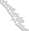
Fig 10. Relationships of Abdounodus and paenungulates.
Most parsimonious tree resulting from the unweigheted analysis with ordered features. Details, Bremer index and distribution of synapomorphies are provided in S2 Text (part III.1). Tree length: 735. Retention index: 53. Consistency Index: 37.
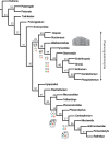
Fig 11. Relationships of Abdounodus and paenungulates.
Most parsimonious tree resulting from analysis with implied weighting and ordered features. This tree is in this work our reference topology for the discussion of the relationships of Abdounodus and Ocepeia and of the distribution of the characters. Main upper molar patterns are outlined at the node of lophodont taxa (incl. early stages) to show the convergent development of the pseudohypocone in the Paenungulatomorpha and the hypocone in the Perissodactyla and other lophodont euungulates such as Anthracobunidae. See Fig 9 for caption of symbols of dental structures. Details, Bremer index and distribution of synapomorphies of this tree are provided in S2 Fig and S2 Text (part III.2). The clades numbers are reported above the nodes. Tree length: 746. Retention index: 52. Consistency Index: 33.
Similar articles
- Petrosal and bony labyrinth morphology of the stem paenungulate mammal (Paenungulatomorpha) Ocepeia daouiensis from the Paleocene of Morocco.
Gheerbrant E, Schmitt A, Billet G. Gheerbrant E, et al. J Anat. 2022 Apr;240(4):595-611. doi: 10.1111/joa.13255. Epub 2020 Jul 31. J Anat. 2022. PMID: 32735727 Free PMC article. - Ocepeia (Middle Paleocene of Morocco): the oldest skull of an afrotherian mammal.
Gheerbrant E, Amaghzaz M, Bouya B, Goussard F, Letenneur C. Gheerbrant E, et al. PLoS One. 2014 Feb 26;9(2):e89739. doi: 10.1371/journal.pone.0089739. eCollection 2014. PLoS One. 2014. PMID: 24587000 Free PMC article. - Early African Fossils Elucidate the Origin of Embrithopod Mammals.
Gheerbrant E, Schmitt A, Kocsis L. Gheerbrant E, et al. Curr Biol. 2018 Jul 9;28(13):2167-2173.e2. doi: 10.1016/j.cub.2018.05.032. Epub 2018 Jun 28. Curr Biol. 2018. PMID: 30008332 - Mammal madness: is the mammal tree of life not yet resolved?
Foley NM, Springer MS, Teeling EC. Foley NM, et al. Philos Trans R Soc Lond B Biol Sci. 2016 Jul 19;371(1699):20150140. doi: 10.1098/rstb.2015.0140. Philos Trans R Soc Lond B Biol Sci. 2016. PMID: 27325836 Free PMC article. Review. - The chromosomes of Afrotheria and their bearing on mammalian genome evolution.
Svartman M, Stanyon R. Svartman M, et al. Cytogenet Genome Res. 2012;137(2-4):144-53. doi: 10.1159/000341387. Epub 2012 Aug 3. Cytogenet Genome Res. 2012. PMID: 22868637 Review.
Cited by
- Developmental influence on evolutionary rates and the origin of placental mammal tooth complexity.
Couzens AMC, Sears KE, Rücklin M. Couzens AMC, et al. Proc Natl Acad Sci U S A. 2021 Jun 8;118(23):e2019294118. doi: 10.1073/pnas.2019294118. Proc Natl Acad Sci U S A. 2021. PMID: 34083433 Free PMC article. - A new early Eocene deperetellid tapiroid illuminates the origin of Deperetellidae and the pattern of premolar molarization in Perissodactyla.
Bai B, Meng J, Mao FY, Zhang ZQ, Wang YQ. Bai B, et al. PLoS One. 2019 Nov 8;14(11):e0225045. doi: 10.1371/journal.pone.0225045. eCollection 2019. PLoS One. 2019. PMID: 31703104 Free PMC article. - Stripe and spot selection in cusp patterning of mammalian molar formation.
Morita W, Morimoto N, Otsu K, Miura T. Morita W, et al. Sci Rep. 2022 Jun 14;12(1):9149. doi: 10.1038/s41598-022-13539-w. Sci Rep. 2022. PMID: 35701484 Free PMC article. - Petrosal and bony labyrinth morphology of the stem paenungulate mammal (Paenungulatomorpha) Ocepeia daouiensis from the Paleocene of Morocco.
Gheerbrant E, Schmitt A, Billet G. Gheerbrant E, et al. J Anat. 2022 Apr;240(4):595-611. doi: 10.1111/joa.13255. Epub 2020 Jul 31. J Anat. 2022. PMID: 32735727 Free PMC article. - Improvements in the fossil record may largely resolve current conflicts between morphological and molecular estimates of mammal phylogeny.
Beck RMD, Baillie C. Beck RMD, et al. Proc Biol Sci. 2018 Dec 19;285(1893):20181632. doi: 10.1098/rspb.2018.1632. Proc Biol Sci. 2018. PMID: 30963896 Free PMC article.
References
- Waddell PJ, Okada N, Hasegawa M. Towards resolving the interordinal relationships of placental mammals. Syst. Biol. 1999; 48: 1–5. - PubMed
- Madsen O, Scally M, Douady CJ, Kao DJ et al. Parallel adaptive radiations in two major clades of placental mammals. Nature 2001; 409:610–614. - PubMed
- Murphy WJ, Eizirik E, Johnson WE, Zhang YP, Ryder OA, O’brien SJ. Molecular phylogenetics and the origin of placental mammals. Nature 2001; 409: 614–618. - PubMed
- Springer MS, Stanhope J, Madsen O, Dejong WW. Molecules consolidate the placental mammal tree. Trends Ecol. Evol. 2004; 19: 430–438. - PubMed
MeSH terms
Grants and funding
These authors have no support or funding to report.
LinkOut - more resources
Full Text Sources
Other Literature Sources
