Cutting Edge: IL-4, IL-21, and IFN-γ Interact To Govern T-bet and CD11c Expression in TLR-Activated B Cells - PubMed (original) (raw)
. 2016 Aug 15;197(4):1023-8.
doi: 10.4049/jimmunol.1600522. Epub 2016 Jul 18.
Arpita Myles 1, Daniel P Beiting 2, Kenneth J Roberts 3, Lucas Dawson 2, Ramin Sedaghat Herati 4, Bertram Bengsch 5, Susanne L Linderman 6, Erietta Stelekati 5, Rosanne Spolski 7, E John Wherry 5, Christopher Hunter 2, Scott E Hensley 6, Warren J Leonard 7, Michael P Cancro 8
Affiliations
- PMID: 27430719
- PMCID: PMC4975960
- DOI: 10.4049/jimmunol.1600522
Cutting Edge: IL-4, IL-21, and IFN-γ Interact To Govern T-bet and CD11c Expression in TLR-Activated B Cells
Martin S Naradikian et al. J Immunol. 2016.
Abstract
T-bet and CD11c expression in B cells is linked with IgG2c isotype switching, virus-specific immune responses, and humoral autoimmunity. However, the activation requisites and regulatory cues governing T-bet and CD11c expression in B cells remain poorly defined. In this article, we reveal a relationship among TLR engagement, IL-4, IL-21, and IFN-γ that regulates T-bet expression in B cells. We find that IL-21 or IFN-γ directly promote T-bet expression in the context of TLR engagement. Further, IL-4 antagonizes T-bet induction. Finally, IL-21, but not IFN-γ, promotes CD11c expression independent of T-bet. Using influenza virus and Heligmosomoides polygyrus infections, we show that these interactions function in vivo to determine whether T-bet(+) and CD11c(+) B cells are formed. These findings suggest that T-bet(+) B cells seen in health and disease share the common initiating features of TLR-driven activation within this circumscribed cytokine milieu.
Copyright © 2016 by The American Association of Immunologists, Inc.
Figures
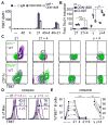
Figure 1. IL4 and IL21 act in a cell intrinsic manner to regulate TBET expression in vitro
Magnetically enriched CD23+ splenic B cells were cultured in vitro with various combinations of α-Ig-μ (IgM), α-CD40 (40), IL4 (4), IL21 (21), and IFNγ (γ). Mouse data are representative of 3 independent experiments. (A) WT or _Cd19_cre/+_Tbx21_f/f B cells treated for 48hrs and probed for TBET (ΔMFI=WT-mutant). (B) Tbx21 mRNA levels in WT cells treated for 20hrs. (C) WT, _Il21r_−/−, or _Stat6_−/− B cells were labeled with either CFSE (green plots) or Violet Cell Trace (VCT, purple plots), treated with ODN1826 and indicated cytokines for 48h, then stained for CD11c and TBET. (D) Magnetically enriched CD27−CD19+ human B cells were labeled with CFSE, treated for 108h, and probed for TBET on live, CFSE− cells. (E) Frequency of TBET+ B cells from each treatment across 6 healthy, adult donors.
Figure 2. TBET+CD11c+ cells delineate a BMEM cell subset and accumulate in _Il21_tg mice
(A–B) GC B and BMEM cells were analyzed for TBET and CD11c expression by FACS. GC B and BMEM cell gating strategies are in Supplemental Fig. 1G. All panels are representative of 3 independent experiments with ≥ 3 mice per strain. (A) TBET staining on GC B cells from C57BL/6 (B6, n=14) or BALB/c (n=23) mice with frequency enumeration. (B) TBET and CD11c staining on BMEM cells from B6 mice. (C) TBET and CD11c staining on splenic B-2 cells from WT and _Il21_tg mice. (D) Serum IgG1 or IgG2a/c (IgG2a + IgG2c) levels in WT and _Il21_tg mice were determined by ELISA. Values are means ± S.E.M. from 5 WT and 7 _Il21_tg mice.
Figure 3. Influenza virus infection drives TBET+CD11c+ BMEM cell formation in the absence of both IFNγ and IL4
Splenocytes were harvested from non-infected (−) or day 10 post i.n. 30 TCID50 PR8 infection (+) WT (n=21, black bars), _Ifng_−/− (n=10, white bars), or _Il4_−/−_Ifng_−/− (n=13, gray bars) mice across 3–7 experiments with ≥3 mice per group. GC B, BMEM, and TFH cell gating strategies are in Supplemental Figures 1G & 1J. (A) Enumeration of GC B cells. (B) TBET staining on GC B cells. (C) Il4 and (D) Il21 mRNA levels from sorted naïve CD62L+ CD4 T (TN, n=9) or TFH cells. (E) Proportions and (F) numbers of TBET+CD11c+ BMEM cells.
Figure 4. Activated B cells express T-BET independent of IFNγ in IL4 limiting conditions
Splenocytes and sera were harvested from non-infected (−) or day 14 post oral gavage (+) of 200 H. polygyrus in WT (n=20, black bars), _Il4_−/− (n=24, white bars), or _Il4_−/−_Ifng_−/− (n=11, gray bars) mice across 3–6 experiments with ≥3 mice per group. GC B, BMEM, and TFH cell gating strategies are in Supplemental Figures 1G & 1J. (A) Enumeration of GC B cells. (B) TBET staining on GC B cells. (C) Serum concentrations of IgG1 and IgG2c + IgG2b. (D) Il21 mRNA levels from sorted TFH cells. (E) Proportions and (F) numbers of TBET+CD11c+ BMEM cells.
Similar articles
- CD11c+ T-bet+ B Cells Require IL-21 and IFN-γ from Type 1 T Follicular Helper Cells and Intrinsic Bcl-6 Expression but Develop Normally in the Absence of T-bet.
Levack RC, Newell KL, Popescu M, Cabrera-Martinez B, Winslow GM. Levack RC, et al. J Immunol. 2020 Aug 15;205(4):1050-1058. doi: 10.4049/jimmunol.2000206. Epub 2020 Jul 17. J Immunol. 2020. PMID: 32680956 Free PMC article. - Regulation of B-cell function and expression of CD11c, T-bet, and FcRL5 in response to different activation signals.
Kleberg L, Courey-Ghaouzi AD, Lautenbach MJ, Färnert A, Sundling C. Kleberg L, et al. Eur J Immunol. 2024 Aug;54(8):e2350736. doi: 10.1002/eji.202350736. Epub 2024 May 3. Eur J Immunol. 2024. PMID: 38700378 - T-box transcription factor T-bet, a key player in a unique type of B-cell activation essential for effective viral clearance.
Rubtsova K, Rubtsov AV, van Dyk LF, Kappler JW, Marrack P. Rubtsova K, et al. Proc Natl Acad Sci U S A. 2013 Aug 20;110(34):E3216-24. doi: 10.1073/pnas.1312348110. Epub 2013 Aug 6. Proc Natl Acad Sci U S A. 2013. PMID: 23922396 Free PMC article. - CD11c+ T-bet+ memory B cells: Immune maintenance during chronic infection and inflammation?
Winslow GM, Papillion AM, Kenderes KJ, Levack RC. Winslow GM, et al. Cell Immunol. 2017 Nov;321:8-17. doi: 10.1016/j.cellimm.2017.07.006. Epub 2017 Jul 19. Cell Immunol. 2017. PMID: 28838763 Free PMC article. Review. - Role of CD11c+ T-bet+ B cells in human health and disease.
Karnell JL, Kumar V, Wang J, Wang S, Voynova E, Ettinger R. Karnell JL, et al. Cell Immunol. 2017 Nov;321:40-45. doi: 10.1016/j.cellimm.2017.05.008. Epub 2017 Jul 11. Cell Immunol. 2017. PMID: 28756897 Review.
Cited by
- Hyper-metabolic B cells in the spleens of old mice make antibodies with autoimmune specificities.
Frasca D, Romero M, Garcia D, Diaz A, Blomberg BB. Frasca D, et al. Immun Ageing. 2021 Feb 27;18(1):9. doi: 10.1186/s12979-021-00222-3. Immun Ageing. 2021. PMID: 33639971 Free PMC article. - The deficiency in Th2-like Tfh cells affects the maturation and quality of HIV-specific B cell response in viremic infection.
Noto A, Suffiotti M, Joo V, Mancarella A, Procopio FA, Cavet G, Leung Y, Corpataux JM, Cavassini M, Riva A, Stamatatos L, Gottardo R, McDermott AB, Koup RA, Fenwick C, Perreau M, Pantaleo G. Noto A, et al. Front Immunol. 2022 Aug 24;13:960120. doi: 10.3389/fimmu.2022.960120. eCollection 2022. Front Immunol. 2022. PMID: 36091040 Free PMC article. - Rare SH2B3 coding variants in lupus patients impair B cell tolerance and predispose to autoimmunity.
Zhang Y, Morris R, Brown GJ, Lorenzo AMD, Meng X, Kershaw NJ, Kiridena P, Burgio G, Gross S, Cappello JY, Shen Q, Wang H, Turnbull C, Lea-Henry T, Stanley M, Yu Z, Ballard FD, Chuah A, Lee JC, Hatch AM, Enders A, Masters SL, Headley AP, Trnka P, Mallon D, Fletcher JT, Walters GD, Šestan M, Jelušić M, Cook MC, Athanasopoulos V, Fulcher DA, Babon JJ, Vinuesa CG, Ellyard JI. Zhang Y, et al. J Exp Med. 2024 Apr 1;221(4):e20221080. doi: 10.1084/jem.20221080. Epub 2024 Feb 28. J Exp Med. 2024. PMID: 38417019 Free PMC article. - Age/autoimmunity-associated B cells in inflammatory arthritis: An emerging therapeutic target.
Li ZY, Cai ML, Qin Y, Chen Z. Li ZY, et al. Front Immunol. 2023 Jan 24;14:1103307. doi: 10.3389/fimmu.2023.1103307. eCollection 2023. Front Immunol. 2023. PMID: 36817481 Free PMC article. Review. - Involvement of age-associated B cells in EBV-triggered autoimmunity.
Sachinidis A, Garyfallos A. Sachinidis A, et al. Immunol Res. 2022 Aug;70(4):546-549. doi: 10.1007/s12026-022-09291-y. Epub 2022 May 16. Immunol Res. 2022. PMID: 35575824 Free PMC article.
References
- Gerth AJ, Lin L, Peng SL. T-bet regulates T-independent IgG2a class switching. Int Immunol. 2003;15:937–944. - PubMed
- Liu N, Ohnishi N, Ni L, Akira S, Bacon KB. CpG directly induces T-bet expression and inhibits IgG1 and IgE switching in B cells. Nat Immunol. 2003;4:687–693. - PubMed
- Szabo SJ, Kim ST, Costa GL, Zhang X, Fathman CG, Glimcher LH. A novel transcription factor, T-bet, directs Th1 lineage commitment. Cell. 2000;100:655–669. - PubMed
MeSH terms
Substances
Grants and funding
- R01 AG030227/AG/NIA NIH HHS/United States
- T32 AI055428/AI/NIAID NIH HHS/United States
- R01 AI118691/AI/NIAID NIH HHS/United States
- R01 AI108686/AI/NIAID NIH HHS/United States
- K08 AI114852/AI/NIAID NIH HHS/United States
- R01 AI113047/AI/NIAID NIH HHS/United States
- T32 CA009171/CA/NCI NIH HHS/United States
LinkOut - more resources
Full Text Sources
Other Literature Sources
Research Materials


