The mammalian dynein-dynactin complex is a strong opponent to kinesin in a tug-of-war competition - PubMed (original) (raw)
. 2016 Sep;18(9):1018-24.
doi: 10.1038/ncb3393. Epub 2016 Jul 25.
Affiliations
- PMID: 27454819
- PMCID: PMC5007201
- DOI: 10.1038/ncb3393
The mammalian dynein-dynactin complex is a strong opponent to kinesin in a tug-of-war competition
Vladislav Belyy et al. Nat Cell Biol. 2016 Sep.
Abstract
Kinesin and dynein motors transport intracellular cargos bidirectionally by pulling them in opposite directions along microtubules, through a process frequently described as a 'tug of war'. While kinesin produces 6 pN of force, mammalian dynein was found to be a surprisingly weak motor (0.5-1.5 pN) in vitro, suggesting that many dyneins are required to counteract the pull of a single kinesin. Mammalian dynein's association with dynactin and Bicaudal-D2 (BICD2) activates its processive motility, but it was unknown how this affects dynein's force output. Here, we show that formation of the dynein-dynactin-BICD2 (DDB) complex increases human dynein's force production to 4.3 pN. An in vitro tug-of-war assay revealed that a single DDB successfully resists a single kinesin. Contrary to previous reports, the clustering of many dyneins is not required to win the tug of war. Our work reveals the key role of dynactin and a cargo adaptor protein in shifting the balance of forces between dynein and kinesin motors during intracellular transport.
Conflict of interest statement
The authors declare no competing financial interests.
Figures
Figure 1. Activation of human dynein motility by dynactin, BICD2N, and artificial cargos
(a) Schematic depiction of the DDB complex on a microtubule. The putative orientation of the p150 subunit of dynactin is shown with a semi-transparent outline and omitted from illustrations elsewhere in the manuscript. (b) Sample kymographs represent the motility of individual dynein alone and DDB motors on axonemes. (c) Denaturing SDS–PAGE gel of purified dynein fractions. Bands corresponding to all dynein subunits can be observed. (d) Denaturing SDS–PAGE gel of purified BICD2N. Full gel scans can be found in Supplementary Fig. 5. (e) Illustration of dynein with a quantum dot attached to the tail (top) and sample kymograph (bottom). The experiment was repeated 4 times. (f) Illustration of dynein with a 200 nm bead attached to the tail (top) and sample kymograph (bottom). The experiment was repeated 3 times. (g) Illustration of dynein with an optically trapped 860 nm bead attached to the tail (top) and sample trajectory of dynein pulling the bead against a constant 0.4 pN hindering force in force feedback mode (bottom). The experiment was repeated 4 times. (h) Effect of cargo size and type on dynein velocity in comparison to the DDB complex. N = 51, 83, 21, 19, 15 runs from 3 independent experiments in order from top to bottom. Vertical bars represent median values and quartiles. Median values, from top to bottom, are 373, 188, 213, 29, and 56 nm/s. Mean ± s.e.m., from top to bottom, are 513 ± 58, 257 ± 26, 200 ± 23, 49 ± 11, and 79 ± 11 nm/s.
Figure 2. Effect of dynactin and BICD2N on dynein force production
(a) (Top) The optically trapped bead, represented with a force arrow, is attached to GFP on dynein’s tail via an anti-GFP antibody. (Middle) Trace showing a typical stall of a bead driven by GFP-dynein in a fixed-trap assay. Red arrowhead represents the detachment of the motor from a microtubule after the stall. (Bottom) The histogram of observed stalls reveals the mean stall force (mean ± s.e.m., N = 50 stalls from 12 beads in 4 independent experiments). (b) Stall force of GFP-dynein with the addition of 5× molar excess of dynactin (N = 41 stalls from 10 beads in 4 independent experiments). (c) Stall force measurement of GFP-dynein with the addition of 5× molar excess of dynactin and 2× molar excess of BICD2N. The underlying distribution of the observed stalls is fitted to two Gaussians (blue curve; N = 195 stalls from 47 beads in 19 independent experiments) using a Gaussian Mixture Model. A three-Gaussian fit, shown with a dotted line, is not statistically warranted as determined by the Bayes Information Criterion (see Supplementary Fig. 2). (d) Stall force measurement of dynein with the addition of 5× molar excess of dynactin and 2× molar excess of BICD2N with a C-terminal GFP (BICD2N-GFP). The BICD2N-GFP is the attachment point (N = 45 stalls from 14 beads in 4 independent experiments). (e) Stall force measurement of kinesin-1 with a C-terminal GFP fusion as the attachment point (N = 37 stalls from 15 beads in 4 independent experiments).
Figure 3. Processive motility of DDB complexes is driven by single dynein dimers
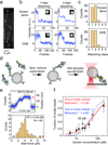
(a) GFP-dynein molecules tightly bound to microtubules in the absence of ATP. (b) Intensity traces of GFP-dynein alone in the presence and absence of dynactin and BICD2N show one- and two-step photobleaching. (c) Histograms showing the number of photobleaching steps of dynein in the absence and presence of dynactin and BICD2N. N = 127 for dynein alone and N = 192 for DDB. The experiment was repeated 3 times with dynein from two independent preparations. (d) Schematic depiction of the modified sample preparation for optical trapping. Dynein is mixed with beads and excess dynein is removed by centrifugation. BICD2N and dynactin are added after the removal of free motors to rule out dynactin- and BICD2N-dependent aggregation of dynein motors on beads. (e) Representative stall trace of a DDB complex and distribution of stall forces of the DDB complexes (mean ± s.e.m.). The experiment was repeated 3 times. (f) Fraction of dynein-coated beads moving as a function of dynein concentration. Values are represented as the mean ± the square root of F(1 − F)/N, with N being the number of beads tested. The solid red line represents a fit to the Poisson probability 1 − e−λC that the bead is carried by one or more motors, where C is dynein concentration and λ is the fit parameter (reduced χ2 = 0.26). The dashed blue line represents a fit to the probability 1 − e−λC − (λC)e−λC that the bead is carried by two or more motors (reduced χ2 = 4.62). For each data point, from left to right, N = 23, 25, 10, 482, 28, 35, 20, 24 beads from 3 independent experiments, except for the 20 pM data point, which was obtained from 19 independent experiments. The mean values, from left to right, are 0.087, 0.16, 0.2, 0.22, 0.22, 0.33, 0.61, 0.88.
Figure 4. In vitro competition experiment between human dynein and human kinesin
(a) Kinesin-1 is labeled with a short DNA oligo and TMR, while dynein is separately labeled with a complementary oligo and Alexa647. The two motors are connected through DNA hybridization. (b) The fraction of kinesin motors labeled with DNA was determined from an SDS-PAGE denaturing gel. (c) The fraction of dynein motors labeled with DNA was determined by separately imaging total protein quantity and fraction of DNA-free motors in an SDS-PAGE gel. Higher DNA:dynein ratios resulting in lower Alexa647 fluorescence (bottom), since the DNA occupied larger fractions of SNAPf binding sites, making them unavailable for labeling with Alexa647-BG-GLA. Full gel scans can be found in Supplementary Fig. 5. (d) Kymographs of dynein-Alexa647 (red) and kinesin-TMR (cyan) motility in the absence of dynactin and BICD2N on microtubules. Colocalizers are identified with black arrows. (e) Velocity distribution of kinesin only, dynein only, and kinesin-dynein colocalizers. Positive values correspond to plus end-directed velocities. From top to bottom, N = 655, 44, 59 runs from 3 independent experiments. Vertical bars represent median values and quartiles. Median values, from top to bottom, are 590, −76, and 460 nm/s. (f) Kymographs of dynein and kinesin motility in the presence of dynactin and BICD2N. The black arrows indicate colocalizers. Colocalizers in the top row are walking towards the minus-end of the microtubule. The red and cyan channels are laterally offset by five pixels to enhance the visibility of the colocalizers. The experiment was repeated 4 times. (g) Representative trace of DDB slowly walking towards the plus end in response to a 6 pN pulling force exerted by the optical trap. The experiment was repeated 3 times. (h) Velocity distribution of kinesin only, DDB only, kinesin-DDB colocalizers, and DDB walking against a plus end-directed 6 pN force. From top to bottom, N = 513, 79, 55, 35 runs from 3 independent experiments. Vertical bars represent median values and quartiles. Median values, from top to bottom, are 680, −176, 26, and 10 nm/s.
Similar articles
- Kinesin-1, -2, and -3 motors use family-specific mechanochemical strategies to effectively compete with dynein during bidirectional transport.
Gicking AM, Ma TC, Feng Q, Jiang R, Badieyan S, Cianfrocco MA, Hancock WO. Gicking AM, et al. Elife. 2022 Sep 20;11:e82228. doi: 10.7554/eLife.82228. Elife. 2022. PMID: 36125250 Free PMC article. - Synthetic cargo adaptors reveal molecular features that can enhance dynein activation.
Siva A, Gillies JP, de Borchgrave A, Garrott SR, Mishra R, Jbeily RE, Clarke RS, Conklin C, Gibson D, Zang JL, DeSantis ME. Siva A, et al. bioRxiv [Preprint]. 2025 Jun 7:2025.06.06.658359. doi: 10.1101/2025.06.06.658359. bioRxiv. 2025. PMID: 40501990 Free PMC article. Preprint. - Lis1 activates dynein motility by modulating its pairing with dynactin.
Elshenawy MM, Kusakci E, Volz S, Baumbach J, Bullock SL, Yildiz A. Elshenawy MM, et al. Nat Cell Biol. 2020 May;22(5):570-578. doi: 10.1038/s41556-020-0501-4. Epub 2020 Apr 27. Nat Cell Biol. 2020. PMID: 32341547 Free PMC article. - Fat traffic control: S-acylation in axonal transport.
Doerksen AH, Herath NN, Sanders SS. Doerksen AH, et al. Mol Pharmacol. 2025 Jun;107(6):100039. doi: 10.1016/j.molpha.2025.100039. Epub 2025 Apr 16. Mol Pharmacol. 2025. PMID: 40349611 Review. - Home treatment for mental health problems: a systematic review.
Burns T, Knapp M, Catty J, Healey A, Henderson J, Watt H, Wright C. Burns T, et al. Health Technol Assess. 2001;5(15):1-139. doi: 10.3310/hta5150. Health Technol Assess. 2001. PMID: 11532236
Cited by
- Resolving cargo-motor-track interactions with bifocal parallax single-particle tracking.
Cheng X, Chen K, Dong B, Filbrun SL, Wang G, Fang N. Cheng X, et al. Biophys J. 2021 Apr 20;120(8):1378-1386. doi: 10.1016/j.bpj.2020.11.2278. Epub 2020 Dec 25. Biophys J. 2021. PMID: 33359832 Free PMC article. - Systems mapping of bidirectional endosomal transport through the crowded cell.
Jongsma MLM, Bakker N, Voortman LM, Koning RI, Bos E, Akkermans JJLL, Janssen L, Neefjes J. Jongsma MLM, et al. Curr Biol. 2024 Oct 7;34(19):4476-4494.e11. doi: 10.1016/j.cub.2024.08.026. Epub 2024 Sep 13. Curr Biol. 2024. PMID: 39276769 Free PMC article. - Kinesin and dynein use distinct mechanisms to bypass obstacles.
Ferro LS, Can S, Turner MA, ElShenawy MM, Yildiz A. Ferro LS, et al. Elife. 2019 Sep 9;8:e48629. doi: 10.7554/eLife.48629. Elife. 2019. PMID: 31498080 Free PMC article. - Time-dependent alterations in the rat nigrostriatal system after intrastriatal injection of fibrils formed by α-Syn and tau fragments.
Yang X, Wang J, Zeng W, Zhang X, Yang X, Xu Y, Xu Y, Cao X. Yang X, et al. Front Aging Neurosci. 2022 Nov 28;14:1049418. doi: 10.3389/fnagi.2022.1049418. eCollection 2022. Front Aging Neurosci. 2022. PMID: 36518823 Free PMC article. - Lis1 slows force-induced detachment of cytoplasmic dynein from microtubules.
Kusakci E, Htet ZM, Zhao Y, Gillies JP, Reck-Peterson SL, Yildiz A. Kusakci E, et al. Nat Chem Biol. 2024 Apr;20(4):521-529. doi: 10.1038/s41589-023-01464-6. Epub 2023 Nov 2. Nat Chem Biol. 2024. PMID: 37919547 Free PMC article.
References
- Burgess SA, Walker ML, Sakakibara H, Knight PJ, Oiwa K. Dynein structure and power stroke. Nature. 2003;421:715–718. - PubMed
- Torisawa T, et al. Autoinhibition and cooperative activation mechanisms of cytoplasmic dynein. Nat. Cell Biol. 2014;16:1118–1124. - PubMed
- Wang Z, Sheetz MP. One-dimensional diffusion on microtubules of particles coated with cytoplasmic dynein and immunoglobulins. Cell Struct. Funct. 1999;24:373–383. - PubMed
MeSH terms
Substances
LinkOut - more resources
Full Text Sources
Other Literature Sources
Miscellaneous


