Cellular tagging as a neural network mechanism for behavioural tagging - PubMed (original) (raw)
Figure 1. Novel context exploration (NCE) training in a narrow time window leads to NOR–LTM formation.
(a) Novel object recognition (NOR) set-up. Mice were habituated to the NOR arena in the absence of objects for 6 min per day for 4 consecutive days, after which they were exposed for 5 min (weak training) or 15 min (strong training) in the same arena to two objects (A and B). Then, after 30 min or 24 h, they underwent a 5 min retention test in which one of the objects (B) was replaced with a novel object (C). (b–e) Animals' exploration preference for the familiar or novel object in a memory retention test (percent of time exploring each object). Bars above each graph indicate the time interval between training and test. An asterisk indicates a significant difference between the two preferences. Data are presented as mean±s.e.m. VEH, vehicle; ANI, anisomycin. (b) NOR–LTM was formed by training for 15 min (_n_=5; _t_-test, _t_8=−4.402, _P_=0.002). (c) NOR–LTM was not formed by training for 5 min (_n_=5; _t_-test, _t_8=−1.203, _P_=0.263). (d) NOR–STM was formed by training for 5 min (_n_=9; _t_-test, _t_16=−3.694, _P_=0.001). (e) NOR–STM formed by training for 5 min was not affected by protein synthesis inhibitor (VEH: _n_=10; _t_-test, _t_18=−6.799, _P_=2.3E-06; ANI: _n_=7; _t_-test, _t_12=−8.034, _P_=3.6E-06; VEH versus ANI for exploration preference for C in test: _t_-test, _t_15=0.551, _P_=0.589). There was no significant difference between any of the groups in total exploration time at training (_t_-test, _t_15=−0.071, P<0.943). (f) Effect of NCE on the retention of NOR–LTM. Left, behavioural tag-training scheme. Right, exploration preference for familiar and novel objects in test sessions (Pre 180 min: _n_=8; _t_-test, _t_14=−0.953, _P_=0.350; Pre 60 min: _n_=5; _t_-test, _t_8=−3.683, _P_=0.006; Control: _n_=25; _t_-test, _t_48=0.304, _P_=0.762; Post 30 min: _n_=8: _t_-test, _t_14=−6.880, _P_=7.5E-06; Post 60 min: _n_=9: t test, t16=−6.160, _P_=1.3E-05; Post 180 min: _n_=14; _t_-test, _t_26=−1.290, _P_=0.208). Control, no NCE training control. There was no significant difference between any of the groups for total exploration time at training (one-way ANOVA: _F_5,68=0.673, _P_=0.645). The number sign (#) and asterisk indicate a significant difference (*P<0.05, #P<0.05). Data are presented as mean±s.e.m. Error bars indicate s.e.m. n, number of animals.
Figure 2. Place novelty promotes NOR–LTM dependently of hippocampal protein synthesis.
(a–b) Exploration preferences for familiar (A) and novel (C) objects in test session. Behavioural tag-training involved NCE (10 min) and then, after 60 min, NOR training (5 min). (a) Left, behavioural paradigm. ANI was injected into the hippocampus immediately after the NCE or NOR training. Right, effect of ANI on NOR–LTM (VEH after novelty: _n_=9; _t_-test, _t_16=−8.637, _P_=2E-07. ANI after novelty: _n_=9; _t_-test, _t_16=0.661, _P_=0.517. VEH after NOR: _n_=10; _t_-test, _t_18=−4.150, _P_=0.0006; ANI after NOR, _n_=9, _t_-test _t_16=−5.837, _P_=2.5E-05; two-way ANOVA of the preference for object C during the retention test: drug, _F_1,33=3.518, _P_=0.069; time, _F_1,33=3.716, _P_=0.062; Drug versus time interaction, _F_1,33=10.39, _P_=0.002 (Tukey–Kramer post hoc test), * P<0.05). There was no significant difference between any of the groups for total exploration time at training (two-way ANOVA of total exploration time at training: drug, _F_1,33=0.411, _P_=0.525; time, _F_1,33=0.388, _P_=0.537; Drug versus time interaction, _F_1,33=1.687, _P_=0.203). (b) Left, behavioural paradigm. Mice were familiarized with the square chamber for 6 min per day for 4 consecutive days before the onset of behavioural tag-training. Right, effect of the familiarization on NOR–LTM (familiarized: _n_=10; _t_-test, _t_18=−0.330, _P_=0.744; novelty: _n_=9; _t_-test, _t_16=−5.219, _P_=8.4E-05; _t_-test for exploration preference for C between familiarized and novelty, _t_17=3.462, _P_=0.002). There was no significant difference between any of the groups for total exploration time at training (_t_-test _t_17=0.929, _P_=0.380). Data are presented as mean±s.e.m. of the percentage of time exploring a particular object out of the total time of object exploration. # and * indicate a significant difference (*P<0.05, #P<0.05). Error bars indicate s.e.m. n, number of animals.
Figure 3. Cell ensemble analyses after behavioural tag-training in hippocampal subregions.
(a) catFISH experiment scheme. Control mice were familiarized to the square chamber and subjected to behavioural tag-training. Mice were killed 5 min after the behavioural session. Although non-behavioural tag group done outside the temporal proximity critical to tagging (that is, 180 min) may also serve as a proper control, the cytoplasmic Arc RNA signal disappears 60 min after transcription, which made it impossible to carry out the catFISH experiment outside the temporal proximity critical to tagging. (b) Scheme of intracellular localization of Arc RNA. (c–f) Detection of cells activated during behavioural tag-training. Left panels, representative photomicrographs of Arc signals in slices from CA1 (c) and CA3 (e) of home cage, familiarized and novelty groups. The Arc RNA signal and DAPI nuclear staining are shown in green and blue, respectively. Scale bar, 25 μm. Middle graphs show the percentages of cells containing cytoplasmic (yellow arrowheads) or nuclear (white arrowheads) Arc RNA in DAPI-positive cells in CA1 (c) and CA3 (e). Right panels show the percentages of cytoplasmic and nuclear Arc RNA double-positive cells (red arrowheads) in CA1 (c) and CA3 (e). Chance levels for double-positive cells are also shown in the graph (grey diamonds). Familiarized and novelty groups showed higher percentages of cytoplasmic or nuclear Arc RNA-positive cells in CA1 and CA3 than did the home cage group, but both percentages were comparable between familiarized and novelty groups (home cage, _n_=9; familiarized, _n_=11; novelty, _n_=12; one-way ANOVA, % CA1 cytoplasmic: _F_2,31=13.04, _P_=9.1E-05; % CA1 nuclear: _F_2,31=15.38, _P_=2.8E-05; % CA3 cytoplasmic: _F_2,31=27.55, _P_=2E-07; % CA3 nuclear: _F_2,31=13.45, _P_=7.3E-05, with Tukey–Kramer post hoc tests, * P<0.05). # indicates a significant difference between percentage of overlap and its chance level in each group (CA1: home cage, _t_-test _t_16=1.954, _P_=0.068; familiarized, _t_-test _t_20=4.567, _P_=0.0001; novelty, _t_-test _t_22=2.781, _P_=0.010; CA3: home cage, _t_-test _t_16=1.720, _P_=0.104; familiarized, _t_-test _t_20=4.699, _P_=0.0001; novelty, _t_-test _t_22=4.734, _P_=0.0001, #P<0.05). The size of each circle reflects the cell number. n, number of animals; three sections were analysed from each animal. Data are presented as mean±s.e.m. Error bars indicate s.e.m. NS, not significant. Venn diagram showing percent overlap between NOR and NCE (place memory ensemble) in CA1 (d) and CA3 (f).
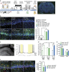
Figure 4. Optogenetic silencing of the cell ensembles corresponding to the initial place impairs NOR memory retrieval.
(a) Left, optogenetic experimental scheme. The activity-dependent targeting of neurons in c-fos-tetracycline transactivator (c-fos::tTA) mice treated with a lentivirus (LV) harbouring a TRE upstream of an archaerhodopsin-T and enhanced yellow fluorescent protein (ArchT–EYFP) fusion gene. The horizontal blue line indicates the presence of Dox. Twelve days after LV infection, c-fos::tTA mice underwent behavioural tag-training consisting of NOR, followed by NCE. After 2 days of OFF-Dox, mice were subjected to context-exposure session consisting of exposure to a square or circular chamber under OFF-Dox conditions. ArchT–EYFP expression was induced in activated cells (green). On the next day, mice were subjected to the NOR test session under ON-Dox conditions, with or without laser illumination in the hippocampus, and then were killed for immunohistochemistry. Right, representative ArchT–EYFP labelling pattern of CA1 cells in a LV-injected c-fos::tTA mouse that had been exposed to the square chamber OFF-Dox. The EYFP (green) and DAPI (blue) signals were visualized by immunostaining for EYFP followed by fluorescence microscopy. Scale bar, 1 mm. (b) Activity-dependent and OFF-Dox-dependent labelling of cells with ArchT–EYFP in the CA1 region. Left, representative photomicrographs showing ArchT–EYFP labelling patterns in CA1 of LV-injected c-fos::tTA mice obtained in different conditions of context-exposure session (square, circular or home cage) and illumination (Laser ON and Laser OFF). Square, square chamber; circle, circular chamber. Scale bar, 50 μm. Right, percentages of ArchT–EYFP-positive cells in the CA1 region normalized to DAPI-positive cells. Exposure to the chambers without Dox treatment induced the larger ArchT–EYFP expression compared with either the home cage or ON-Dox conditions (ON-Dox home cage, _n_=5; ON-Dox square, _n_=3; OFF-Dox home cage, _n_=4; OFF-Dox square laser OFF, _n_=9; OFF-Dox square laser ON, _n_=11; OFF-Dox circle laser ON, _n_=11; two-way ANOVA for the percentages of ArchT–EYFP-positive cells: Dox treatment versus context-exposure interaction, _F_1,39=18.44, _P_=0.0001; Dox treatment, _F_1,39=18.91, _P_=0.0001; Context-exposure, _F_1,39=10.33, _P_=0.002 (with the Tukey–Kramer post hoc test); * P<0.05). n, number of animals; three sections were analysed from each animal. Data are presented as mean±s.e.m. Error bars indicate s.e.m. (c) Hyperpolarization and suppression of spiking by light-emitting diode illumination in a CA1 neuron of LV-injected c-fos::tTA mice. Left, low and high magnification images of the slice and the neuron recorded (inset). The arrowhead indicates the tip of the recording pipette. Scale bar, 200 μm, 10 μm (inset). Right, the membrane potentials of the neuron (above) and the injected current (below). The yellow boxes indicate the periods of yellow light-emitting diode illumination. Horizontal dotted line shows −60 mV. (d) Silencing of initial chamber-related cell ensembles impairs NOR–LTM retrieval (square laser OFF, _n_=9, _t_-test _t_16=−8.780, _P_=1.6E-07; square laser ON, _n_=11, _t_-test _t_20=1.364, _P_=0.187; circle laser ON, _n_=11, _t_-test _t_20=−7.330, _P_=4.4E-07; one-way ANOVA, _F_2,30=16.85, _P_=2E-05, with Tukey–Kramer post hoc tests, *P<0.05). (e) Optogenetic manipulation did not affect the exploration behaviour (one-way ANOVA, in training: _F_2,30=0.444, _P_=0.646; in test: _F_2,30=1.167, _P_=0.326). (f) Laser illumination inhibited Egr1 expression in ArchT–EYFP-positive cells 60 min after NOR memory retrieval. Left, representative photomicrographs of triple immunofluorescence for EYFP (green), Egr1 (red) and DAPI (blue). Yellow arrowheads indicate Egr1 and EYFP double-positive cells. Scale bar, 50 μm. Right, percentages of Egr1 and EYFP double-positive cells normalized to EYFP-positive cells in the CA1 region (one-way ANOVA, _F_2,30=6.439, _P_=0.005, with Tukey–Kramer post hoc test, *P<0.05). # and * indicate significant differences (* P<0.05, # P<0.05). Error bars indicate s.e.m. NS, not significant.
Figure 5. Cell ensemble analyses in CA1 after the retrieval test.
(a) catFISH experiment scheme. Two days after behavioural tag-training, mice were exposed to a square or circular chamber followed by NOR testing at an interval of 30 min (Square-NOR or Circle-NOR, respectively), and then killed 5 min after the behavioural session. (b) Scheme of intracellular Arc RNA localization after the retrieval test. (c,d) Detection of cells activated during the behavioural session. (c) Representative photomicrographs of Arc signals in slices from the CA1 of Square-NOR or Circle-NOR groups. The Arc RNA signal and DAPI nuclear staining are shown in green and blue, respectively. Scale bar, 25 μm. (d) Left graph shows the percentage of cells containing cytoplasmic (yellow arrowheads in c) or nuclear (white arrowheads in c) Arc RNA in DAPI-positive cells. Right graph shows the percentage of cytoplasmic and nuclear Arc RNA-double-positive cells (red arrowheads in c). Chance level for double-positive cells (grey diamonds) is also shown. The percentages of cells positive for cytoplasmic or nuclear Arc RNA were comparable between Square-NOR and Circle-NOR groups (Square-NOR, _n_=9; Circle-NOR, _n_=9; % cytoplasmic, _t_-test _t_16=0.709, _P_=0.488; % nuclear, _t_-test _t_16=0.981, _P_=0.341). # indicates significant difference between the percentage of overlap and the chance level in each group (Square-NOR, _t_-test _t_16=3.820, _P_=0.001; Circle-NOR, _t_-test _t_16=4.157, _P_=0.0007). * indicates a significant difference (P<0.05) with unpaired Student's _t_-test. n, number of animals; three sections were analysed from each animal. Data are presented as mean±s.e.m. Error bars indicate s.e.m. NS, not significant. (e) Venn diagram showing percent overlap between NOR and place memory ensemble in CA1. Size of each circle reflects the cell number.
Figure 6. Blockade of dopamine D1/D5 receptor during behavioural tag-training affects the NOR–LTM and the ratio of overlapping cells in CA1.
(a) Left, behavioural experimental scheme. Dopamine D1/D5 receptor antagonist SCH23390 was intraperitoneally injected into the mice immediately after the NOR training, followed by NCE at an interval of 30 min. Right, effect of the SCH23390 on NOR–LTM. Exploration preferences for familiar (A) and novel (C) objects in a test session (VEH, _n_=7, _t_-test _t_12=−10.78, _P_=1.5E-07; SCH23390, _n_=9, _t_-test _t_16=−2.816, _P_=0.012). There was no significant difference between any of the groups for total exploration time at training (_t_-test _t_14=−0.135, _P_=0.894). Data are presented as mean±s.e.m. of the percentage of time exploring a particular object over the total time of object exploration. #, significant difference between familiar and novel objects in each group. *, significant difference between groups in exploration preference for C in test session. Error bars indicate s.e.m. (b) catFISH experiment scheme. Mice were subjected to behavioural tag-training as shown in a and then killed 5 min after the behavioural session. (c) Scheme of intracellular Arc RNA localization after behavioural tag-training. (d) Representative photomicrographs of Arc signals in slices from the CA1 of VEH or SCH23390 group. The Arc RNA signal and DAPI nuclear staining are shown in green and blue, respectively. Scale bar, 25 μm. (e) Left graph shows the percentages of cells containing cytoplasmic (yellow arrowheads in d) or nuclear (white arrowheads in d) Arc RNA in DAPI-positive cells. Right graph shows the percentage of cytoplasmic and nuclear Arc RNA double-positive cells (red arrowheads in d) in the CA1 region. Chance level for double-positive cells (grey diamonds) is also shown. The percentages of cells positive for cytoplasmic or nuclear Arc RNA were comparable between the VEH and SCH23390 groups (VEH, _n_=7; SCH23390, _n_=7; % cytoplasmic, _t_-test _t_12=0.318, _P_=0.755; % nuclear, _t_-test _t_12=−0.522, _P_=0.610). # indicates a significant difference (P<0.05) between the percentage overlap and the chance level in each group (VEH, _t_-test _t_12=8.117, _P_=3.2E-06; SCH23390, _t_-test _t_12=2.852, _P_=0.014). *indicates a significant difference (P<0.05) with unpaired Student's _t_-test. n, number of animals; three sections were analysed from each animal. Data are presented as mean±s.e.m. Error bars indicate s.e.m. NS, not significant. (f) Venn diagram showing percent overlap between NOR and NCE (place memory ensemble) in CA1. Size of each circle reflects the cell number.


