An overview of kidney stone imaging techniques - PubMed (original) (raw)
Review
An overview of kidney stone imaging techniques
Wayne Brisbane et al. Nat Rev Urol. 2016 Nov.
Abstract
Kidney stone imaging is an important diagnostic tool and initial step in deciding which therapeutic options to use for the management of kidney stones. Guidelines provided by the American College of Radiology, American Urological Association, and European Association of Urology differ regarding the optimal initial imaging modality to use to evaluate patients with suspected obstructive nephrolithiasis. Noncontrast CT of the abdomen and pelvis consistently provides the most accurate diagnosis but also exposes patients to ionizing radiation. Traditionally, ultrasonography has a lower sensitivity and specificity than CT, but does not require use of radiation. However, when these imaging modalities were compared in a randomized controlled trial they were found to have equivalent diagnostic accuracy within the emergency department. Both modalities have advantages and disadvantages. Kidney, ureter, bladder (KUB) plain film radiography is most helpful in evaluating for interval stone growth in patients with known stone disease, and is less useful in the setting of acute stones. MRI provides the possibility of 3D imaging without exposure to radiation, but it is costly and currently stones are difficult to visualize. Further developments are expected to enhance each imaging modality for the evaluation and treatment of kidney stones in the near future. A proposed algorithm for imaging patients with acute stones in light of the current guidelines and a randomized controlled trial could aid clinicians.
Conflict of interest statement
Competing interests statement
The authors declare no competing interests.
Figures
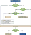
Figure 1. A proposed algorithm for imaging patients with acute stone disease in the emergency department
Initial stratification is based on age; American Urological Association (AUA) guidelines delineate patients <14 years old as paediatric patients. Adult patients are stratified based on whether they are pregnant and BMI. Ultrasonography should also be considered first in adult patients, especially those with a normal BMI and adults in whom a reasonable suspicion of stone disease exists. In such cases the sensitivity and specificity will be sufficiently high to augment the patient’s pretest probability of having a kidney stone without considerably increasing the risk of missing a alternative diagnosis. low-dose CT, noncontrast CT with <3 mSv of radiation exposure.
Figure 2. A coronal demonstration of bilateral 8 mm nephrolithiasis on noncontrast CT
These stones are clearly visible using this imaging modality. Additional anatomical detail can be obtained by reconstructing the images in an axial plane. a | This coronal CT image clearly demonstrates a left-sided obstructing stone. b | Posterior coronal CT view of panel a demonstrating a lower-pole nonobstructing stone. An excellent level of anatomical detail can be seen here and can be further increased by reconstructing the image in an axial plane.
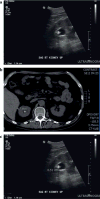
Figure 3. Comparison of stone size estimates by B-mode ultrasonography and CT
a | B-mode ultrasonographic stone sizing on a longitudinal view of the kidney. b | CT scan image with an estimate of stone size, which is about half of the estimated size according to ultrasonography. c | Measurement of the stone shadow using B-mode ultrasonography provides a much closer estimate to that estimated using CT imaging.
Figure 4. Comparison of ultrasonography in B-mode or the novel ‘S-mode’ in humans
a | Conventional B-mode and b | novel S-mode. S-Mode combines enhanced B-mode and enhanced Doppler detection based on the twinkling artefact to make the stone particularly evident in the image. The bright opaque green on the bright white stone increases the contrast:background ratio, enabling easy of identification of the stone.
Figure 5. Two plain films of the abdomen before and after treatment for an obstructing left-ureteral stone
This patient also underwent CT imaging (FIG. 2). a | The left obstructing stone is clearly visible before treatment. b | After treatment the stone is no longer visible. Stones overlaying a bony structure, <5 mm or shaded by a bowel gas loop can easily be missed.
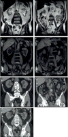
Figure 6. Image sequence demonstrating coronal cuts on MRI
a,b | Images obtained using T2 and c,d | images obtained using T1 sequences, clearly demonstrating hydronephrosis but distal pathology is not clear. Scan performed for cancer surveillance — differential included metastasis, extrinsic ureteral compression and stones. e-g | CT images showing the hydronephrosis and hydroureter; a distal right ureteral stone is now easily visualized enabling diagnosis and considerably altering this patient’s treatment course.
Similar articles
- Lifetime Radiation Exposure in Patients with Recurrent Nephrolithiasis.
Elkoushy MA, Andonian S. Elkoushy MA, et al. Curr Urol Rep. 2017 Sep 12;18(11):85. doi: 10.1007/s11934-017-0731-6. Curr Urol Rep. 2017. PMID: 28900827 Review. - The Utility of the Kidneys-ureters-bladder Radiograph as the Sole Imaging Modality and Its Combination With Ultrasonography for the Detection of Renal Stones.
Kanno T, Kubota M, Funada S, Okada T, Higashi Y, Yamada H. Kanno T, et al. Urology. 2017 Jun;104:40-44. doi: 10.1016/j.urology.2017.03.019. Epub 2017 Mar 21. Urology. 2017. PMID: 28341578 - Renal Imaging in Stone Disease: Which Modality to Choose?
Kaul I, Moore S, Barry E, Pareek G. Kaul I, et al. R I Med J (2013). 2023 Dec 1;106(11):31-35. R I Med J (2013). 2023. PMID: 38015782 - Renal tract calculi: comparison of stone size on plain radiography and noncontrast spiral CT scan.
Dundee P, Bouchier-Hayes D, Haxhimolla H, Dowling R, Costello A. Dundee P, et al. J Endourol. 2006 Dec;20(12):1005-9. doi: 10.1089/end.2006.20.1005. J Endourol. 2006. PMID: 17206892 - Imaging in diagnosis, treatment, and follow-up of stone patients.
Dhar M, Denstedt JD. Dhar M, et al. Adv Chronic Kidney Dis. 2009 Jan;16(1):39-47. doi: 10.1053/j.ackd.2008.10.005. Adv Chronic Kidney Dis. 2009. PMID: 19095204 Review.
Cited by
- Feasibility of non-linear beamforming ultrasound methods to characterize and size kidney stones.
Hsi RS, Schlunk SG, Tierney JE, Dei K, Jones R, George M, Karve P, Duddu R, Byram BC. Hsi RS, et al. PLoS One. 2018 Aug 28;13(8):e0203138. doi: 10.1371/journal.pone.0203138. eCollection 2018. PLoS One. 2018. PMID: 30153279 Free PMC article. - Clinical Low Dose Photon Counting CT for the Detection of Urolithiasis: Evaluation of Image Quality and Radiation Dose.
Niehoff JH, Carmichael AF, Woeltjen MM, Boriesosdick J, Lopez Schmidt I, Michael AE, Große Hokamp N, Piechota H, Borggrefe J, Kroeger JR. Niehoff JH, et al. Tomography. 2022 Jun 23;8(4):1666-1675. doi: 10.3390/tomography8040138. Tomography. 2022. PMID: 35894003 Free PMC article. - A Tale From the Early Stone Age: Pediatric Ureterolithiasis as Appendicitis Mimic - A Case Report and Management Overview.
Larson NP, Bridwell RE, Yoo MJ. Larson NP, et al. Cureus. 2020 Sep 24;12(9):e10637. doi: 10.7759/cureus.10637. Cureus. 2020. PMID: 33123450 Free PMC article. - "Renal emergencies: a comprehensive pictorial review with MR imaging".
Gopireddy DR, Mahmoud H, Baig S, Le R, Bhosale P, Lall C. Gopireddy DR, et al. Emerg Radiol. 2021 Apr;28(2):373-388. doi: 10.1007/s10140-020-01852-8. Epub 2020 Sep 25. Emerg Radiol. 2021. PMID: 32974867 Review. - Prevalence of Urolithiasis by Ultrasonography Among Patients with Gout: A Cross-Sectional Study from the UP-Philippine General Hospital.
Tee M, Lustre Ii C, Abrilla A, Afos IE, Cañal JP. Tee M, et al. Res Rep Urol. 2020 Sep 25;12:423-431. doi: 10.2147/RRU.S268700. eCollection 2020. Res Rep Urol. 2020. PMID: 33062621 Free PMC article.
References
- Stamatelou KK, Francis ME, Jones CA, Nyberg LM, Curhan GC. Time trends in reported prevalence of kidney stones in the United States: 1976–1994. Kidney Int. 2003;63:1817–1823. - PubMed
- Preminger GM, et al. 2007 guideline for the management of ureteral calculi. J Urol. 2007;178:2418–2434. - PubMed
Publication types
MeSH terms
LinkOut - more resources
Full Text Sources
Other Literature Sources
Medical


