How We Read Oncologic FDG PET/CT - PubMed (original) (raw)
Review
How We Read Oncologic FDG PET/CT
Michael S Hofman et al. Cancer Imaging. 2016.
Abstract
18F-fluorodeoxyglucose (FDG) PET/CT is a pivotal imaging modality for cancer imaging, assisting diagnosis, staging of patients with newly diagnosed malignancy, restaging following therapy and surveillance. Interpretation requires integration of the metabolic and anatomic findings provided by the PET and CT components which transcend the knowledge base isolated in the worlds of nuclear medicine and radiology, respectively. In the manuscript we detail our approach to reviewing and reporting a PET/CT study using the most commonly used radiotracer, FDG. This encompasses how we display, threshold intensity of images and sequence our review, which are essential for accurate interpretation. For interpretation, it is important to be aware of benign variants that demonstrate high glycolytic activity, and pathologic lesions which may not be FDG-avid, and understand the physiologic and biochemical basis of these findings. Whilst FDG PET/CT performs well in the conventional imaging paradigm of identifying, counting and measuring tumour extent, a key paradigm change is its ability to non-invasively measure glycolytic metabolism. Integrating this "metabolic signature" into interpretation enables improved accuracy and characterisation of disease providing important prognostic information that may confer a high management impact and enable better personalised patient care.
Keywords: Fluorodeoxyglucose FDG; Medical oncology; Positron-emission tomography; Radiology.
Figures
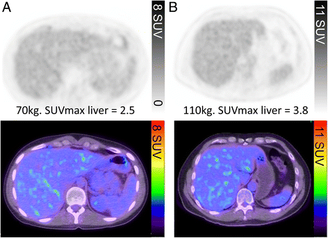
Fig. 1
The PET window intensity is adjusted so that the liver appears light to mid-grey on the grey scale, corresponding to flecks of green in the liver on the rainbow colour scale. Despite the difference in SUVmax of the liver secondary to differences in weights of the two patients (a and b), the liver intensity this appears the same in both patients
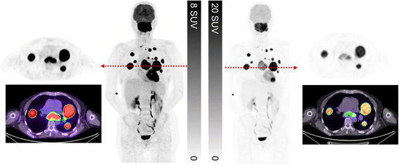
Fig. 2
This patient presented with suspected metastatic nasopharyngeal cancer. Initial workup with endoscopic ultrasound and biopsy of the subcarinal node was non-diagnostic with necrotic tissue. FDG PET/CT demonstrates very intense uptake at all sites with lower uptake in the subcarinal node, only evident when widening the PET window. The findings suggest a different tumour biology at this site with necrosis. When feasible, we recommend biopsy of the most FDG-avid lesion which likely represents the site of most aggressive disease and least likely to be non-diagnostic. In summary, the PET study windowed narrowly is primed for sensitivity whereas a wider window enables superior characterisation
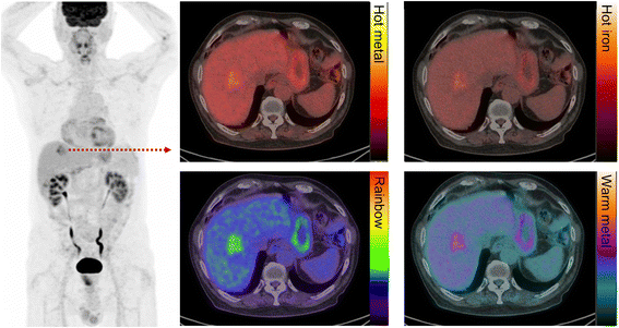
Fig. 3
Patient with metastatic colorectal carcinoma and hepatic metastasis. The fused image is presented in different colour scales. We recommend using the “rainbow” scale owing to the superior tumour-to-liver contrast compared to other commonly used colour maps
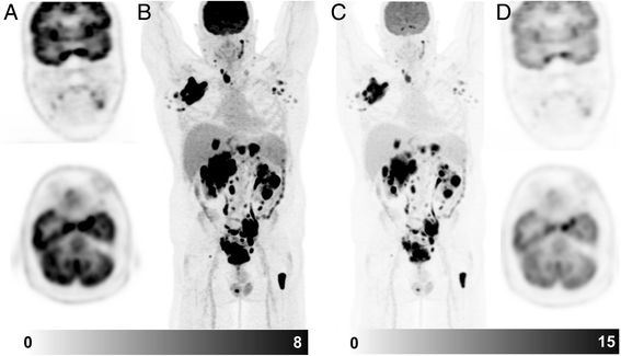
Fig. 4
Patient with diffuse large B cell lymphoma. On the standard windowing, no abnormality is readily identified in the brain (a coronal & axial slice, b MIP image). By increasing the upper SUV threshold, abnormal uptake becomes readily becomes visible (c MIP image, d coronal & axial slice). This corresponded to a MRI abnormality which was not reported prospectively but identified following targeted review after the PET scan. Changing the PET window so that abnormalities can be identified above physiologic brain activity should be a routine component of image review
Fig. 5
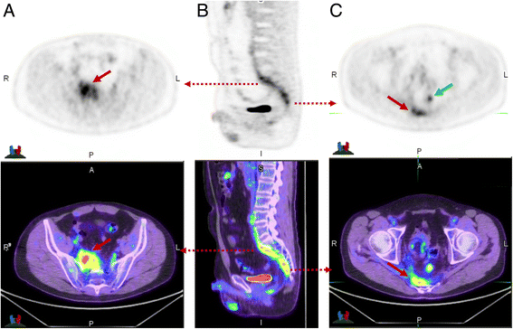
This patient had suspicion of pelvic recurrence in the setting of prior surgical excision for rectal carcinoma. There was intense uptake in the known pre-sacral soft tissue thickening (a) and (c) (red arrow) with SUVmax of 11. The linear morphology on the coronal image (b) suggested this was more likely inflammatory than malignant. A separate linear tract of metabolic activity was also seen (green arrow) extending from the pre-sacral abnormality to the peri-anal region (not shown). All abnormalities resolved following antiobiotic therapy confirming inflammatory aetiology
Fig. 6
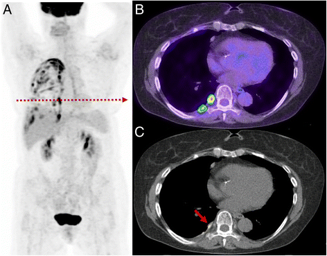
Patient with prior lung malignancy presents for surveillance. The study demonstrates a typical appearance of inflammatory change post talc pleurodesis with intense multi-focal uptake evident throughout the pleural surface (a). On the axial PET/CT (b) and CT (c) the high focal uptake correlates with a site of talc on CT recognised by its high density. Such change can persistent for many years after pleurodesis
Fig. 7
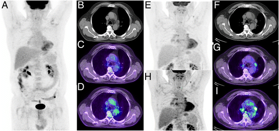
Patient with non-small cell lung cancer treated with curative intent radiotherapy. Post treatment restaging PET/CT demonstrated a complete metabolic response (a–d, c upper SUV threshold adjusted to liver background as detailed above, d upper SUV threshold of 5). Follow-up CT 9 months later demonstrated enlargement of multiple mediastinal nodes considered likely to represent malignant aetiology. Repeat PET/CT (e–i) demonstrated low-to-moderate uptake in these nodes. Given the symmetry of distribution in hilar and mediastinal nodes the aetiology was considered inflammatory, which was confirmed by resolution on follow-up. Thresholding the PET with a SUV threshold of 5 (h–i) might lead to erroneous description of intense uptake and interpretation as malignant in aetiology
Fig. 8
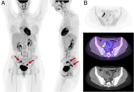
Appearance of physiologic adnexal uptake observed mid-cycle. Although the metabolic activity is high, on the rotating MIP images (a anterior and lateral) the activity is bilateral and curvilinear, characteristic of fallopian tube activity (b). Unilateral focal ovarian follicular activity is frequently seen in association with this finding
Fig. 9
Patient with HPV-p16 positive cervical squamous cell carcinoma presents for staging. FDG PET (a) demonstrates subtle uptake in an enlarged right external node (b) which would be difficult to discern without knowledge of the CT findings. Correlation with prior contrast-enhanced CT (c) demonstrates the node has rim enhancement and central necrosis consistent with malignant aetiology. The rim of viable tumour is thin and below the resolution of PET imaging explaining the absence of significant uptake. Integration of CT morphology is critical in this case for accurate interpretation
Fig. 10
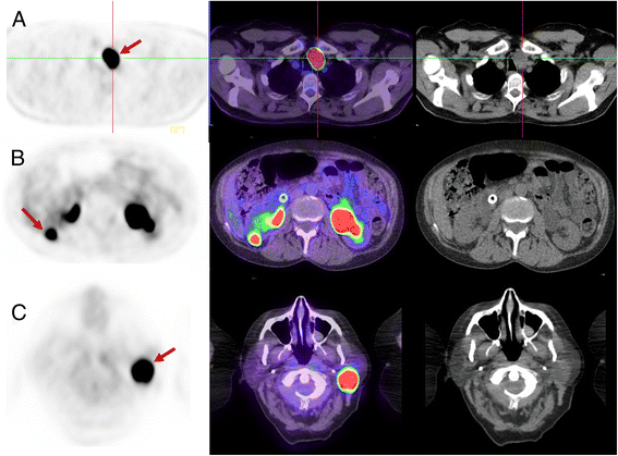
Three different patients with (a) Hurthle cell adenoma (thyroid oncocytoma), (b) renal oncocytoma and (c) Parotid Warthin’s tumour (parotid oncocytoma). Each has high SUVmax of 45, 22 and 35, respectively. In each case, the abnormality was present on imaging more than one year prior and unchanged in size. The very intense FDG uptake could be interpreted as suspicious for aggressive malignancy but the lack of temporal change was inconsistent with this. The lack of progression in a thyroid, renal or parotid lesion with very intense uptake is pathognomonic of benign oncocytomas
Similar articles
- The role of 18F-FDG PET/CT in staging and restaging primary bone lymphoma.
Liu Y. Liu Y. Nucl Med Commun. 2017 Apr;38(4):319-324. doi: 10.1097/MNM.0000000000000652. Nucl Med Commun. 2017. PMID: 28225435 - Diagnostic imaging using positron emission tomography for gynecological malignancy.
Tsuyoshi H, Yoshida Y. Tsuyoshi H, et al. J Obstet Gynaecol Res. 2017 Nov;43(11):1687-1699. doi: 10.1111/jog.13436. Epub 2017 Sep 8. J Obstet Gynaecol Res. 2017. PMID: 28884883 Review. - Role of Optimal Quantification of FDG PET Imaging in the Clinical Practice of Radiology.
Ziai P, Hayeri MR, Salei A, Salavati A, Houshmand S, Alavi A, Teytelboym OM. Ziai P, et al. Radiographics. 2016 Mar-Apr;36(2):481-96. doi: 10.1148/rg.2016150102. Radiographics. 2016. PMID: 26963458 Review.
Cited by
- Venous thromboembolism detected by FDG-PET/CT in cancer patients: a common, yet life-threatening observation.
Kaghazchi F, Borja AJ, Hancin EC, Bhattaru A, Detchou DKE, Seraj SM, Rojulpote C, Hess S, Nardo L, Gabriel PE, Damrauer SM, Werner TJ, Alavi A, Revheim ME. Kaghazchi F, et al. Am J Nucl Med Mol Imaging. 2021 Apr 15;11(2):99-106. eCollection 2021. Am J Nucl Med Mol Imaging. 2021. PMID: 34079639 Free PMC article. - Factors associated with increased diagnostic yield of endobronchial ultrasound-guided transbronchial needle aspiration: an observational single center study.
Kim BK, Choi H, Kim CY. Kim BK, et al. J Thorac Dis. 2024 Jan 30;16(1):439-449. doi: 10.21037/jtd-23-1369. Epub 2024 Jan 12. J Thorac Dis. 2024. PMID: 38410574 Free PMC article. - Oncocytic Adrenocortical Carcinoma With Low 18F-FDG Uptake and the Absence of Glucose Transporter 1 Expression.
Babaya N, Noso S, Hiromine Y, Taketomo Y, Niwano F, Monobe K, Imamura S, Ueda K, Yamazaki Y, Sasano H, Ikegami H. Babaya N, et al. J Endocr Soc. 2021 Aug 23;5(11):bvab143. doi: 10.1210/jendso/bvab143. eCollection 2021 Nov 1. J Endocr Soc. 2021. PMID: 34514280 Free PMC article. - Determinants of Survival with Combined HER2 and PD-1 Blockade in Metastatic Esophagogastric Cancer.
Maron SB, Chatila W, Walch H, Chou JF, Ceglia N, Ptashkin R, Do RKG, Paroder V, Pandit-Taskar N, Lewis JS, Biachi De Castria T, Sabwa S, Socolow F, Feder L, Thomas J, Schulze I, Kim K, Elzein A, Bojilova V, Zatzman M, Bhanot U, Nagy RJ, Lee J, Simmons M, Segal M, Ku GY, Ilson DH, Capanu M, Hechtman JF, Merghoub T, Shah S, Schultz N, Solit DB, Janjigian YY. Maron SB, et al. Clin Cancer Res. 2023 Sep 15;29(18):3633-3640. doi: 10.1158/1078-0432.CCR-22-3769. Clin Cancer Res. 2023. PMID: 37406106 Free PMC article. Clinical Trial. - Imaging for Response Assessment in Cancer Clinical Trials.
Sorace AG, Elkassem AA, Galgano SJ, Lapi SE, Larimer BM, Partridge SC, Quarles CC, Reeves K, Napier TS, Song PN, Yankeelov TE, Woodard S, Smith AD. Sorace AG, et al. Semin Nucl Med. 2020 Nov;50(6):488-504. doi: 10.1053/j.semnuclmed.2020.05.001. Epub 2020 Jun 10. Semin Nucl Med. 2020. PMID: 33059819 Free PMC article.
References
- Binns DS, Pirzkall A, Yu W, Callahan J, Mileshkin L, Conti P, Scott AM, Macfarlane D, Fine BM, Hicks RJ, Team OSS Compliance with PET acquisition protocols for therapeutic monitoring of erlotinib therapy in an international trial for patients with non-small cell lung cancer. Eur J Nucl Med Mol Imaging. 2011;38(4):642–650. doi: 10.1007/s00259-010-1665-0. - DOI - PubMed
Publication types
MeSH terms
LinkOut - more resources
Full Text Sources
Other Literature Sources
