Eomeshi NK Cells in Human Liver Are Long-Lived and Do Not Recirculate but Can Be Replenished from the Circulation - PubMed (original) (raw)
Eomeshi NK Cells in Human Liver Are Long-Lived and Do Not Recirculate but Can Be Replenished from the Circulation
Antonia O Cuff et al. J Immunol. 2016.
Abstract
Human liver contains an Eomeshi population of NK cells that is not present in the blood. In this study, we show that these cells are characterized by a molecular signature that mediates their retention in the liver. By examining liver transplants where donors and recipients are HLA mismatched, we distinguish between donor liver-derived and recipient-derived leukocytes to show that Eomeslo NK cells circulate freely whereas Eomeshi NK cells are unable to leave the liver. Furthermore, Eomeshi NK cells are retained in the liver for up to 13 y. Therefore, Eomeshi NK cells are long-lived liver-resident cells. We go on to show that Eomeshi NK cells can be recruited from the circulation during adult life and that circulating Eomeslo NK cells are able to upregulate Eomes and molecules mediating liver retention under cytokine conditions similar to those in the liver. This suggests that circulating NK cells are a precursor of their liver-resident counterparts.
Copyright © 2016 The Authors.
Conflict of interest statement
The authors have no conflicting financial interests.
Figures
FIGURE 1.
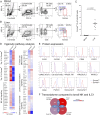
Eomeshi NK cells are present in human liver and have a distinct phenotype. (A) PBLs were isolated from healthy volunteers and NK cells identified by gating on live cells, scatter, CD3−, and CD56+. NK cells were examined for their expression of Eomes and T-bet. (B) Leukocytes were isolated from the perfusion fluid of healthy human livers destined for transplantation and stained as in (A). (C) Summary data showing Eomeshi NK cells as a percentage of total NK cells across 11 samples. Significance was determined using a Mann–Whitney U test. (D) RNASeq data from Eomeslo and Eomeshi NK cells sorted from the perfusion fluid from n = 5 healthy livers. Differentially expressed genes were identified using paired t tests. The top 10 most differentially expressed genes and the top most significantly enriched canonical pathways are shown. (E) Flow cytometry for key proteins expected to differ between Eomeslo (blue dashed line) and Eomeshi (red solid line) liver NK cells. Histograms are representative of n = 4 independent samples. (F) Overlap between top upregulated genes in Eomeslo and Eomeshi liver NK cells, compared with tonsil NK cells and ILC1 (23).
FIGURE 2.
Eomeslo NK cells exit the liver but Eomeshi NK cells do not. (A and B) Example of staining from a single transplant in which an HLA-A3–negative liver (A) was transplanted into an HLA-A3–positive recipient. Twenty-four hours after transplant, PBLs were isolated from the recipient’s blood and circulating liver-derived cells were distinguished from recipient-derived cells on the basis of HLA-A3 staining (B). NK cells were examined for their expression of Eomes and T-bet. (C) Summary data from n = 7 transplants mismatched at either HLA-A2 or HLA-A3. Significance was determined using a paired t test. (D and E) Example staining from a single transplant in which an HLA-A2–positive liver (D) was transplanted into an HLA-A2–negative recipient. Three hours posttransplant, a second biopsy was taken from the liver and liver-derived cells were distinguished from recipient-derived cells on the basis of HLA-A2 staining (E). (F) Summary data from n = 3 transplants, all mismatched at HLA-A2. The gates used to produce the summary data (C and F) are highlighted.
FIGURE 3.
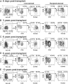
Eomeshi NK cells are long-lived in the liver, but they can be recruited from the circulation during adult life. (A) An HLA-A2–positive liver was transplanted into an HLA-A2–negative recipient and removed 8 d later. Liver leukocytes were isolated from a biopsy and liver-derived cells were distinguished from recipient-derived cells on the basis of HLA-A2 staining. Liver- and recipient-derived NK cells present in the liver were examined for their expression of Eomes and T-bet. (B) An HLA-A2–positive liver was transplanted into an HLA-A2–negative recipient and removed 3 y later. (C) An HLA-A2–positive liver was transplanted into an HLA-A2–negative recipient and removed 6 y later. (D) An HLA-A3–positive liver was transplanted into an HLA-A3–negative recipient and removed 6 y later. (E) An HLA-A2–positive liver was transplanted into an HLA-A2–negative recipient and removed 13 y later.
FIGURE 4.
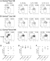
Eomeslo NK cells can become Eomeshi. (A and B) NK cells were sorted from perfusion fluid [(A), Eomeslo; (B), Eomeshi)] and cultured for 7 d in the indicated conditions. At the end of the culture period, the cells were examined for their expression of Eomes and T-bet. (C) Sorted blood NK cells were cultured as above. (D–F) Summary data showing the percentage of Eomeshi NK cells in n = 4 independent experiments, starting with Eomeslo liver NK cells (D), Eomeshi liver NK cells (E), or Eomeslo peripheral blood NK cells (F). Groups that are significantly different (p < 0.05 by one-way ANOVA) are indicated by different letters.
FIGURE 5.
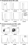
Eomes upregulation is associated with increased expression of mediators of tissue retention. (A) Sorted blood NK cells were cultured for 7 d in IL-15 and TGF-β to produce in vitro–derived Eomeshi NK cells. Eomeslo NK cell expression of the indicated proteins before culture (gray dashed line) and Eomeshi NK cell expression after culture (black solid line) is shown. (B) NKL cells were transduced with Eomes or vector control and gated on transduced (dsRed+) cells. Expression of the indicated proteins in vector-transduced cells (gray dashed line) and Eomes-transduced cells (black solid line) is shown. The histograms show a single experiment, representative of n = 4.
Similar articles
- CXCR6 marks a novel subset of T-bet(lo)Eomes(hi) natural killer cells residing in human liver.
Stegmann KA, Robertson F, Hansi N, Gill U, Pallant C, Christophides T, Pallett LJ, Peppa D, Dunn C, Fusai G, Male V, Davidson BR, Kennedy P, Maini MK. Stegmann KA, et al. Sci Rep. 2016 May 23;6:26157. doi: 10.1038/srep26157. Sci Rep. 2016. PMID: 27210614 Free PMC article. - Tissue-resident Eomes(hi) T-bet(lo) CD56(bright) NK cells with reduced proinflammatory potential are enriched in the adult human liver.
Harmon C, Robinson MW, Fahey R, Whelan S, Houlihan DD, Geoghegan J, O'Farrelly C. Harmon C, et al. Eur J Immunol. 2016 Sep;46(9):2111-20. doi: 10.1002/eji.201646559. Eur J Immunol. 2016. PMID: 27485474 - Liver-Derived TGF-β Maintains the EomeshiTbetlo Phenotype of Liver Resident Natural Killer Cells.
Harmon C, Jameson G, Almuaili D, Houlihan DD, Hoti E, Geoghegan J, Robinson MW, O'Farrelly C. Harmon C, et al. Front Immunol. 2019 Jul 3;10:1502. doi: 10.3389/fimmu.2019.01502. eCollection 2019. Front Immunol. 2019. PMID: 31333651 Free PMC article. - Liver-Resident NK Cells: The Human Factor.
Male V. Male V. Trends Immunol. 2017 May;38(5):307-309. doi: 10.1016/j.it.2017.02.008. Epub 2017 Mar 16. Trends Immunol. 2017. PMID: 28318877 Review. - T-bet and Eomes govern differentiation and function of mouse and human NK cells and ILC1.
Zhang J, Marotel M, Fauteux-Daniel S, Mathieu AL, Viel S, Marçais A, Walzer T. Zhang J, et al. Eur J Immunol. 2018 May;48(5):738-750. doi: 10.1002/eji.201747299. Epub 2018 Feb 28. Eur J Immunol. 2018. PMID: 29424438 Review.
Cited by
- The role of IL-22 in the resolution of sterile and nonsterile inflammation.
Alabbas SY, Begun J, Florin TH, Oancea I. Alabbas SY, et al. Clin Transl Immunology. 2018 Apr 18;7(4):e1017. doi: 10.1002/cti2.1017. eCollection 2018. Clin Transl Immunology. 2018. PMID: 29713472 Free PMC article. Review. - Tissue-resident lymphocytes: from adaptive to innate immunity.
Sun H, Sun C, Xiao W, Sun R. Sun H, et al. Cell Mol Immunol. 2019 Mar;16(3):205-215. doi: 10.1038/s41423-018-0192-y. Epub 2019 Jan 11. Cell Mol Immunol. 2019. PMID: 30635650 Free PMC article. Review. - Natural killer cell homing and trafficking in tissues and tumors: from biology to application.
Ran GH, Lin YQ, Tian L, Zhang T, Yan DM, Yu JH, Deng YC. Ran GH, et al. Signal Transduct Target Ther. 2022 Jun 29;7(1):205. doi: 10.1038/s41392-022-01058-z. Signal Transduct Target Ther. 2022. PMID: 35768424 Free PMC article. Review. - NK Cell-Mediated Recall Responses: Memory-Like, Adaptive, or Antigen-Specific?
Stary V, Stary G. Stary V, et al. Front Cell Infect Microbiol. 2020 May 14;10:208. doi: 10.3389/fcimb.2020.00208. eCollection 2020. Front Cell Infect Microbiol. 2020. PMID: 32477964 Free PMC article. Review. - IL-12 and IL-15 induce the expression of CXCR6 and CD49a on peripheral natural killer cells.
Hydes T, Noll A, Salinas-Riester G, Abuhilal M, Armstrong T, Hamady Z, Primrose J, Takhar A, Walter L, Khakoo SI. Hydes T, et al. Immun Inflamm Dis. 2018 Mar;6(1):34-46. doi: 10.1002/iid3.190. Epub 2017 Sep 27. Immun Inflamm Dis. 2018. PMID: 28952190 Free PMC article.
References
- Hanna J., Goldman-Wohl D., Hamani Y., Avraham I., Greenfield C., Natanson-Yaron S., Prus D., Cohen-Daniel L., Arnon T. I., Manaster I., et al. 2006. Decidual NK cells regulate key developmental processes at the human fetal-maternal interface. Nat. Med. 12: 1065–1074. - PubMed
Publication types
MeSH terms
Substances
Grants and funding
- 10/4001/11/DH_/Department of Health/United Kingdom
- HTD 556/DH_/Department of Health/United Kingdom
- MR/M020126/1/MRC_/Medical Research Council/United Kingdom
- WT105677 /WT_/Wellcome Trust/United Kingdom
LinkOut - more resources
Full Text Sources
Other Literature Sources
Molecular Biology Databases
Research Materials


