Cell-Intrinsic Barriers of T Cell-Based Immunotherapy - PubMed (original) (raw)
Review
Cell-Intrinsic Barriers of T Cell-Based Immunotherapy
Hazem E Ghoneim et al. Trends Mol Med. 2016 Dec.
Abstract
Prolonged exposure of CD8+ T cells to their cognate antigen can result in exhaustion of effector functions enabling the persistence of infected or transformed cells. Recent advances in strategies to rejuvenate host effector function using Immune Checkpoint Blockade have resulted in tremendous success towards the treatment of several cancers. However, it is unclear if T cell rejuvenation results in long-lived antitumor functions. Emerging evidence suggests that T cell exhaustion may also represent a significant impediment in sustaining long-lived antitumor activity by chimeric antigen receptor T cells. Here, we discuss current findings regarding transcriptional regulation during T cell exhaustion and address the hypothesis that epigenetics may be a potential barrier to achieving the maximum benefit of T cell-based immunotherapies.
Keywords: DNA methylation.; T cell exhaustion; chimeric antigen receptor T cells; epigenetics; histone modification; immune checkpoint blockade.
Copyright © 2016 Elsevier Ltd. All rights reserved.
Figures
Key Figure, Figure 1. Epigenetic Barriers to Immune Checkpoint Blockade-Mediated Rejuvenation of T Cells
(A) The schematic depicts the repression in developmental plasticity acquired by antigen-specific T cells during clonal expansion in response to acute or chronic antigen exposure. Upon acute antigen exposure, naive CD8+ T cells clonally expand and differentiate into effector cells. During chronic high-level antigen exposure, persistent T cell receptor (TCR) stimulation can cause T cell exhaustion. Effector function is progressively repressed during the development of T cell exhaustion, thereby leading to a heterogeneous population of T cells at various levels of exhaustion. Partially exhausted T cells are phenotypically defined by sustained expression of a minimal number of immune inhibitory receptors (IRs) and characterized by differential expression of T-bet and Eomes (T-bethigh Eomeslow T cells), whereas fully exhausted T cells are marked by co-expression of multiple IRs. The immune checkpoint blockade (ICB) obstructs inhibitory signals from immune IRs, resulting in rejuvenation of partially exhausted T cells. (B) The schematic depicts the effect of progressive epigenetic programming that reinforces silencing of genes involved in effector function, proliferation, and metabolic fitness of exhausted T cells. Partially exhausted T cells may retain a degree of epigenetic plasticity at exhaustion-specific silenced genes manifested by partially methylated DNA and deposition of fewer repressive, more permissive histone marks. Terminally differentiated exhausted T cells may be more epigenetically restrained through fully methylated DNA, the deposition of more repressive histone marks, and the loss of permissive histone marks. (C) The diagram models the potential reprogramming of exhaustion-associated epigenetic programs that could be done to remove cell-intrinsic barriers to achieving a better, possibly durable rejuvenation response of fully exhausted T cells following ICB therapy.
Figure 2. Genetically-Engineered T cell Immunotherapeutic Approaches
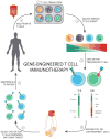
The schematic depicts the multiuse process of generating genetically engineered human T cells targeted against specific tumor antigens. (Step 1) Autologous T cells can be isolated from a patient’s peripheral blood mononuclear cells or an excised tumor. (Step 2) T cell pools or specific subsets can be stimulated with (1) activating CD3 antibody (OKT3), (2) anti-CD3/CD28–coated beads, or (3) allogeneic feeder cell lines. (Step 3) Specific T cell subsets can be sorted or enriched according to their differentiation status to exploit proliferative and effector functions. (Step 4) Engineered T cells can be generated by cloning either T cell receptors (TCRs) or chimeric antigen receptors (CARs) and transducing a patient’s T cells with retro- or lenti-viruses, thus redirecting recognition toward tumor-associated antigens. (Step 5) Engineered T cells can then be expanded in vitro in the presence of conditioning cytokines (e.g., IL-2 or IL-15) to increase the frequency of tumor-specific T cells generated. (Step 6) Engineered T cells specific for an antigen (target) of interest are selected. (Step 7) Engineered T cells can then be reinfused into the patient (usually post-lymphodepletion).
Similar articles
- Genetic engineering of T cells for adoptive immunotherapy.
Varela-Rohena A, Carpenito C, Perez EE, Richardson M, Parry RV, Milone M, Scholler J, Hao X, Mexas A, Carroll RG, June CH, Riley JL. Varela-Rohena A, et al. Immunol Res. 2008;42(1-3):166-81. doi: 10.1007/s12026-008-8057-6. Immunol Res. 2008. PMID: 18841331 Free PMC article. Review. - Treatment of solid tumors with chimeric antigen receptor-engineered T cells: current status and future prospects.
Di S, Li Z. Di S, et al. Sci China Life Sci. 2016 Apr;59(4):360-9. doi: 10.1007/s11427-016-5025-6. Epub 2016 Mar 11. Sci China Life Sci. 2016. PMID: 26968709 Review. - Regional Infusion of Chimeric Antigen Receptor T Cells to Overcome Barriers for Solid Tumor Immunotherapy.
Hardaway JC, Prince E, Arepally A, Katz SC. Hardaway JC, et al. J Vasc Interv Radiol. 2018 Jul;29(7):1017-1021.e1. doi: 10.1016/j.jvir.2018.03.001. J Vasc Interv Radiol. 2018. PMID: 29935783 No abstract available. - Spotlight on chimeric antigen receptor engineered T cell research and clinical trials in China.
Luo C, Wei J, Han W. Luo C, et al. Sci China Life Sci. 2016 Apr;59(4):349-59. doi: 10.1007/s11427-016-5034-5. Epub 2016 Mar 24. Sci China Life Sci. 2016. PMID: 27009301 Review. - Chimeric antigen receptor engineering: a right step in the evolution of adoptive cellular immunotherapy.
Figueroa JA, Reidy A, Mirandola L, Trotter K, Suvorava N, Figueroa A, Konala V, Aulakh A, Littlefield L, Grizzi F, Rahman RL, Jenkins MR, Musgrove B, Radhi S, D'Cunha N, D'Cunha LN, Hermonat PL, Cobos E, Chiriva-Internati M. Figueroa JA, et al. Int Rev Immunol. 2015 Mar;34(2):154-87. doi: 10.3109/08830185.2015.1018419. Int Rev Immunol. 2015. PMID: 25901860 Review.
Cited by
- Pan-cancer and single-cell analysis reveal dual roles of lymphocyte activation gene-3 (LAG3) in cancer immunity and prognosis.
Wang Y, Zhao Y, Zhang G, Lin Y, Fan C, Wei H, Chen S, Guan L, Liu K, Yu S, Fu L, Zhang J, Yuan Y, He J, Cai H. Wang Y, et al. Sci Rep. 2024 Oct 15;14(1):24203. doi: 10.1038/s41598-024-74808-4. Sci Rep. 2024. PMID: 39406840 Free PMC article. - Predictive Biomarkers and Resistance Mechanisms of Checkpoint Inhibitors in Malignant Solid Tumors.
Pavelescu LA, Enache RM, Roşu OA, Profir M, Creţoiu SM, Gaspar BS. Pavelescu LA, et al. Int J Mol Sci. 2024 Sep 6;25(17):9659. doi: 10.3390/ijms25179659. Int J Mol Sci. 2024. PMID: 39273605 Free PMC article. Review. - Chronic viral infection alters PD-1 locus subnuclear localization in cytotoxic CD8+ T cells.
Sacristán C, Youngblood BA, Lu P, Bally APR, Xu JX, McGary K, Hewitt SL, Boss JM, Skok JA, Ahmed R, Dustin ML. Sacristán C, et al. Cell Rep. 2024 Aug 27;43(8):114547. doi: 10.1016/j.celrep.2024.114547. Epub 2024 Jul 30. Cell Rep. 2024. PMID: 39083377 Free PMC article. - 5-Hydroxymethylcytosine in Cell-Free DNA Predicts Immunotherapy Response in Lung Cancer.
Shao J, Xu Y, Olsen RJ, Kasparian S, Sun K, Mathur S, Zhang J, He C, Chen SH, Bernicker EH, Li Z. Shao J, et al. Cells. 2024 Apr 19;13(8):715. doi: 10.3390/cells13080715. Cells. 2024. PMID: 38667328 Free PMC article. - Assessment of CTLA-4 Gene Expression Levels on CD8+ T Cells in Renal Transplant Patients and Relation with Serum sCTLA-4 Levels.
Çerçi Alkaç B, Soyöz M, Pehlivan M, Kılıçaslan Ayna T, Tatar E, Karahan Çöven Hİ, Tanrısev M, Pirim İ. Çerçi Alkaç B, et al. Biochem Genet. 2024 Mar 11. doi: 10.1007/s10528-024-10723-7. Online ahead of print. Biochem Genet. 2024. PMID: 38467886
References
- Moskophidis D, et al. Virus persistence in acutely infected immunocompetent mice by exhaustion of antiviral cytotoxic effector T cells. Nature. 1993;362:758–761. - PubMed
- Fuller MJ, Zajac AJ. Ablation of CD8 and CD4 T cell responses by high viral loads. J Immunol. 2003;170:477–486. - PubMed
Publication types
MeSH terms
Substances
LinkOut - more resources
Full Text Sources
Other Literature Sources
Research Materials

