Andreas Vesalius: Celebrating 500 years of dissecting nature - PubMed (original) (raw)
Review
Andreas Vesalius: Celebrating 500 years of dissecting nature
Fabio Zampieri et al. Glob Cardiol Sci Pract. 2015.
No abstract available
Figures
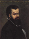
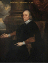
Figure 1.
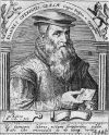
Andreas Vesalius portrait in the “Hall of Medicine” at the Bo Palace of the University of Padua.
Figure 2.
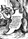
Coat of arms of the Vesalius family, showing three weasels, as appears at the top of the frontispiece of his masterpiece: De humani corporis fabrica libri septem (Vesalius 1543a).
Figure 3.
Seal of the Catholic University of Leuven.
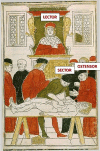
Figure 4.
Lesson of anatomy from the “Fasciculo de medicina” (Ketham 1494) following the medieval method. The professor on the chair is thought to be Mondino de’ Liuzzi. The professor was called lector, the barber dissecting the body was called sector, while the assistant of the professor, in this scene aside the sector, was called ostensor.
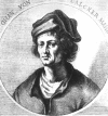
Figure 5.
Portrait of Jan van Calcar.
Figure 6.
On the left: drawing of the liver and vena cava made by Vitus Tritonius Athesinus, friend of Vesalius and student of Medicine in Padua, from a manuscript preserved at the National Library of Vienna (O'Malley 1958); on the right: close-up of the Tabula II of Vesalius’ Tabulae anatomicae sex (Vesalius 1538), from which it is possible to appreciate the strict similitude with Vitus’ drawing.
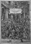
Figure 7.
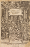
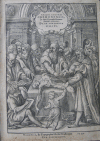
Frontispiece of the first edition of Vesalius’ De humani corporis fabrica libri septem (1543), where Vesalius is making a dissection in a crowded anatomical amphitheater.
Figure 8.
Engraving depicting Johannes Oporinus, editor of Vesalius’ most important works.
Figure 9.
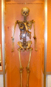
The skeleton prepared by Vesalius still now preserved at the Anatomy Museum of the Institute of Anatomy of the University of Basel.
Figure 10.
Frontispiece of the second edition of Vesalius’ Fabrica (1555).
Figure 11.
Engraving from the collection Quarante Tableaux, published in France between 1569 and 1570 by the engravers Jean Perrissin and Jacques Tortorel, showing where Henri II was cured and died, where also Andreas Vesalius and Antoine Parè were present. Vesalius and Parè are probably the two physicians discussing together at the center of the room, in front of the table.
Figure 12.
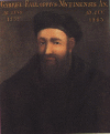
Gabriele Falloppia's portrait in the “Hall of Medicine” at the Bo Palace of the University of Padua.
Figure 13.
Close-up of the sixth table of the Tabulae anatomicae sex, where von Calcar is attributed as editor of the publication.
Figure 14.
Close-up of the III table on Arteria magna (the aorta) of Vesalius’ Tabulae anatomicae sex (Vesalius 1538). The heart is defined as “source of vital spirit and the arterial system”; the pulmonary vein is the structure which “brings air from lungs to the left ventricle” (letter “Q” of the caption on the left). All of these ideas were strictly Galenic.
Figure 15.
On the left: close-up from the Tabula III of Vesalius’ Tabulae anatomicae sex (Vesalius 1538), showing the heart and the structure of the carotids; on the right: structure of the carotids in human and simian, demonstrating that Vesalius represented in man the structure typical of simian, following Galen's anatomy (picture taken from: Singer & Rabin 1946).
Figure 16.
Close-up of the frontispiece of De humani corporis fabrica (Vesalus 1543a), depicting Vesalius personifying all the three figures of the previous method for teaching anatomy: he was lector, because professor of anatomy, ostensor, because he indicated, with his left hand, what he was explaining during the dissection, and sector, because he made the dissection himself with his right hand.
Figure 17.
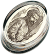
Paperweight celebrating the 500 years from the birth of Andreas Vesalius (1514–2014), produced by the University of Padua, where Vesalius is Scholae medicorum patavinae professor, as Vesalius defined himself in the Fabrica (Vesalius 1543a).
Figure 18.
Illustration in Vesalius’ Fabrica of a table for animal vivisection with anatomical instruments both for human anatomy and animal vivisection (Vesalius 1543a).
Figure 19.
Capital letters of the Fabrica showing methods of preparing bones specimens. On top left: capital letter “O” of the 1543 edition, showing the method of boiling bodies to extract bones. On top right, capital letter “C” of the 1543 edition, showing three men who carry a casket full of holes about to be immersed in a river. After having removed the majority of soft tissue, Vesalius covered the specimen with lime and placed it in a perforated wooden casket for a time. Then the casket was firmly tied down at the bottom of a river, where the current gradually removed the remaining soft tissue as it flowed through the casket (Olry 1998). On bottom left: capital letter “P” of 1543 edition, showing cherubs reconstructing the skeleton from the bones obtained with the methods previously described. On bottom right: capital letter “P” of 1555 edition, showing the same scene.
Figure 20.
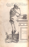
One of the Vesalius’ plates on the human skeleton, with the figure in an allegoric pose, thinking with a skull in his right hand, and a Latin inscription in the base where the skull is posed: “Vivitur ingenio, caetera mortis erunt”. Which means: genius lives on, all else is mortal.
Figure 21.
Frontispiece of Realdo Colombo's De re anatomica (1559) with a portrait of Colombo, at the center, doing the dissection.
Figure 22.
One of the most famous portraits of William Harvey, now displayed at the Royal College of Physicians in London.
Similar articles
- Centennial ties: Harvey Cushing (1869-1939) and William Osler (1849-1919) on Andreas Vesalius (1514-1564).
Toodayan N. Toodayan N. J Med Biogr. 2017 Aug;25(3):197-207. doi: 10.1177/0967772015601585. Epub 2015 Aug 25. J Med Biogr. 2017. PMID: 26307412 - The Flemish anatomist Andreas Vesalius (1514-1564) and the kidney.
DeBroe ME, Sacré D, Snelders ED, De Weerdt DL. DeBroe ME, et al. Am J Nephrol. 1997;17(3-4):252-60. doi: 10.1159/000169110. Am J Nephrol. 1997. PMID: 9189243 - Andreas Vesalius' 500th Anniversary: First Description of the Mammary Suspensory Ligaments.
Brinkman RJ, Hage JJ. Brinkman RJ, et al. World J Surg. 2016 Sep;40(9):2144-8. doi: 10.1007/s00268-016-3481-6. World J Surg. 2016. PMID: 26943658 - Andreas Vesalius, the Predecessor of Neurosurgery: How his Progressive Scientific Achievements Affected his Professional Life and Destiny.
Splavski B, Rotim K, Lakičević G, Gienapp AJ, Boop FA, Arnautović KI. Splavski B, et al. World Neurosurg. 2019 Sep;129:202-209. doi: 10.1016/j.wneu.2019.06.008. Epub 2019 Jun 13. World Neurosurg. 2019. PMID: 31201946 Review. - Andreas Vesalius (1515-1564) on animal cognition.
Brinkman RJ, Hage JJ, Oostra RJ, van der Horst CM. Brinkman RJ, et al. Psychon Bull Rev. 2019 Oct;26(5):1588-1595. doi: 10.3758/s13423-019-01643-4. Psychon Bull Rev. 2019. PMID: 31368024 Review.
Cited by
- Transcriptomic analysis of the 12 major human breast cell types reveals mechanisms of cell and tissue function.
Del Toro K, Sayaman R, Thi K, Licon-Munoz Y, Hines WC. Del Toro K, et al. PLoS Biol. 2024 Nov 5;22(11):e3002820. doi: 10.1371/journal.pbio.3002820. eCollection 2024 Nov. PLoS Biol. 2024. PMID: 39499736 Free PMC article. - Quantitative medicine: Tracing the transition from holistic to reductionist approaches. A new "quantitative holism" is possible?
Saba L, Tagliagambe S. Saba L, et al. J Public Health Res. 2023 Jun 20;12(2):22799036231182271. doi: 10.1177/22799036231182271. eCollection 2023 Apr. J Public Health Res. 2023. PMID: 37361238 Free PMC article. - Current Understanding of the Anatomy, Physiology, and Magnetic Resonance Imaging of Neurofluids: Update From the 2022 "ISMRM Imaging Neurofluids Study group" Workshop in Rome.
Agarwal N, Lewis LD, Hirschler L, Rivera LR, Naganawa S, Levendovszky SR, Ringstad G, Klarica M, Wardlaw J, Iadecola C, Hawkes C, Carare RO, Wells J, Bakker ENTP, Kurtcuoglu V, Bilston L, Nedergaard M, Mori Y, Stoodley M, Alperin N, de Leon M, van Osch MJP. Agarwal N, et al. J Magn Reson Imaging. 2024 Feb;59(2):431-449. doi: 10.1002/jmri.28759. Epub 2023 May 4. J Magn Reson Imaging. 2024. PMID: 37141288 Free PMC article. Review. - Anatomy Nights: An international public engagement event increases audience knowledge of brain anatomy.
Sanders KA, Philp JAC, Jordan CY, Cale AS, Cunningham CL, Organ JM. Sanders KA, et al. PLoS One. 2022 Jun 9;17(6):e0267550. doi: 10.1371/journal.pone.0267550. eCollection 2022. PLoS One. 2022. PMID: 35679263 Free PMC article. - The rich heritage of anatomical texts during Renaissance and thereafter: a lead up to Henry Gray's masterpiece.
Ghosh SK, Kumar A. Ghosh SK, et al. Anat Cell Biol. 2019 Dec;52(4):357-368. doi: 10.5115/acb.19.102. Epub 2019 Nov 12. Anat Cell Biol. 2019. PMID: 31949973 Free PMC article. Review.
References
- Bacon F. Novum organum scientiarum. Leiden: Ex officina A. Wyngaerden & F. Moiardus; 1620.
- Berengario da Carpi. Isagogae breves perlucide ac uberrime in anatomia humani corporis […] Bologna: Per Benedictum Hectoris Bibliopolam Bononiensem; 1523.
- Colombo R. De re anatomica. Venice: Ex typographia Nicolai Bevilacquae; 1559.
- Cushing W. A bio-bibliography of Andreas Vesalius. New York: Archon Books; 1962.
- Eftekhari K, Choe CH, Vagefi MR, Eckstein LA. The last ride of Henry II of France: Orbital injury and a king's demise. Surv Ophthalmol. 2015;60:274–278. - PubMed
Publication types
LinkOut - more resources
Full Text Sources
Other Literature Sources
