Control of muscle formation by the fusogenic micropeptide myomixer - PubMed (original) (raw)
Control of muscle formation by the fusogenic micropeptide myomixer
Pengpeng Bi et al. Science. 2017.
Abstract
Skeletal muscle formation occurs through fusion of myoblasts to form multinucleated myofibers. From a genome-wide clustered regularly interspaced short palindromic repeats (CRISPR) loss-of-function screen for genes required for myoblast fusion and myogenesis, we discovered an 84-amino acid muscle-specific peptide that we call Myomixer. Myomixer expression coincides with myoblast differentiation and is essential for fusion and skeletal muscle formation during embryogenesis. Myomixer localizes to the plasma membrane, where it promotes myoblast fusion and associates with Myomaker, a fusogenic membrane protein. Myomixer together with Myomaker can also induce fibroblast-fibroblast fusion and fibroblast-myoblast fusion. We conclude that the Myomixer-Myomaker pair controls the critical step in myofiber formation during muscle development.
Copyright © 2017, American Association for the Advancement of Science.
Figures
Fig. 1. Myomixer is a small-membrane protein that is essential for myoblast fusion
(A) Western blot showing Myomixer, Myosin, and Gapdh expression in WTor Myomixer KO primary myoblast cultures after 4 days in DM. (B) WT and Myomixer KO primary myoblasts differentiated for 1 week and stained with MY32 antibody (myosin) and Hoechst show the requirement of Myomixer for fusion. Scale bar, 50 μm. (C) Quantification of fusion in WT and Myomixer KO primary myoblast cultures (n = 3 pairs). (D) Amino acid sequence of Myomixer and cross-species homology. Basic residues are blue, acidic residues are red, cysteines are green, and leucines are yellow. H, helix; C, coil. (E) Live-cell staining of Myomixer-transfected C2C12 myoblasts showing cell-surface localization of Myomixer with a C-terminal FLAG tag. Laminin was stained after FLAG staining and permeabilization. (F) Western blot analysis of cytosolic (C) and membrane (M) fractions of retrovirus-Myomixer–infected 293 cells and WT C2C12 cells (day 4 after differentiation). Gapdh blot was used as a positive control of cytosolic proteins. N-Cadherin and epidermal growth factor (EGF) receptor blots were used as positive controls of membrane proteins. *P < 0.05, ***P < 0.001, Student’s t test. Data are mean ± SEM.
Fig. 2. Myomixer expression during muscle development and regeneration in mice
(A) Western blot showing Myomixer, Myosin, and Gapdh expression during C2C12 differentiation. (B) Western blot showing Myomixer and Gapdh expression in the indicated tissues. SKM, skeletal muscle. (C) In situ hybridization showing Myomixer transcript expression in transverse sections of mouse embryos at E12.5 and E15.5. Scale bar, 250 μm (a), 500 μm (b), and 200 μm (c). Image c is a magnification of the boxed area shown in image b. H, heart; R, radius. (D) Myomixer mRNA expression in CTX-injured skeletal muscle from 1-month-old WT mice, as determined with quantitative PCR. (n = 3 mice/time point). (E) Up-regulation of Myomixer mRNA expression in mdx compared with WT muscle, as detected with quantitative PCR (n = 4 pairs). Data are mean ± SEM.
Fig. 3. Myomixer is essential for muscle development in mice
(A) WT and Myomixer KO embryos at E17.5 dissected and skinned to reveal the lack of muscle in Myomixer KO limbs. (B) Hematoxylin and eosin (H&E) staining of E17.5 limb muscles shows lack of muscle fibers in Myomixer KO embryos. Scale bar, 50 μm. (C) Immunohistochemistry of E17.5 limb muscles using MY32 (myosin) antibody. WT myofibers show multinucleation, which is absent in Myomixer KO sections. Scale bar, 50 μm. (D) Western blot analysis of Myomixer and Gapdh in forelimb tissues of E17.5 WT and Myomixer KO embryos.
Fig. 4. Myomixer binds Myomaker and synergizes to induce cell fusion
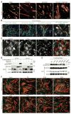
(A) MY32 (myosin) immunostaining of C2C12 cells infected with retroviruses expressing Myomaker, Myomixer, or both and differentiated for 4 days. Nuclei were counterstained with Hoechst and pseudocolored green. Scale bar, 50 μm. (B and C) Fluorescence images of GFP, mCherry, and Hoechst to counterstain nucleus. Scale bars, 100 μm (B), 50 μm (C). Arrows point to multinucleated GFP+/mCherry+ cells. (D and E) Coimmunoprecipitation assays were performed by using 10T1/2 cells or C2C12 myoblasts infected with retroviruses expressing Myomixer and/or FLAG-Myomaker. IP, immunoprecipitation; IB, immunoblot. (F) MY32 (myosin) immunostaining of C2C12 cells infected with retroviruses expressing Myomaker, together with WTor mutated versions of Myomixer, differentiated for 1 week. Nuclei were counterstained with Hoechst and pseudocolored green. Scale bar, 50 μm. R, arginine; E, glutamic acid; C, cysteine; L, leucine; A, alanine; EEEAA, R34E-R38E-R46E-C52A-L54A.
Similar articles
- Requirement of the fusogenic micropeptide myomixer for muscle formation in zebrafish.
Shi J, Bi P, Pei J, Li H, Grishin NV, Bassel-Duby R, Chen EH, Olson EN. Shi J, et al. Proc Natl Acad Sci U S A. 2017 Nov 7;114(45):11950-11955. doi: 10.1073/pnas.1715229114. Epub 2017 Oct 23. Proc Natl Acad Sci U S A. 2017. PMID: 29078404 Free PMC article. - Fusogenic micropeptide Myomixer is essential for satellite cell fusion and muscle regeneration.
Bi P, McAnally JR, Shelton JM, Sánchez-Ortiz E, Bassel-Duby R, Olson EN. Bi P, et al. Proc Natl Acad Sci U S A. 2018 Apr 10;115(15):3864-3869. doi: 10.1073/pnas.1800052115. Epub 2018 Mar 26. Proc Natl Acad Sci U S A. 2018. PMID: 29581287 Free PMC article. - The regulatory role of Myomaker and Myomixer-Myomerger-Minion in muscle development and regeneration.
Chen B, You W, Wang Y, Shan T. Chen B, et al. Cell Mol Life Sci. 2020 Apr;77(8):1551-1569. doi: 10.1007/s00018-019-03341-9. Epub 2019 Oct 23. Cell Mol Life Sci. 2020. PMID: 31642939 Free PMC article. Review. - Myomaker and Myomixer Characterization in Gilthead Sea Bream under Different Myogenesis Conditions.
Perelló-Amorós M, Otero-Tarrazón A, Jorge-Pedraza V, García-Pérez I, Sánchez-Moya A, Gabillard JC, Moshayedi F, Navarro I, Capilla E, Fernández-Borràs J, Blasco J, Chillarón J, García de la Serrana D, Gutiérrez J. Perelló-Amorós M, et al. Int J Mol Sci. 2022 Nov 24;23(23):14639. doi: 10.3390/ijms232314639. Int J Mol Sci. 2022. PMID: 36498967 Free PMC article. - [Molecular regulation mechanism of Myomaker and Myomerger in myoblast fusion].
Zhu Y, Zhang JW, Qi J, Li XW, Chen L, Li MZ, Ma JD. Zhu Y, et al. Yi Chuan. 2019 Dec 20;41(12):1110-1118. doi: 10.16288/j.yczz.19-232. Yi Chuan. 2019. PMID: 31857282 Review. Chinese.
Cited by
- Suppressing PDGFRβ Signaling Enhances Myocyte Fusion to Promote Skeletal Muscle Regeneration.
Xue S, Benvie AM, Blum JE, Kolba NJ, Cosgrove BD, Thalacker-Mercer A, Berry DC. Xue S, et al. bioRxiv [Preprint]. 2024 Oct 18:2024.10.15.618247. doi: 10.1101/2024.10.15.618247. bioRxiv. 2024. PMID: 39464006 Free PMC article. Preprint. - Satellite cell-derived TRIM28 is pivotal for mechanical load- and injury-induced myogenesis.
Lin KH, Hibbert JE, Flynn CG, Lemens JL, Torbey MM, Steinert ND, Flejsierowicz PM, Melka KM, Lindley GT, Lares M, Setaluri V, Wagers AJ, Hornberger TA. Lin KH, et al. EMBO Rep. 2024 Sep;25(9):3812-3841. doi: 10.1038/s44319-024-00227-1. Epub 2024 Aug 14. EMBO Rep. 2024. PMID: 39143258 Free PMC article. - Skeletal muscle: molecular structure, myogenesis, biological functions, and diseases.
Feng LT, Chen ZN, Bian H. Feng LT, et al. MedComm (2020). 2024 Jul 10;5(7):e649. doi: 10.1002/mco2.649. eCollection 2024 Jul. MedComm (2020). 2024. PMID: 38988494 Free PMC article. Review. - The IRE1α/XBP1 signaling axis drives myoblast fusion in adult skeletal muscle.
Joshi AS, Tomaz da Silva M, Roy A, Koike TE, Wu M, Castillo MB, Gunaratne PH, Liu Y, Iwawaki T, Kumar A. Joshi AS, et al. EMBO Rep. 2024 Aug;25(8):3627-3650. doi: 10.1038/s44319-024-00197-4. Epub 2024 Jul 9. EMBO Rep. 2024. PMID: 38982191 Free PMC article. - Spatial transcriptomics in embryonic mouse diaphragm muscle reveals regional gradients and subdomains of developmental gene expression.
Kaplan MM, Zeidler M, Knapp A, Hölzl M, Kress M, Fritsch H, Krogsdam A, Flucher BE. Kaplan MM, et al. iScience. 2024 May 17;27(6):110018. doi: 10.1016/j.isci.2024.110018. eCollection 2024 Jun 21. iScience. 2024. PMID: 38883818 Free PMC article.
References
- Buckingham M, Rigby PWJ. Dev Cell. 2014;28:225–238. - PubMed
Publication types
MeSH terms
Substances
Grants and funding
- R01 DK099653/DK/NIDDK NIH HHS/United States
- R01 HL130253/HL/NHLBI NIH HHS/United States
- R01 AR067294/AR/NIAMS NIH HHS/United States
- U54 HD087351/HD/NICHD NIH HHS/United States
- P50 HD087351/HD/NICHD NIH HHS/United States
LinkOut - more resources
Full Text Sources
Other Literature Sources
Molecular Biology Databases
Miscellaneous


