Single-cell RNA-seq reveals new types of human blood dendritic cells, monocytes, and progenitors - PubMed (original) (raw)
. 2017 Apr 21;356(6335):eaah4573.
doi: 10.1126/science.aah4573.
Rahul Satija 3 4 5, Gary Reynolds 6, Siranush Sarkizova 3, Karthik Shekhar 3, James Fletcher 6, Morgane Griesbeck 7, Andrew Butler 4 5, Shiwei Zheng 4 5, Suzan Lazo 8, Laura Jardine 6, David Dixon 6, Emily Stephenson 6, Emil Nilsson 9, Ida Grundberg 9, David McDonald 6, Andrew Filby 6, Weibo Li 3 2, Philip L De Jager 3 10, Orit Rozenblatt-Rosen 3, Andrew A Lane 3 8, Muzlifah Haniffa 11 12, Aviv Regev 1 13 14, Nir Hacohen 1 2
Affiliations
- PMID: 28428369
- PMCID: PMC5775029
- DOI: 10.1126/science.aah4573
Single-cell RNA-seq reveals new types of human blood dendritic cells, monocytes, and progenitors
Alexandra-Chloé Villani et al. Science. 2017.
Abstract
Dendritic cells (DCs) and monocytes play a central role in pathogen sensing, phagocytosis, and antigen presentation and consist of multiple specialized subtypes. However, their identities and interrelationships are not fully understood. Using unbiased single-cell RNA sequencing (RNA-seq) of ~2400 cells, we identified six human DCs and four monocyte subtypes in human blood. Our study reveals a new DC subset that shares properties with plasmacytoid DCs (pDCs) but potently activates T cells, thus redefining pDCs; a new subdivision within the CD1C+ subset of DCs; the relationship between blastic plasmacytoid DC neoplasia cells and healthy DCs; and circulating progenitor of conventional DCs (cDCs). Our revised taxonomy will enable more accurate functional and developmental analyses as well as immune monitoring in health and disease.
Copyright © 2017, American Association for the Advancement of Science.
Figures
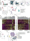
Figure 1. Human blood DC heterogeneity delineated by single-cell RNA-sequencing
(A) Workflow of experimental strategy: (i) isolation of human PBMC from blood; (ii) sorting single DC (8×96-well plates) and monocytes (4×96-well plates) into single wells using an antibody cocktail to enrich for cell fractions; (iii) single cell transcriptome profiling. (B) Gating strategy for single-cell sorting: DCs were defined as live, LIN(CD3, CD19, CD56)−CD14−HLA-DR+ cells. Three loose overlapping gates were drawn as an enrichment strategy to ensure a comprehensive and even sampling of all populations: CD11C+CD141+ (CD141; turquoise), CD11C+CD1C+ (CD1C; orange), CD11C+CD141−CD1C− (“double negative”; blue), and CD11C−CD123+ plasmacytoid DCs (pDCs; purple). 24 single cells from these four gates were sorted per 96-well plate. A fifth gate (CD11C−CD123−; red dashed) was subsequently investigated (see Fig. 6). (C) _t_-SNE analysis of DCs (n = 742). Numbers of successfully profiled single cells per cluster: DC1 (n =166); DC2 (_n_=105); DC3 (_n_=95); DC4 (n =175); DC5 (_n_=30); DC6 (n =171). The number of discriminative genes with AUC cutoff ≥ 0.85 is reported in bracket next to each cluster ID. Up to five top discriminators are listed next to each cluster; number in bracket refers to AUC value. Colors indicate unbiased DC classification via graph-based clustering. Each dot represents an individual cell. (D) Heatmap reports scaled expression [log TPM (transcripts per million) values] of discriminative gene sets for each cluster defined in Fig. 1C with AUC cutoff ≥0.85. Color scheme is based on z-score distribution from −2.5 (purple) to 2.5 (yellow). Right margin color bars highlight gene sets specific to the respective DC subset.
Figure 2. Definition and validation of CD1C+ DC subsets
(A) Heatmap showing scaled expression (log TPM values) of discriminative gene sets defining each CD1C+ DC subset with AUC cutoff ≥ 0.75. Color scheme is based on z-score distribution, from −2.5 (purple) to 2.5 (yellow). Violin plots illustrate expression distribution of candidate genes across subsets on the x-axis (orange for CD1C_A/DC2; green for CD1C_B/DC3). In red are three markers used for subsequent enrichment strategy: CD163, CD36 and FCGR2B/CD32B (AUC =0.63). (B) Enrichment gating strategy of CD1C+ DC subsets [LIN(CD3, CD19, CD56)−HLA-DR+CD14−CD1C+CD11C+]. The CD1C_A/DC2 subset was further enriched by sorting on the 10% brightest CD32B+ cells (orange gate); the CD1C_B/DC3 subset was enriched by sorting on CD32B−CD163+CD36+ cells (green gate) or on CD32B−CD163+. Right: Overlay of the two sorted CD1C+ DC populations; 47 single cells were sorted from the green and orange gates in a 96-well plate for profiling. (C) Heatmap reporting scaled expression (log TPM values) of scRNAseq data from three cell subsets defined by CD1C+CD32B+, CD1C+CD36+CD163+, and CD1C+CD163+. Either CD1C+CD36+CD163+ or CD1C+CD163+ population recapitulated the CD1C_B/DC3 signature. (D) Proliferation of allogeneic CD4+ and CD8+ T cells 5 days after co-culture with CD14+ monocytes, pDCs, CD1C_A/DC2 DCs (CD1C+CD32B+), and CD1C_B DC3 (CD1C+CD163+). Left: Representative pseudocolor dot plot. Right: Bar graphs of composite data (n=3, mean ± SEM, *P<0.05, paired t-test).
Figure 3. Human blood monocyte heterogeneity
(A) Gating strategy for monocyte single cell sorting. Monocytes were enriched by first gating on LIN(CD3, CD19, CD56)−CD14+/lo, followed by three loose overlapping gates defined by relative expression of CD14 and CD16 for comprehensive sampling of CD14++CD16− (yellow), CD14++CD16+ (purple), and CD14+CD16++ (blue); 32 cells from each gate were sorted per 96-well plate profiled. Bottom right: Dot plot shows overlay of the sorted populations. (B) _t_-SNE analysis incorporating monocytes (_n_=337 successfully profiled) and DCs (_n_=742). Number of successfully profiled single monocytes per transcriptionally defined clusters includes Mono1 (_n_=148), Mono2 (_n_=137), Mono3 (_n_=31), and Mono4 (_n_=21). The number of discriminative genes with AUC cutoff ≥ 0.85 (combined analysis of DC and monocyte datasets) is reported in bracket next to cluster ID. Up to 5 top discriminators are listed next to each cluster; the number in bracket next to each gene refers to AUC value. Colors indicate unbiased DC and monocyte clustering from graph-based clustering. Each dot represents an individual cell. (C) Heatmap reporting scaled expression (log TPM values) of discriminative gene sets for each monocyte subsets with AUC cutoff ≥ 0.85 (see fig. S4B for detailed heatmap). Color scheme is based on _z_-score distribution, from −2.5 (purple) to 2.5 (yellow). Color bars in right margin highlight gene sets of interest.
Figure 4. Identification of AXL+SIGLEC6+ DCs (AS DCs)
(A) Violin plots showing expression distribution of surface markers AXL and SIGLEC6. Other populations are depicted on the x axis; each dot represents an individual cell. (B) Flow cytometry gating strategy to identify AXL+SIGLEC6+ cells within human blood LIN(CD3, CD19, CD20, CD161)− and HLA-DR+ mononuclear fraction. AXL+SIGLEC6+ cells were further distinguished by the relative expression of IL3RA/CD123 and ITGAX/CD11C [1 = CD123+CD11c−/lo (pink); 2 = CD123loCD11c+ (blue)]. Data shown are a representative analysis of 10 healthy individuals. (C) _t_-SNE analysis of all DCs (_n_=742), along with prospectively profiled AXL+SIGLEC6+ single cells (_n_=105), using gating strategy in (B) (sorted from purple gate). Newly isolated AS DCs overlap with the originally identified DC5 cluster (_n_=30), indicated by purple dashed circle. (D) Heatmap reporting scaled expression (log TPM values) of discriminative gene sets (AUC cutoff ≥ 0.75), highlighting the expression continuum of AS DCs. Top bar graph defines the AS DCs population purity score based on the top 10 most discriminative genes (i.e., AXL, PPP1R14A, SIGLEC6, CD22, DAB2, S100A10, FAM105A, MED12L, ALDH2, and LTK). (E) Heatmap reporting scaled expression (log TPM values) of prospectively enriched AS DCs populations (_n_=90) isolated by relative ITGAX/CD11C and IL3RA/CD123 expression levels [red in (**D)**]; 43 single AXL+SIGLEC6+CD11C− [pink gate in (B)] and 47 single AXL+SIGLEC6+CD11C+ [blue gate in (B)] were sequenced. The average expression values of the original CD1C+ (combined DC2 and DC3), CD141+/CLEC9A+ (DC1) and pDC (DC6) single cells were used as reference to highlight enrichment of cDC-like and pDC-like gene sets. Top bar graph represents AS DC purity score. (F) Frequency (% mean ±SEM) of AXL+SIGLEC6+CD123+CD11C−/lo [population 1 (pink): 0.1 ± 0.014] and AXL+SIGLEC6+CD123loCD11C+ [population 2 (blue): 0.04 ± 0.01] as a percentage of LIN(CD3, CD19, CD20, CD161)−HLA-DR+ PBMCs. Scatter plot includes data from nine healthy individuals. (G) _t_-SNE analysis of flow cytometry data for LIN(CD3,CD19,CD20,CD161)−HLA-DR+CD14−CD16− PBMCs based on the protein expression levels of AXL, SIGLEC6, CD1C, CD11C, CD22, CD33, CD34, CD45RA, CD100, CD123, CD303 and HLA-DR (see Fig. 6 for CD100hiCD34int population). Overlay of populations defined by conventional flow cytometry gating on clusters derived by _t_-SNE analysis shown in the legend.
Figure 5. Phenotypic and functional characterization of AS DCs and “pure” pDCs
(A) Heatmap reporting scaled expression (log TPM values) of gene sets common between AS DCs (DC5) and cDCs (clusters DC1 to DC4), and genes uniquely expressed in pDCs (DC6). Gene sets were generated through K-means clustering using the doKmeans function in the Seurat package. (B) Morphology of pDCs, CD1C+ DCs, CLEC9A+ DCs, AXL+SIGLEC6+CD123+CD11C−/lo and AXL+SIGLEC6+CD123loCD11C+ by Giemsa-Wright stain. Scale bar, 10μm. (C) IFNα (left panel) and IL-12p70 (right panel) concentration in culture supernatant 24 hours after CpG and LPS stimulation (_n_=8) or after LPS, R848 and poly(I:C) stimulation (_n_=4) of CD14++CD16− monocytes, pDCs, CLEC9A+ DCs, CD1C+ DCs, AXL+SIGLEC6+CD123+CD11C−/lo (1, pink), AXL+SIGLEC6+CD123loCD11C+ (2, blue), and CD100hiCD34int cells (3, beige). Composite data from four to eight donors is shown (mean ±SEM; **P<0.01, *** P<0.001, Mann-Whitney U test). (D) Proliferation of allogeneic CD4+ and CD8+ T cells 5 days after co-culture with pDCs contaminated with AXL+SIGLEC6+ cells compared with pDCs devoid of AXL+SIGLEC6+cells, in the context of LPS or LPS+R848 stimulation. Top: Representative pseudocolor dot plot. Bottom: Bar graphs of composite data (_n_=4, mean ± SEM, *P<0.05, paired t-test). (E) Proliferation of allogeneic CD4+ and CD8+ T cells 5 days after co-culture with CD14++CD16− monocytes, pDCs, CLEC9A+ DCs, CD1C+ DCs, AXL+SIGLEC6+CD123+CD11C−/lo (1, pink), AXL+SIGLEC6+CD123loCD11C+ (2, blue) cells, and CD100hiCD34int (3, beige) cells. Top: Representative pseudocolor dot plot. Bottom: Bar graphs of composite data (_n_=7, mean ±SEM, **P<0.01, paired t-test). (F) Top: Immunohistochemical staining of human tonsil with AXL (brown), CD123 (purple) and CD3 (green). Brown arrows depict AXL+CD123+ cells adjacent to CD3+ T cells. Data shown are representative of four donors. Scale bar, 50μm. Middle: Frequency of AXL+SIGLEC6+CD123+ and CD123lo/− cells in human tonsil determined by flow cytometry analysis, as a percentage of CD45+LIN(CD3,CD19,CD20,CD56,CD161)−HLA-DR+ cells (mean ± SEM of three donors shown; AXL+SIGLEC6+CD123+, 0.7 ± 0.2%; AXL+SIGLEC6+CD123lo/−, 1.7 ± 0.2%). Bottom: Representative pseudocolor dot plot of AXL+SIGLEC6+CD123+ (pop. 1, pink) and AXL+SIGLEC6+CD123lo/− (pop. 2, blue) cells in human tonsil by flow cytometry analysis (_n_=3).
Figure 6. Identification and characterization of circulating CD100hiCD34int cDC progenitor
(A) Flow cytometry gating strategy to isolate DC subsets: CLEC9A+ DCs (red), CD1C+ DCs (blue), pDCs (green), AXL+SIGLEC6+ cells (purple), and CD123−CD11C− cells (red) for differentiation assays. Data shown are a representative analysis of at least 10 healthy individuals. (B) Differentiation assay readout (flow cytometry for CLEC9A+ DCs, CD1C+ DCs and pDC; scRNA-seq profiling of CD45+ cells) after 7 days of co-culturing LIN(CD3,CD19,CD20,CD161)−HLA-DR+CD14−CD16−AXL−SIGLEC6−CD123−CD11C− cells on MS5 stromal cell line supplemented with GM-CSF, SCF and FLT3LG. Top: Representative overlay dot plots. Overlay of pDC (green) and output cells (gray) for CD123 and CD303 expression is shown at far right (in green). Population 3 (in beige) represents CD100hiCD34int at day 0. Top right: Composite bar graphs for CLEC9A+ and CD1C+ DCs differentiated from culture by flow cytometry analysis (_n_=4, mean ± SEM). Heatmap in bottom panel reports scaled expression (log TPM values) signature from culture output by scRNA-seq (_n_=132), confirming differentiated CLEC9A+ (red) and CD1C+ (blue) DC transcriptional identities. Original transcriptional signatures from DC1 (CD141+/CLEC9A+ DC), DC2 (CD1C_A subset), and DC3 (CD1C_B subset) clusters are used as reference set. (C) Top: Flow cytometry gating strategy used to identify the CD100hiCD34int subset. All cell fractions in dashed gate were tested for differentiation potential (see fig. S7, A to F). Bottom: Output from CD100hiCD34int fraction (population 3, beige gate). (D) Frequency of CD100hiCD34int subset as of LIN(CD3,CD19,CD20,CD161)−HLA-DR+ PBMCs (_n_=9 healthy donors). Morphology of CD100hiCD34int cell by Giemsa-Wright stain. Scale bar, 10μm. (E) Proliferative capacity of peripheral blood Cell Trace Violet (CTV)-labeled CD34+ HSCs (purple), CD100hiCD34int (3, beige), AXL+SIGLEC6+CD123+CD11C−/lo (1, pink), and AXL+SIGLEC6+CD123loCD11C+ (2, blue), as measured by CTV dilution after 5 days in culture on MS5 stromal cell line supplemented with GM-CSF, SCF and FLT3LG. Left: Representative overlay histogram. Right: Composite bar graphs illustrating percentage of proliferated cells and number of proliferations undergone from three donors shown (*P<0.05, paired t-test). (F) Output from differentiation assays seeded with CLEC9A+ DCs, CD1C+ DCs, pDCs, and AXL+SIGLEC6+cells isolated using gating strategy in (A). AXL+SIGLEC6+x2 = double FLT3L concentration. Also shown in (C) and (F) are representative culture outputs on day 7 and composite bar graphs (mean ± SEM; _n_=6 donors). (G) PCA analysis incorporating monocytes (_n_=339), DCs (_n_=742), and four BPDCN patient samples (_n_=174) using the R software package Seurat. PC1 versus PC2 demonstrates the close transcriptional proximity between all four BPDCN samples and pDCs (dashed black circle); black bracket indicates overlapping cells. PC1 and PC2 variance is 3.8%. Each dot represents an individual cell; colored legend for each subset is shown at the right.
Comment in
- Single-cell sequencing made simple.
Perkel JM. Perkel JM. Nature. 2017 Jul 3;547(7661):125-126. doi: 10.1038/547125a. Nature. 2017. PMID: 28682345 No abstract available. - Human dendritic cell subset 4 (DC4) correlates to a subset of CD14dim/-CD16++ monocytes.
Calzetti F, Tamassia N, Micheletti A, Finotti G, Bianchetto-Aguilera F, Cassatella MA. Calzetti F, et al. J Allergy Clin Immunol. 2018 Jun;141(6):2276-2279.e3. doi: 10.1016/j.jaci.2017.12.988. Epub 2018 Jan 31. J Allergy Clin Immunol. 2018. PMID: 29366702 No abstract available.
Similar articles
- Phenotypic and Transcriptomic Analysis of Peripheral Blood Plasmacytoid and Conventional Dendritic Cells in Early Drug Naïve Rheumatoid Arthritis.
Cooles FAH, Anderson AE, Skelton A, Pratt AG, Kurowska-Stolarska MS, McInnes I, Hilkens CMU, Isaacs JD. Cooles FAH, et al. Front Immunol. 2018 May 9;9:755. doi: 10.3389/fimmu.2018.00755. eCollection 2018. Front Immunol. 2018. PMID: 29867920 Free PMC article. - Dendritic Cell Subsets and Effector Function in Idiopathic and Connective Tissue Disease-Associated Pulmonary Arterial Hypertension.
van Uden D, Boomars K, Kool M. van Uden D, et al. Front Immunol. 2019 Jan 22;10:11. doi: 10.3389/fimmu.2019.00011. eCollection 2019. Front Immunol. 2019. PMID: 30723471 Free PMC article. Review. - Circulating CD1c(+) DCs are superior at activating Th2 responses upon Phl p stimulation compared with basophils and pDCs.
Rydnert F, Lundberg K, Greiff L, Lindstedt M. Rydnert F, et al. Immunol Cell Biol. 2014 Jul;92(6):557-60. doi: 10.1038/icb.2014.23. Epub 2014 Apr 1. Immunol Cell Biol. 2014. PMID: 24687020 Clinical Trial. - Human in vivo-generated monocyte-derived dendritic cells and macrophages cross-present antigens through a vacuolar pathway.
Tang-Huau TL, Gueguen P, Goudot C, Durand M, Bohec M, Baulande S, Pasquier B, Amigorena S, Segura E. Tang-Huau TL, et al. Nat Commun. 2018 Jul 2;9(1):2570. doi: 10.1038/s41467-018-04985-0. Nat Commun. 2018. PMID: 29967419 Free PMC article. - Antigen presentation by mouse monocyte-derived cells: Re-evaluating the concept of monocyte-derived dendritic cells.
Coillard A, Segura E. Coillard A, et al. Mol Immunol. 2021 Jul;135:165-169. doi: 10.1016/j.molimm.2021.04.012. Epub 2021 Apr 23. Mol Immunol. 2021. PMID: 33901761 Review.
Cited by
- Therapeutic cancer vaccines.
Saxena M, van der Burg SH, Melief CJM, Bhardwaj N. Saxena M, et al. Nat Rev Cancer. 2021 Jun;21(6):360-378. doi: 10.1038/s41568-021-00346-0. Epub 2021 Apr 27. Nat Rev Cancer. 2021. PMID: 33907315 Review. - A machine learning method for the discovery of minimum marker gene combinations for cell type identification from single-cell RNA sequencing.
Aevermann B, Zhang Y, Novotny M, Keshk M, Bakken T, Miller J, Hodge R, Lelieveldt B, Lein E, Scheuermann RH. Aevermann B, et al. Genome Res. 2021 Oct;31(10):1767-1780. doi: 10.1101/gr.275569.121. Epub 2021 Jun 4. Genome Res. 2021. PMID: 34088715 Free PMC article. - Genetic models of human and mouse dendritic cell development and function.
Anderson DA 3rd, Dutertre CA, Ginhoux F, Murphy KM. Anderson DA 3rd, et al. Nat Rev Immunol. 2021 Feb;21(2):101-115. doi: 10.1038/s41577-020-00413-x. Epub 2020 Sep 9. Nat Rev Immunol. 2021. PMID: 32908299 Free PMC article. Review. - Fcγ receptors and immunomodulatory antibodies in cancer.
Galvez-Cancino F, Simpson AP, Costoya C, Matos I, Qian D, Peggs KS, Litchfield K, Quezada SA. Galvez-Cancino F, et al. Nat Rev Cancer. 2024 Jan;24(1):51-71. doi: 10.1038/s41568-023-00637-8. Epub 2023 Dec 7. Nat Rev Cancer. 2024. PMID: 38062252 Review. - The Role of Macrophages in the Pathogenesis of SARS-CoV-2-Associated Acute Respiratory Distress Syndrome.
Kosyreva A, Dzhalilova D, Lokhonina A, Vishnyakova P, Fatkhudinov T. Kosyreva A, et al. Front Immunol. 2021 May 10;12:682871. doi: 10.3389/fimmu.2021.682871. eCollection 2021. Front Immunol. 2021. PMID: 34040616 Free PMC article. Review.
References
Publication types
MeSH terms
Grants and funding
- P30 DK043351/DK/NIDDK NIH HHS/United States
- T32 HG002295/HG/NHGRI NIH HHS/United States
- DP2 HG009623/HG/NHGRI NIH HHS/United States
- U19 AI082630/AI/NIAID NIH HHS/United States
- P50 HG006193/HG/NHGRI NIH HHS/United States
- WT_/Wellcome Trust/United Kingdom
- P30 AI060354/AI/NIAID NIH HHS/United States
- MR/N005872/1/MRC_/Medical Research Council/United Kingdom
- RM1 HG006193/HG/NHGRI NIH HHS/United States
- HHMI/Howard Hughes Medical Institute/United States
LinkOut - more resources
Full Text Sources
Other Literature Sources


