Age-Related Effects and Sex Differences in Gray Matter Density, Volume, Mass, and Cortical Thickness from Childhood to Young Adulthood - PubMed (original) (raw)
Age-Related Effects and Sex Differences in Gray Matter Density, Volume, Mass, and Cortical Thickness from Childhood to Young Adulthood
Efstathios D Gennatas et al. J Neurosci. 2017.
Abstract
Developmental structural neuroimaging studies in humans have long described decreases in gray matter volume (GMV) and cortical thickness (CT) during adolescence. Gray matter density (GMD), a measure often assumed to be highly related to volume, has not been systematically investigated in development. We used T1 imaging data collected on the Philadelphia Neurodevelopmental Cohort to study age-related effects and sex differences in four regional gray matter measures in 1189 youths ranging in age from 8 to 23 years. Custom T1 segmentation and a novel high-resolution gray matter parcellation were used to extract GMD, GMV, gray matter mass (GMM; defined as GMD × GMV), and CT from 1625 brain regions. Nonlinear models revealed that each modality exhibits unique age-related effects and sex differences. While GMV and CT generally decrease with age, GMD increases and shows the strongest age-related effects, while GMM shows a slight decline overall. Females have lower GMV but higher GMD than males throughout the brain. Our findings suggest that GMD is a prime phenotype for the assessment of brain development and likely cognition and that periadolescent gray matter loss may be less pronounced than previously thought. This work highlights the need for combined quantitative histological MRI studies.SIGNIFICANCE STATEMENT This study demonstrates that different MRI-derived gray matter measures show distinct age and sex effects and should not be considered equivalent but complementary. It is shown for the first time that gray matter density increases from childhood to young adulthood, in contrast with gray matter volume and cortical thickness, and that females, who are known to have lower gray matter volume than males, have higher density throughout the brain. A custom preprocessing pipeline and a novel high-resolution parcellation were created to analyze brain scans of 1189 youths collected as part of the Philadelphia Neurodevelopmental Cohort. A clear understanding of normal structural brain development is essential for the examination of brain-behavior relationships, the study of brain disease, and, ultimately, clinical applications of neuroimaging.
Keywords: MRI; T1-weighted imaging; brain structure; cortical thickness; gray matter density; gray matter volume.
Copyright © 2017 the authors 0270-6474/17/375065-09$15.00/0.
Figures
Figure 1.
T1 preprocessing and high-resolution gray matter parcellation. A, Raw T1 MPRAGE volumes were first corrected for field inhomogeneity and then skull stripped by transforming the MNI brain mask to native space. Gray matter segmentation was performed without the use of tissue priors to produce unbiased estimates of GMD. B, The GMD maps of an age- and sex-balanced subsample of 240 subjects were averaged and smoothed; 1 − the gradient of the resulting image was calculated and passed to a 3D watershed algorithm, resulting in 1625 regions covering the whole-brain gray matter.
Figure 2.
Density increases in adolescence while other measures largely decrease. Females have higher density and lower volume. Plots show fitted values of whole-brain gray matter measures against age for the two sexes. GMD and CT were averaged across the brain (weighted by N voxels in each parcel), and GMV and GMM were summed. To make results comparable across measures, they are plotted as percentages: 100% is defined as the fitted value for males at 8 years of age. Shaded bands correspond to ±2 × SE of the fit (∼95% confidence interval).
Figure 3.
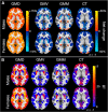
Percentage net change and variance explained by sex and modality. A, For each parcel, the percentage net change was calculated as follows: (fitted value at 23 − fitted value at 8)/(fitted value at 8) × 100%. GMD increased virtually throughout the brain, while the other modalities show mostly decreases. Females showed a greater increase in density than males throughout the brain. B, Percentage variance of each measure explained by age. GMD showed the highest _R_2 values, followed by CT. High bilateral symmetry on all maps suggests biological plausibility. Interactive movies including all axial slices in this figure are available on-line at
https://egenn.github.io/gmdvdev
.
Figure 4.
Sex differences by modality by MNI label against age. The difference of male and female fitted values for each modality for each MNI label was calculated at each year from 8 to 23 years of age. This plot highlights qualitatively how sex differences vary with age, in most cases in a nonlinear fashion (a constant sex difference in any measure would appear as a horizontal line). Note that only in CT the direction of the difference changes in frontal and occipital lobes as well as the bilateral insula from a male to a female advantage.
Figure 5.
Intermodal correlations averaged by MNI label. Pairwise spearman correlations (rho) were estimated between the fitted values of model 3 (top row) of all gray matter measures to summarize the similarity of age-related effects among modalities and between their residuals (bottom row). Brain slices with these results are available on-line at
https://egenn.github.io/gmdvdev/imcor.html
. D, Gray matter density; V, gray matter volume; M, gray matter mass; T, cortical thickness.
Similar articles
- Cortical and Subcortical Gray Matter Volume in Youths With Conduct Problems: A Meta-analysis.
Rogers JC, De Brito SA. Rogers JC, et al. JAMA Psychiatry. 2016 Jan;73(1):64-72. doi: 10.1001/jamapsychiatry.2015.2423. JAMA Psychiatry. 2016. PMID: 26650724 Review. - Structural brain development between childhood and adulthood: Convergence across four longitudinal samples.
Mills KL, Goddings AL, Herting MM, Meuwese R, Blakemore SJ, Crone EA, Dahl RE, Güroğlu B, Raznahan A, Sowell ER, Tamnes CK. Mills KL, et al. Neuroimage. 2016 Nov 1;141:273-281. doi: 10.1016/j.neuroimage.2016.07.044. Epub 2016 Jul 22. Neuroimage. 2016. PMID: 27453157 Free PMC article. - Emotional and behavioral problems change the development of cerebellar gray matter volume, thickness, and surface area from childhood to adolescence: A longitudinal cohort study.
Wang Y, Ma L, Chen R, Liu N, Zhang H, Li Y, Wang J, Hu M, Zhao G, Men W, Tan S, Gao JH, Qin S, He Y, Dong Q, Tao S. Wang Y, et al. CNS Neurosci Ther. 2023 Nov;29(11):3528-3548. doi: 10.1111/cns.14286. Epub 2023 Jun 8. CNS Neurosci Ther. 2023. PMID: 37287420 Free PMC article. - A voxel-based morphometric magnetic resonance imaging study of the brain detects age-related gray matter volume changes in healthy subjects of 21-45 years old.
Bourisly AK, El-Beltagi A, Cherian J, Gejo G, Al-Jazzaf A, Ismail M. Bourisly AK, et al. Neuroradiol J. 2015 Oct;28(5):450-9. doi: 10.1177/1971400915598078. Epub 2015 Aug 25. Neuroradiol J. 2015. PMID: 26306927 Free PMC article. - The effects of childhood maltreatment on cortical thickness and gray matter volume: a coordinate-based meta-analysis.
Yang W, Jin S, Duan W, Yu H, Ping L, Shen Z, Cheng Y, Xu X, Zhou C. Yang W, et al. Psychol Med. 2023 Apr;53(5):1681-1699. doi: 10.1017/S0033291723000661. Epub 2023 Mar 22. Psychol Med. 2023. PMID: 36946124 Review.
Cited by
- More similarity than difference: comparison of within- and between-sex variance in early adolescent brain structure.
Torgerson C, Bottenhorn K, Ahmadi H, Choupan J, Herting MM. Torgerson C, et al. bioRxiv [Preprint]. 2024 Aug 19:2024.08.15.608129. doi: 10.1101/2024.08.15.608129. bioRxiv. 2024. PMID: 39229144 Free PMC article. Preprint. - The Speed of Development of Adolescent Brain Age Depends on Sex and Is Genetically Determined.
Brouwer RM, Schutte J, Janssen R, Boomsma DI, Hulshoff Pol HE, Schnack HG. Brouwer RM, et al. Cereb Cortex. 2021 Jan 5;31(2):1296-1306. doi: 10.1093/cercor/bhaa296. Cereb Cortex. 2021. PMID: 33073292 Free PMC article. - Longitudinal associations of physical activity and pubertal development with academic achievement in adolescents.
Haapala EA, Haapala HL, Syväoja H, Tammelin TH, Finni T, Kiuru N. Haapala EA, et al. J Sport Health Sci. 2020 May;9(3):265-273. doi: 10.1016/j.jshs.2019.07.003. Epub 2019 Jul 12. J Sport Health Sci. 2020. PMID: 32444151 Free PMC article. - Brain functional and structural characteristics of patients with seizure recurrence following drug withdrawal.
Tan G, Li X, Chen D, Wang H, Gong Q, Liu L. Tan G, et al. Neuroradiology. 2021 Dec;63(12):2087-2097. doi: 10.1007/s00234-021-02755-2. Epub 2021 Jul 1. Neuroradiology. 2021. PMID: 34195875 - Regulation of the Cerebral Circulation During Development.
Koehler RC. Koehler RC. Compr Physiol. 2021 Sep 23;11(4):2371-2432. doi: 10.1002/cphy.c200028. Compr Physiol. 2021. PMID: 34558670 Free PMC article.
References
Publication types
MeSH terms
LinkOut - more resources
Full Text Sources
Other Literature Sources
Medical


