Rationally Designed Influenza Virus Vaccines That Are Antigenically Stable during Growth in Eggs - PubMed (original) (raw)
Rationally Designed Influenza Virus Vaccines That Are Antigenically Stable during Growth in Eggs
Alfred T Harding et al. mBio. 2017.
Abstract
Influenza virus vaccine production is currently limited by the ability to grow circulating human strains in chicken eggs or in cell culture. To facilitate cost-effective growth, vaccine strains are serially passaged under production conditions, which frequently results in mutations of the major antigenic protein, the viral hemagglutinin (HA). Human vaccination with an antigenically drifted strain is known to contribute to poor vaccine efficacy. To address this problem, we developed a replication-competent influenza A virus (IAV) with an artificial genomic organization that allowed the incorporation of two independent and functional HA proteins with different growth requirements onto the same virion. Vaccination with these viruses induced protective immunity against both strains from which the HA proteins were derived, and the magnitude of the response was as high as or higher than vaccination with either of the monovalent parental strains alone. Dual-HA viruses also displayed remarkable antigenic stability; even when using an HA protein known to be highly unstable during growth in eggs, we observed high-titer virus amplification without a single adaptive mutation. Thus, the viral genomic design described in this work can be used to grow influenza virus vaccines to high titers without introducing antigenic mutations.IMPORTANCE Influenza A virus (IAV) is a major public health threat, and vaccination is currently the best available strategy to prevent infection. While there have been many advances in influenza vaccine production, the fact that we cannot predict the growth characteristics of a given strain under vaccine production conditions a priori introduces fundamental uncertainty into the process. Clinically relevant IAV strains frequently grow poorly under vaccine conditions, and this poor growth can result in the delay of vaccine production or the exchange of the recommended strain for one with favorable growth properties. Even in strains that grow to high titers, adaptive mutations in the antigenic protein hemagglutinin (HA) that make it antigenically dissimilar to the circulating strain are common. The genomic restructuring of the influenza virus described in this work offers a solution to the problem of uncertain or unstable growth of IAV during vaccine production.
Keywords: antigenic instability; genetic engineering; influenza A virus; influenza B virus; vaccines.
Copyright © 2017 Harding et al.
Figures
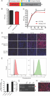
FIG 1
Encoding a red fluorescent reporter protein in segment 4 without leaving residual tags on the viral HA protein. (A) Diagram of the genomic segment 4 HA-based fluorescent reporter virus. (B) Endpoint titer of the mRuby2-HA virus compared to wild-type PR8 after 72-h incubation in eggs. (C) Multicycle growth kinetics of the mRuby2-HA virus on MDCK cells compared to WT. (D) Fluorescence microscopy time course of a single-cycle infection on MDCK cells comparing red fluorescence between wild-type and mRuby2-HA viruses. (E) Flow cytometry of mRuby2-HA- and mNeon-HA-infected cells (red or green, respectively) and uninfected cells (gray) represented as a histogram. (F) Quantification of the brightness of mRuby2-HA- and mNeon-HA-infected cells. (G) Viral segment 4 RT-PCR from wild-type PR8 and passages 0 and 4 of mRuby2-HA. The red arrowhead indicates the presence of the reporter gene; the black arrowhead indicates no reporter. The molecular weights of the DNA ladder bands are indicated in white. (H) Fluorescence microscopy of cells 24 h postinfection at an MOI of 1 with the passage 4 mRuby2-HA virus. *, P ≤ 0.05; **, P ≤ 0.001; bars, 100 μm (all panels).
FIG 2
Encoding a green fluorescent reporter protein in segment 6 without leaving residual tags on the viral NA protein. (A) Diagram of the genomic segment 6 NA-based fluorescent reporter virus. (B) Endpoint titer of the NA-furin-mNeon virus compared to WT PR8 after 72-h incubation in eggs. (C) Multicycle growth kinetics of the NA-furin-mNeon virus on MDCK cells compared to WT. (D) Flow cytometry of NA-furin-mNeon-infected cells (green) and uninfected cells (gray) represented as a histogram. (E) Quantification of brightness of fluorescence in the NA-furin-mNeon-infected cells. (F) Comparison of neuraminidase activity of purified FLAG-tagged neuraminidase from WT PR8 and the NA-furin-mNeon virus. (G) Viral segment RT-PCR from wild-type PR8 and passages 0 and 4 of the NA-furin-mNeon virus. The green arrowhead indicates the presence of the reporter gene; the black arrowhead indicates no reporter. The molecular weights of the DNA ladder bands are indicated in white. (H) Fluorescence microscopy of cells 24 h postinfection at an MOI of 1 with the passage 4 NA-furin-mNeon virus. *, P ≤ 0.05; **, P ≤ 0.001; bars, 100 μm (all panels).
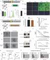
FIG 3
Expression of the HA and NA glycoproteins from a single segment allows the generation of a replication-competent H1/H3 dual-HA virus. (A) Diagram of the virus expressing both HA and NA glycoproteins in the genomic segment 4 and ZsGreen in the genomic segment 6. (B) Fluorescence microscopy time course of a single-cycle infection on MDCK cells comparing green fluorescence between wild-type and segment 4 NA/HA, segment 6 ZsGreen viruses. (C) Endpoint titer of the segment 4 NA/HA, segment 6 ZsGreen virus compared to wild-type PR8 after 72-h incubation in 10-day-old eggs. (D) Flow cytometry of the segment 4 NA/HA, segment 6 ZsGreen virus-infected (green) and uninfected (gray) cells represented as a histogram. (E) Quantification of the fold induction of fluorescence in infected cells over that in uninfected cells, caused by the segment 4 NA/HA, segment 6 ZsGreen virus. (F) Diagram of the H1/H3 dual-HA virus expressing both the subtype 1 HA and NA from genomic segment 4 and the subtype 3 HA from genomic segment 6. (G) Endpoint titer of the segment 4 NA/HA, segment 6 A/Hong Kong/1968 HA virus compared to wild-type PR8 after 72-h incubation in 11-day-old eggs. (H) Multicycle growth curve of the H1/H3 virus compared to wild-type PR8 after incubation in 11-day-old eggs. (I) Subtype-specific antibody staining of PR8, X31, and H1/H3 virus plaques. (J) Western blot assay of concentrated virus for the subtype 1 and 3 hemagglutinins. (K) ELISA measuring subtype 1 HA content utilizing a virus expressing two subtype 1 HAs from segment 4 and segment 6. (L) Sandwich ELISA of PR8, X31, and H1/H3 virus measuring content of H1 and H3 subtype HAs on the same virion. (M) Plaque reduction assays with subtype-specific H1 (0.1 μg/ml) and H3 (1 μg/ml) monoclonal antibodies. (N) Hemagglutination inhibition (HAI) assays utilizing antibodies to both subtype 1 and subtype 3 hemagglutinins. *, P ≤ 0.05; **, P ≤ 0.001; bars, 100 μm (all panels).
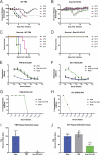
FIG 4
Infection with the live-attenuated H1/H3 dual-HA virus generates high levels of neutralizing antibodies to PR8 and X31. (A and B) Weight-loss curves from infections with the indicated doses of wild-type PR8 (A) or the H1/H3 dual-HA virus (B). (C and D) Survival curves from infections with the indicated doses of wild-type PR8 (C) or the H1/H3 virus (D). (E and F) H1-specific (E) or H3-specific (F) ELISA using sera from infected mice that received the highest dose of each strain and survived (PR8, 101; H1/H3 dual HA, 105). (G and H) HAI assays with subtype 1 (PR8) (G) and subtype 3 (X31) (H) HA viruses with pooled RDE-treated sera from mice that received the highest dose of infection and survived (PR8, 101; H1/H3 dual HA, 105). (I and J) Plaque reduction assays with PR8 (I) or X31 (J) using pooled RDE-treated sera from infected mice diluted 1:25. *, P ≤ 0.05; **, P ≤ 0.001 (all panels).
FIG 5
Vaccination with inactivated H1/H3 dual-HA virus generates high levels of protective antibodies to PR8 and X31. (A and B) H1-specific (A) or H3-specific (B) ELISA from vaccinated mouse serum. (C) Neutralization of the H1/H3 dual-HA influenza virus with polyclonal mouse sera raised against H1-, H3-, or H1/H3-expressing viruses. (D and E) HAI assays with subtype 1 (PR8) (D) and subtype 3 (X31) (E) HA viruses with pooled RDE-treated sera from vaccinated mice. (F and G) Plaque reduction assays with PR8 (F) or X31 (G) using pooled RDE-treated sera from vaccinated mice diluted 1:25. (H and I) Challenge experiments with X31 (H) or PR8 (I) in mice receiving inactivated PR8 or H1/H3 dual-HA virus vaccination. *, P ≤ 0.05; **, P ≤ 0.001 (all panels).
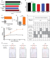
FIG 6
Dual-HA viruses can be generated with a variety of HA proteins and are antigenically stable during growth in eggs. (A) Hemagglutination units of the indicated dual-HA viruses from various IAV and IBV strains (H3, A/Hong Kong/1968; H1, A/Puerto Rico/8/1934; B Yamagata lineage, B/Yamagata/1988; B Victoria lineage, B/Malaysia/2004) relative to the parental PR8 strain. (B) Titers of the viruses from panel A. (C) Hemagglutination units of a dual-HA virus expressing the A/Fujian/411/2002 HA relative to the mono-HA A/Fujian/411/2002 WT. (D) Endpoint titers of the viruses from panel C. (E) Multicycle growth comparing the 6+2 reassortant in the PR8 background with A/Fujian/411/2002 glycoproteins and the dual-HA A/Fujian/411/2002-PR8 viruses. (F) Comparison of the parental A/Fujian/411/2002 sequence with the dual-HA virus after growth in eggs, along with previously published reports of mutations that occur in the HA of A/Fujian/411/2002 which are required to allow egg growth. (G) Sequencing chromatograms of the A/Fujian/411/2002 HA in the bivalent background after egg growth. Red boxes indicate positions that have been previously published to mutate upon egg adaptation. *, P ≤ 0.05; **, P ≤ 0.001; ns, not significant (all panels).
Similar articles
- Generation of a Genetically Stable High-Fidelity Influenza Vaccine Strain.
Naito T, Mori K, Ushirogawa H, Takizawa N, Nobusawa E, Odagiri T, Tashiro M, Ohniwa RL, Nagata K, Saito M. Naito T, et al. J Virol. 2017 Feb 28;91(6):e01073-16. doi: 10.1128/JVI.01073-16. Print 2017 Mar 15. J Virol. 2017. PMID: 28053101 Free PMC article. - Generation of DelNS1 Influenza Viruses: a Strategy for Optimizing Live Attenuated Influenza Vaccines.
Wang P, Zheng M, Lau SY, Chen P, Mok BW, Liu S, Liu H, Huang X, Cremin CJ, Song W, Chen Y, Wong YC, Huang H, To KK, Chen Z, Xia N, Yuen KY, Chen H. Wang P, et al. mBio. 2019 Sep 17;10(5):e02180-19. doi: 10.1128/mBio.02180-19. mBio. 2019. PMID: 31530680 Free PMC article. - Realities and enigmas of human viral influenza: pathogenesis, epidemiology and control.
Hilleman MR. Hilleman MR. Vaccine. 2002 Aug 19;20(25-26):3068-87. doi: 10.1016/s0264-410x(02)00254-2. Vaccine. 2002. PMID: 12163258 Review. - Targeting Hemagglutinin: Approaches for Broad Protection against the Influenza A Virus.
Zhang Y, Xu C, Zhang H, Liu GD, Xue C, Cao Y. Zhang Y, et al. Viruses. 2019 Apr 30;11(5):405. doi: 10.3390/v11050405. Viruses. 2019. PMID: 31052339 Free PMC article. Review.
Cited by
- Improved influenza vaccine responses after expression of multiple viral glycoproteins from a single mRNA.
Leonard RA, Burke KN, Spreng RL, Macintyre AN, Tam Y, Alameh MG, Weissman D, Heaton NS. Leonard RA, et al. Nat Commun. 2024 Oct 8;15(1):8712. doi: 10.1038/s41467-024-52940-z. Nat Commun. 2024. PMID: 39379405 Free PMC article. - Influenza virus strains expressing SARS-CoV-2 receptor binding domain protein confer immunity in K18-hACE2 mice.
Rader NA, Lee KS, Loes AN, Miller-Stump OA, Cooper M, Wong TY, Boehm DT, Barbier M, Bevere JR, Heath Damron F. Rader NA, et al. Vaccine X. 2024 Aug 3;20:100543. doi: 10.1016/j.jvacx.2024.100543. eCollection 2024 Oct. Vaccine X. 2024. PMID: 39221180 Free PMC article. - Vaccines and cardiovascular outcomes: lessons learned from influenza epidemics.
Yedlapati SH, Mendu A, Tummala VR, Maganti SS, Nasir K, Khan SU. Yedlapati SH, et al. Eur Heart J Suppl. 2023 Feb 14;25(Suppl A):A17-A24. doi: 10.1093/eurheartjsupp/suac110. eCollection 2023 Feb. Eur Heart J Suppl. 2023. PMID: 36937374 Free PMC article. - Single-cell genome-wide association reveals that a nonsynonymous variant in ERAP1 confers increased susceptibility to influenza virus.
Schott BH, Wang L, Zhu X, Harding AT, Ko ER, Bourgeois JS, Washington EJ, Burke TW, Anderson J, Bergstrom E, Gardener Z, Paterson S, Brennan RG, Chiu C, McClain MT, Woods CW, Gregory SG, Heaton NS, Ko DC. Schott BH, et al. Cell Genom. 2022 Nov 9;2(11):100207. doi: 10.1016/j.xgen.2022.100207. Cell Genom. 2022. PMID: 36465279 Free PMC article. - A Virion-Based Combination Vaccine Protects against Influenza and SARS-CoV-2 Disease in Mice.
Chaparian RR, Harding AT, Hamele CE, Riebe K, Karlsson A, Sempowski GD, Heaton NS, Heaton BE. Chaparian RR, et al. J Virol. 2022 Aug 10;96(15):e0068922. doi: 10.1128/jvi.00689-22. Epub 2022 Jul 12. J Virol. 2022. PMID: 35862698 Free PMC article.
References
- Shaw ML, Palese P. 2013. Orthomyxoviruses, p 1151–1185. In Knipe DM, Howley PM, Cohen JI, Griffin DE, Lamb RA, Martin MA, Racaniello VR, Roizman B (ed), Fields virology, 6th ed. Lippincott Williams & Wilkins, Philadelphia, PA.
- WHO 2014. Influenza (seasonal): fact sheet no. 211. WHO, Geneva, Switzerland.
Publication types
MeSH terms
Substances
LinkOut - more resources
Full Text Sources
Other Literature Sources
Medical


