A general method to fine-tune fluorophores for live-cell and in vivo imaging - PubMed (original) (raw)
A general method to fine-tune fluorophores for live-cell and in vivo imaging
Jonathan B Grimm et al. Nat Methods. 2017 Oct.
Abstract
Pushing the frontier of fluorescence microscopy requires the design of enhanced fluorophores with finely tuned properties. We recently discovered that incorporation of four-membered azetidine rings into classic fluorophore structures elicits substantial increases in brightness and photostability, resulting in the Janelia Fluor (JF) series of dyes. We refined and extended this strategy, finding that incorporation of 3-substituted azetidine groups allows rational tuning of the spectral and chemical properties of rhodamine dyes with unprecedented precision. This strategy allowed us to establish principles for fine-tuning the properties of fluorophores and to develop a palette of new fluorescent and fluorogenic labels with excitation ranging from blue to the far-red. Our results demonstrate the versatility of these new dyes in cells, tissues and animals.
Conflict of interest statement
Competing Financial Interests Statement
The authors declare competing interests: J.B.G. and L.D.L. have filed patent applications whose value may be affected by this publication.
Figures
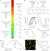
Figure 1. Fine-tuning rhodamine dyes
(a) Comparing coarse-tuning of _λ_abs for dyes 1–4 and fine-tuning observed for azetidinyl rhodamines 5–12. (b) Correlation between calculated (DFT) and experimental _λ_abs values for dyes 1, 5–12; dashed line shows ideal fit. (c) Correlation of experimental _λ_abs vs. inductive Hammett constants (_σ_I) for dyes 1, 5, 8–12. For the geminal disubstituted compounds 5 and 12 the _σ_I of the substituent was doubled. Solid line shows linear regression (R2 = 0.97). (d) Fine-tuning of the lactone–zwitterion equilibrium constant (_K_L–Z) for dyes 1, 5, 9–12. (e) Normalized absorption vs. dielectric constant (εr) for dyes 1, 5, and 9–12; error bars show ± s.e.m; n = 4. (f) Absolute absorbance of 1, 5, 9–12 (5 μM) in 1:1 dioxane:H2O. (g) Correlation of _K_L–Z vs. inductive Hammett constants (_σ_I) for dyes 1, 5, 9–12. For the geminal disubstituted compounds 5 and 12 the _σ_I of the substituent was doubled. Solid line shows linear regression (R2 = 0.91). (h) Chemical structure of JF525–HaloTag ligand 13 and JF549–HaloTag ligand 14. (i) Image of live, washed COS7 cells expressing histone H2B–HaloTag fusions and labeled with ligand 13. Scale bar: 35 μm. (j) Plot of percent labeling of histone H2B–HaloTag fusions in live cells vs. incubation time for ligands 13 (100 nM) and 14 (100 nm); error bars show ± s.e.m; n = 113–248 (see Methods).
Figure 2. Rational fine-tuning of other dyes
(a) Tuning of JF519 (4) to yield JF503 (16). (b) Structure of JF503–HaloTag ligand 17. (c) Image of live, washed COS7 cells expressing histone H2B–HaloTag fusions and labeled with ligand 17. Scale bar: 35 μm. (d) Comparison of the photostability of cells labeled with 17 and cells labeled with 488 nm-excited dyes 18 and 19 (Supplementary Fig. 2d); the initial photobleaching measurements are fitted to a linear regression. (e) Tuning of JF608 (2) to yield JF585 (21). (f) Structure of HaloTag ligands derived from JF608 (22) and JF585 (23). (g) Absorbance of HaloTag ligands 22 and 23 in the presence (+HT) or absence (–HT) of excess HaloTag protein; n = 2. (h,i) Representative images of COS7 cells expressing HaloTag–histone H2B fusion and labeled with 250 nM of HaloTag ligands 22 and 23 for 1 h and imaged directly without washing. The image for each dye pair was taken with identical microscope settings, _λ_ex = 594 nm. Numbers indicate mean signal (nuclear) to background (cytosol) ratio (S/B) in three fields of view. (h) JF608 ligand 22 (S/B from n = 224 areas). (i) JF585 ligand 23 (S/B from n = 235 areas). (j) Tuning of JF646 (3) to yield JF635 (25). (k) Structure of HaloTag ligands derived from JF646 (26) and JF635 (27) (l) Absorbance of HaloTag ligands 26 and 27 in the presence (+HT) or absence (−HT) of excess HaloTag protein (n = 2). (m,n) Representative images of COS7 cells expressing HaloTag–histone H2B fusion and labeled with 250 nM of HaloTag ligands 26 and 27 for 1 h and imaged directly without washing. The image for each dye pair was taken with identical microscope settings, _λ_ex = 647 nm. Numbers indicate mean signal (nuclear) to background (cytosol) ratio (S/B) in three fields of view. (m) JF646 ligand 26 (S/B from n = 175 areas). (n) JF635 ligand 27 (S/B from n = 278 areas). Scale bars for h,i,m,n: 15 μm.
Figure 3. Labeling in tissue and in vivo
(a) SiMView light-sheet microscopy image (3D projection) of the central nervous system of a third instar Drosophila larva expressing myristoylated HaloTag protein in ‘Basin’ neurons (BNs) and stained with JF635–HaloTag ligand (27); LBL: left brain lobe; VNC: ventral nerve cord; RBL: right brain lobe. Scale bar: 100 μm. (b) Zoom in of boxed area in panel a showing individual BN cell bodies. Scale bar: 20 μm. (c) Two-photon fluorescence excitation spectra of HaloTag conjugates (1 μM) from HaloTag ligands 13, 14, 17, 23, 26, and 27 in 10 mM HEPES buffer (pH 7.3). The two-photon excitation spectra for mCherry is shown for reference. (d) Ratio of JF585 fluorescence to GCaMP6s epifluorescence at different time points after a single injection of JF585–HaloTag ligand (23, 100 nmol) either intravenous (IV) or intraperitoneal (IP) into mice expressing HaloTag protein in either layer 4 (L4) or layer 5 (L5) cortical neurons; n = 3 fields of view; error bars show ± s.e.m. (e) Two-photon microscopy images of neurons in layer 5 of the visual cortex coexpressing GCaMP6s (green) and JF585-labeled HaloTag (magenta) after IV injection of ligand 23 (t = 5 h). Scale bar: 100 μm. Yellow circles in the merged image indicate individual neurons as regions of interest (ROIs). (f) Raster plot of spontaneous neuronal activity in different ROIs (n = 61) before and after labeling with JF585–HaloTag ligand (23). (g) Plot of average spontaneous neural activity in each ROI before and after labeling with JF585–HaloTag ligand (23); central line shows mean; error bars show ± s.d.; no significant difference is observed between time points (one-way ANOVA: p = 0.95).
Similar articles
- Synthesis of Janelia Fluor HaloTag and SNAP-Tag Ligands and Their Use in Cellular Imaging Experiments.
Grimm JB, Brown TA, English BP, Lionnet T, Lavis LD. Grimm JB, et al. Methods Mol Biol. 2017;1663:179-188. doi: 10.1007/978-1-4939-7265-4_15. Methods Mol Biol. 2017. PMID: 28924668 - Bright photoactivatable fluorophores for single-molecule imaging.
Grimm JB, English BP, Choi H, Muthusamy AK, Mehl BP, Dong P, Brown TA, Lippincott-Schwartz J, Liu Z, Lionnet T, Lavis LD. Grimm JB, et al. Nat Methods. 2016 Dec;13(12):985-988. doi: 10.1038/nmeth.4034. Epub 2016 Oct 24. Nat Methods. 2016. PMID: 27776112 - Far-red organic fluorophores contain a fluorescent impurity.
Stone MB, Veatch SL. Stone MB, et al. Chemphyschem. 2014 Aug 4;15(11):2240-6. doi: 10.1002/cphc.201402002. Epub 2014 Apr 29. Chemphyschem. 2014. PMID: 24782148 Free PMC article. - Chemical tags: applications in live cell fluorescence imaging.
Wombacher R, Cornish VW. Wombacher R, et al. J Biophotonics. 2011 Jun;4(6):391-402. doi: 10.1002/jbio.201100018. Epub 2011 May 12. J Biophotonics. 2011. PMID: 21567974 Review. - Photostable and photoswitching fluorescent dyes for super-resolution imaging.
Minoshima M, Kikuchi K. Minoshima M, et al. J Biol Inorg Chem. 2017 Jul;22(5):639-652. doi: 10.1007/s00775-016-1435-y. Epub 2017 Jan 12. J Biol Inorg Chem. 2017. PMID: 28083655 Review.
Cited by
- Gentle Rhodamines for Live-Cell Fluorescence Microscopy.
Liu T, Kompa J, Ling J, Lardon N, Zhang Y, Chen J, Reymond L, Chen P, Tran M, Yang Z, Zhang H, Liu Y, Pitsch S, Zou P, Wang L, Johnsson K, Chen Z. Liu T, et al. ACS Cent Sci. 2024 Oct 2;10(10):1933-1944. doi: 10.1021/acscentsci.4c00616. eCollection 2024 Oct 23. ACS Cent Sci. 2024. PMID: 39463828 Free PMC article. - Multiplexed no-wash cellular imaging using BenzoTag, an evolved self-labeling protein.
Lampkin BJ, Goldberg BJ, Kritzer JA. Lampkin BJ, et al. Chem Sci. 2024 Oct 9;15(42):17337-47. doi: 10.1039/d4sc05090h. Online ahead of print. Chem Sci. 2024. PMID: 39430930 Free PMC article. - A modular chemigenetic calcium indicator for multiplexed in vivo functional imaging.
Farrants H, Shuai Y, Lemon WC, Monroy Hernandez C, Zhang D, Yang S, Patel R, Qiao G, Frei MS, Plutkis SE, Grimm JB, Hanson TL, Tomaska F, Turner GC, Stringer C, Keller PJ, Beyene AG, Chen Y, Liang Y, Lavis LD, Schreiter ER. Farrants H, et al. Nat Methods. 2024 Oct;21(10):1916-1925. doi: 10.1038/s41592-024-02411-6. Epub 2024 Sep 20. Nat Methods. 2024. PMID: 39304767 Free PMC article. - Dimensionality reduction simplifies synaptic partner matching in an olfactory circuit.
Lyu C, Li Z, Xu C, Wong KKL, Luginbuhl DJ, McLaughlin CN, Xie Q, Li T, Li H, Luo L. Lyu C, et al. bioRxiv [Preprint]. 2024 Aug 27:2024.08.27.609939. doi: 10.1101/2024.08.27.609939. bioRxiv. 2024. PMID: 39253519 Free PMC article. Preprint. - Lipid- and protein-directed photosensitizer proximity labeling captures the cholesterol interactome.
Becker AP, Biletch E, Kennelly JP, Julio AR, Villaneuva M, Nagari RT, Turner DW, Burton NR, Fukuta T, Cui L, Xiao X, Hong SG, Mack JJ, Tontonoz P, Backus KM. Becker AP, et al. bioRxiv [Preprint]. 2024 Aug 24:2024.08.20.608660. doi: 10.1101/2024.08.20.608660. bioRxiv. 2024. PMID: 39229057 Free PMC article. Preprint.
References
- Xue L, Karpenko IA, Hiblot J, Johnsson K. Imaging and manipulating proteins in live cells through covalent labeling. Nat Chem Biol. 2015;11:917–923. - PubMed
- Liu Z, Lavis LD, Betzig E. Imaging live-cell dynamics and structure at the single-molecule level. Mol Cell. 2015;58:644–659. - PubMed
- Ceresole M. Verfahren zur Darstellung von Farbstoffen aus der Gruppe des Meta-amidophenolphtaleïns. 44002. Germany Patent. 1887
Methods-Only References
- Critchfield FE, Gibson JA, Jr, Hall JL. Dielectric constant for the dioxane–water system from 20 to 35°. J Am Chem Soc. 1953;75:1991–1992.
- Suzuki K, et al. Reevaluation of absolute luminescence quantum yields of standard solutions using a spectrometer with an integrating sphere and a back-thinned CCD detector. Phys Chem Chem Phys. 2009;11:9850–9860. - PubMed
- Frisch MJ, et al. Gaussian 09, revision D. 01. Gaussian, Inc; Wallingford CT: 2009.
- Dreuw A, Weisman JL, Head-Gordon M. Long-range charge-transfer excited states in time-dependent density functional theory require non-local exchange. J Chem Phys. 2003;119:2943–2946.
- Jacquemin D, et al. Assessment of the efficiency of long-range corrected functionals for some properties of large compounds. J Chem Phys. 2007;126:144105. - PubMed
MeSH terms
Substances
LinkOut - more resources
Full Text Sources
Other Literature Sources
Molecular Biology Databases


