Scalable whole-exome sequencing of cell-free DNA reveals high concordance with metastatic tumors - PubMed (original) (raw)
doi: 10.1038/s41467-017-00965-y.
Gavin Ha 3 4 5, Samuel S Freeman 3 5, Atish D Choudhury 4, Daniel G Stover 4 5, Heather A Parsons 4 5, Gregory Gydush 3, Sarah C Reed 3, Denisse Rotem 3, Justin Rhoades 3, Denis Loginov 3 6, Dimitri Livitz 3, Daniel Rosebrock 3 5, Ignaty Leshchiner 3, Jaegil Kim 3, Chip Stewart 3, Mara Rosenberg 3, Joshua M Francis 3 4, Cheng-Zhong Zhang 3 4 5, Ofir Cohen 3 4, Coyin Oh 3, Huiming Ding 6, Paz Polak 3 5 7, Max Lloyd 4, Sairah Mahmud 4, Karla Helvie 4, Margaret S Merrill 4, Rebecca A Santiago 4, Edward P O'Connor 4, Seong H Jeong 4, Rachel Leeson 6, Rachel M Barry 6, Joseph F Kramkowski 4, Zhenwei Zhang 4, Laura Polacek 4, Jens G Lohr 3 4, Molly Schleicher 3, Emily Lipscomb 3, Andrea Saltzman 3, Nelly M Oliver 4, Lori Marini 4, Adrienne G Waks 4 8, Lauren C Harshman 4, Sara M Tolaney 4, Eliezer M Van Allen 3 4 5 8, Eric P Winer 4, Nancy U Lin 4, Mari Nakabayashi 4 5, Mary-Ellen Taplin 4, Cory M Johannessen 3, Levi A Garraway 3 4 5 8 9, Todd R Golub 3 4 5 9, Jesse S Boehm 3, Nikhil Wagle 3 4 5, Gad Getz 10 11 12, J Christopher Love 13 14, Matthew Meyerson 15 16 17 18
Affiliations
- PMID: 29109393
- PMCID: PMC5673918
- DOI: 10.1038/s41467-017-00965-y
Scalable whole-exome sequencing of cell-free DNA reveals high concordance with metastatic tumors
Viktor A Adalsteinsson et al. Nat Commun. 2017.
Abstract
Whole-exome sequencing of cell-free DNA (cfDNA) could enable comprehensive profiling of tumors from blood but the genome-wide concordance between cfDNA and tumor biopsies is uncertain. Here we report ichorCNA, software that quantifies tumor content in cfDNA from 0.1× coverage whole-genome sequencing data without prior knowledge of tumor mutations. We apply ichorCNA to 1439 blood samples from 520 patients with metastatic prostate or breast cancers. In the earliest tested sample for each patient, 34% of patients have ≥10% tumor-derived cfDNA, sufficient for standard coverage whole-exome sequencing. Using whole-exome sequencing, we validate the concordance of clonal somatic mutations (88%), copy number alterations (80%), mutational signatures, and neoantigens between cfDNA and matched tumor biopsies from 41 patients with ≥10% cfDNA tumor content. In summary, we provide methods to identify patients eligible for comprehensive cfDNA profiling, revealing its applicability to many patients, and demonstrate high concordance of cfDNA and metastatic tumor whole-exome sequencing.
Conflict of interest statement
T.R.G., L.A.G., N.W. are consultants and equity holders in Foundation Medicine, Inc. M.M. was previously a consultant and equity holder in Foundation Medicine, Inc. C.-Z.Z. is a consultant and equity holder in Pillar Biosciences. The authors have filed a patent application on methods described in this manuscript. The remaining authors declare no competing financial interests.
Figures
Fig. 1
Copy number and tumor fractions from ULP-WGS. a cfDNA workflow. b Genome-wide copy number from 0.1× ULP-WGS of cfDNA from a healthy donor. c Genome-wide copy number from 25× WGS and 0.1× WGS of cell-free DNA from a metastatic breast cancer patient (MBC_315), and 1× WGS and WES of matched tumors from this patient. SCNA for tumor WES and cfDNA 25× coverage WGS were predicted using TITAN (“Methods”). d Comparison of copy ratios between ULP-WGS of cfDNA with deep (>10×) WGS of the same cfDNA sample, WGS (1×) of matched tumors from 22 metastatic breast cancer (MBC) patients, and WES (average mean target coverage 173×) of matched tumors from 41 MBC and prostate cancer (CRPC) patients. Log2 copy ratios were computed as normalized read coverage for each 1 Mb (WGS/ULP-WGS) and the mean of overlapping 50 kb bins (WES) after adjustment for tumor fraction/purity. The correlation of copy ratios between tumor and cfDNA was computed using Spearman rank correlation (coefficient ρ). _F_-measure (F1) is the harmonic mean of the CNA positive predictive value (precision) and sensitivity (recall) performance. Recall is defined as the proportion of SCNA gain/loss in tumor biopsy also observed in ULP-WGS of cfDNA (“Methods”). e Comparison of tumor fractions estimated from ULP-WGS and WES of cfDNA. Samples (n = 35) with similar tumor ploidy (difference < 0.75 and ploidy ≥1.5) estimated in both ULP-WGS and tumor WES are shown. The correlation between the two data types was calculated using Pearson correlation (coefficient r). Red line denotes y = x. WES tumor fractions were estimated using ABSOLUTE (shown) and TITAN (Supplementary Fig. 15, Supplementary Data 6)
Fig. 2
Comparison of whole-exome sequencing of cfDNA to whole-exome sequencing of matched tumor biopsies. a Fraction of clonal (≥0.9 cancer cell fraction, CCF) and subclonal (<0.9 CCF) SSNVs detected by MuTect in WES of tumor biopsies and confirmed (i.e., supported by ≥3 variant reads) in WES of cfDNA. Sites with <3 reads that had power <0.9 for mutation calling were not included when computing the fraction of SNVs confirmed (“Methods”). b Fraction of clonal and subclonal SSNVs detected in WES of cfDNA and confirmed in WES of tumor biopsies. For 18 patients with WES of cfDNA at a second time point t 2, SSNVs not detected in the matched tumor biopsy but confirmed at t 2 are indicated with black. c Analysis of clonal dynamics in an ER+ breast cancer patient diagnosed with metastatic disease 1.5 years (yrs) prior to biopsy and cfDNA collection (t 1, Day 0). Clustering analysis of CCF for SSNVs between matched tumor biopsy and cfDNA (t 1) is shown in the left panel. The right panel shows the CCF of four mutation clusters, one containing ESR1 L536P (Subclonal Cluster 1, orange) and the other containing ESR1 D538G (Subclonal Cluster 2, light blue), at t 1 and t 2 (51 days apart) from a patient with ER+ metastatic breast cancer being treated with a SERD. The lymph node biopsy was taken at the same time as cfDNA t 1. Mutations were clustered by the CCFs for each pair of samples using Phylogic (“Methods”). Error bars represent the 95% credible interval of the joint posterior density of the clusters. Mutations, excluding indels, having ≥90% estimated power based on coverage in both samples are shown; clusters with fewer than three mutations are excluded. The number of mutations in each cluster is indicated in the legend in parentheses
Fig. 3
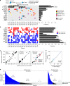
Genomic alterations of known significance and applicability to large cohorts. a, b The alteration status of significantly mutated genes predicted by MutSig2CV, (a), focal SCNAs (b), and known cancer-associated genes are shown for cfDNA and tumor biopsies from 27 metastatic breast cancer (MBC) patients. Mutated genes with MutSig2CV _q_-value < 0.1 are statistically significant. Mutations that were exclusively detected in one sample may be present at low CCF in the other matched sample but were excluded from the frequency calculation. SCNA frequencies were computed for oncogenes (MYC to ERBB2) and tumor suppressors (BRCA1 to ATM) using only amplification and deletion status, respectively. Mutations were predicted using MuTect and SCNAs were predicted using ReCapSeg and ABSOLUTE. Red dot indicates distinct mutations in tumor and cfDNA. c Mutational signatures in whole-exome sequencing of cfDNA and tumor biopsies were predicted using a Bayesian non-negative matrix factorization (NMF) approach (“Methods”). Samples with predicted biallelic inactivation of BRCA1/2 are indicated in red and blue. Black line denotes y = x; blue line denotes model fit using linear least squares regression. d Neoantigen burden, defined as the number of predicted neoantigen SSNVs, was calculated using NetMHCpan (“Methods”). Black line denotes y = x; blue line denotes model fit using linear least squares regression. e Applicability to many patients with metastatic cancer. Tumor fractions estimated from ULP-WGS of cfDNA from 903 blood samples from 391 patients with metastatic breast cancer and 536 blood samples from 129 patients with metastatic prostate cancers. The earliest blood drawn for each patient is shown. Samples with coverage <0.05× were excluded
Similar articles
- Whole-Exome Sequencing of Cell-Free DNA Reveals Temporo-spatial Heterogeneity and Identifies Treatment-Resistant Clones in Neuroblastoma.
Chicard M, Colmet-Daage L, Clement N, Danzon A, Bohec M, Bernard V, Baulande S, Bellini A, Deveau P, Pierron G, Lapouble E, Janoueix-Lerosey I, Peuchmaur M, Corradini N, Defachelles AS, Valteau-Couanet D, Michon J, Combaret V, Delattre O, Schleiermacher G. Chicard M, et al. Clin Cancer Res. 2018 Feb 15;24(4):939-949. doi: 10.1158/1078-0432.CCR-17-1586. Epub 2017 Nov 30. Clin Cancer Res. 2018. PMID: 29191970 - Whole exome sequencing for determination of tumor mutation load in liquid biopsy from advanced cancer patients.
Koeppel F, Blanchard S, Jovelet C, Genin B, Marcaillou C, Martin E, Rouleau E, Solary E, Soria JC, André F, Lacroix L. Koeppel F, et al. PLoS One. 2017 Nov 21;12(11):e0188174. doi: 10.1371/journal.pone.0188174. eCollection 2017. PLoS One. 2017. PMID: 29161279 Free PMC article. - Exome Sequencing of Cell-Free DNA from Metastatic Cancer Patients Identifies Clinically Actionable Mutations Distinct from Primary Disease.
Butler TM, Johnson-Camacho K, Peto M, Wang NJ, Macey TA, Korkola JE, Koppie TM, Corless CL, Gray JW, Spellman PT. Butler TM, et al. PLoS One. 2015 Aug 28;10(8):e0136407. doi: 10.1371/journal.pone.0136407. eCollection 2015. PLoS One. 2015. PMID: 26317216 Free PMC article. - cfSNV: a software tool for the sensitive detection of somatic mutations from cell-free DNA.
Li S, Hu R, Small C, Kang TY, Liu CC, Zhou XJ, Li W. Li S, et al. Nat Protoc. 2023 May;18(5):1563-1583. doi: 10.1038/s41596-023-00807-w. Epub 2023 Feb 27. Nat Protoc. 2023. PMID: 36849599 Free PMC article. Review. - Liquid biopsy: current technology and clinical applications.
Nikanjam M, Kato S, Kurzrock R. Nikanjam M, et al. J Hematol Oncol. 2022 Sep 12;15(1):131. doi: 10.1186/s13045-022-01351-y. J Hematol Oncol. 2022. PMID: 36096847 Free PMC article. Review.
Cited by
- Deep learning model integrating cfDNA methylation and fragment size profiles for lung cancer diagnosis.
Kim M, Park J, Seonghee Oh, Jeong BH, Byun Y, Shin SH, Im Y, Cho JH, Cho EH. Kim M, et al. Sci Rep. 2024 Jun 26;14(1):14797. doi: 10.1038/s41598-024-63411-2. Sci Rep. 2024. PMID: 38926407 Free PMC article. - Integration of multiomics features for blood-based early detection of colorectal cancer.
Gao Y, Cao D, Li M, Zhao F, Wang P, Mei S, Song Q, Wang P, Nie Y, Zhao W, Wang S, Yan H, Wang X, Jiao Y, Liu Q. Gao Y, et al. Mol Cancer. 2024 Aug 22;23(1):173. doi: 10.1186/s12943-024-01959-3. Mol Cancer. 2024. PMID: 39175001 Free PMC article. - MetDecode: methylation-based deconvolution of cell-free DNA for noninvasive multi-cancer typing.
Passemiers A, Tuveri S, Sudhakaran D, Jatsenko T, Laga T, Punie K, Hatse S, Tejpar S, Coosemans A, Van Nieuwenhuysen E, Timmerman D, Floris G, Van Rompuy AS, Sagaert X, Testa A, Ficherova D, Raimondi D, Amant F, Lenaerts L, Moreau Y, Vermeesch JR. Passemiers A, et al. Bioinformatics. 2024 Sep 2;40(9):btae522. doi: 10.1093/bioinformatics/btae522. Bioinformatics. 2024. PMID: 39177091 Free PMC article. - A Systematic Review of the Use of Circulating Cell-Free DNA Dynamics to Monitor Response to Treatment in Metastatic Breast Cancer Patients.
Jongbloed EM, Deger T, Sleijfer S, Martens JWM, Jager A, Wilting SM. Jongbloed EM, et al. Cancers (Basel). 2021 Apr 10;13(8):1811. doi: 10.3390/cancers13081811. Cancers (Basel). 2021. PMID: 33920135 Free PMC article. Review. - Total Number of Alterations in Liquid Biopsies Is an Independent Predictor of Survival in Patients With Advanced Cancers.
Vu P, Khagi Y, Riviere P, Goodman A, Kurzrock R. Vu P, et al. JCO Precis Oncol. 2020 Mar 24;4:PO.19.00204. doi: 10.1200/PO.19.00204. eCollection 2020. JCO Precis Oncol. 2020. PMID: 32923910 Free PMC article.
References
- Russo M, et al. Tumor heterogeneity and lesion-specific response to targeted therapy in colorectal cancer. Cancer Discov. 2015;6:147–153. doi: 10.1158/2159-8290.CD-15-1283. - DOI - PMC - PubMed
Publication types
MeSH terms
Substances
LinkOut - more resources
Full Text Sources
Other Literature Sources


