Optimization of prostate MRI acquisition and post-processing protocol: a pictorial review with access to acquisition protocols - PubMed (original) (raw)
Review
. 2017 Dec 8;6(12):2058460117745574.
doi: 10.1177/2058460117745574. eCollection 2017 Dec.
Affiliations
- PMID: 29242748
- PMCID: PMC5724653
- DOI: 10.1177/2058460117745574
Review
Optimization of prostate MRI acquisition and post-processing protocol: a pictorial review with access to acquisition protocols
Ivan Jambor. Acta Radiol Open. 2017.
Abstract
The aim of this review article is to provide insight into the optimization of 1.5-Testla (T) and 3-T prostate magnetic resonance imaging (MRI). An approach for optimization of data quantification, especially diffusion-weighted imaging (DWI), is provided. Benefits and limitations of various pulse sequences are discussed. Importable MRI protocols and access to imaging datasets is provided. Careful optimization of prostate MR acquisition protocol allows the acquisition of high-quality prostate MRI using clinical 1.5-T/3-T MR scanners with an overall acquisition time < 15 min.
Keywords: Prostate cancer; acquisition protocol; biparametric prostate MRI; diffusion-weighted imaging; magnetic resonance imaging (MRI).
Figures
Fig. 1.
Comparison of the trace DWI b = 2000 s/mm2 image acquired using a TorsoXL coil (a), Philips Medical Systems, Best, The Netherlands, and 32-channel cardiac coil (b), Philips Medical Systems, which demonstrated higher SNRs. Both DWI acquisitions had identical MR acquisitions parameters and image windowing/scaling.
Fig. 2.
Axial T2W image (a), monoexponential apparent diffusion coefficient map (b, calculated using b-values in the range of 0–500 s/mm2), sagittal T2W image (c), trace diffusion-weighted image of b-value 1500 s/mm2 (d), and trace diffusion-weighted image of b-value 2000 s/mm2 (e) acquired using a 1.5-T MR scanner with an acquisition time < 13 min (IMPROD_Siemens_1_5T.pdf/.edx). The suspicious lesion in the apex (write arrows) was interpreted as Likert score 5, PI-RADsv2 4, and DWI score 1 (dominant Gleason grade 4 is probable). Prostate cancer with Gleason score 4 + 3 was found in two cores of the targeted biopsy while no cancer was present in the cores the systematic biopsy. Clinically significant prostate cancer was found only in the targeted biopsy cores, and the prediction of the Gleason score based on DWI was correct. The MR acquisition protocol (IMPROD_Siemens_3T.pdf, IMPROD_Siemens_3T.edx) and reporting system (IMPROD_trial_instructions.pdf) are provided in the supporting material.
Fig. 3.
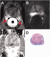
Axial T2W image (a), trace diffusion-weighted image of b-value 2000 s/mm2 (b), monoexponential ADC map (c, calculated using b-values in the range of 0–500 s/mm2) are shown. High SNR is present near to the endorectal coil which is filled with distilled water. (d) Whole mount prostatectomy section (cancer is outlined in blue). Please note the wrong position of receiver coil wire on the right side (a, red arrows).
Fig. 4.
Coronal T2W images (a–c), the same MR acquisition as in Fig. 3, demonstrate multiple air bubbles (black arrows) in outer balloon of the endorectal coil. Instructions on how to fill endorectal coils are provided in the supporting material (Instructions_how_to_fill_ERC.pdf).
Fig. 5.
Axial T2W image (a, black arrow points to wrongly positioned receiver wires of ERC), 1H-MRS 16 × 16 matrix grid overlaid over T2W image (b), trace diffusion-weighted image of b-value 2000 s/mm2 (c), monoexponential ADC map ((d), calculated using b-values in the range of 0–500 s/mm2), whole mount prostatectomy section ((e), cancer is outlined in blue) demonstrate cancer area in peripheral zone. However, the individual spectral obtained using a PRESS sequence ((f), blue square in (b); (g), red square in (b); (h), yellow square in (b); (i), purple square in (b); (j), brown square in (b)) does not demonstrate increase in choline or decrease citrate suggestive of prostate. A real voxel size of the 1H-MRS could be best approximated as a sphere with a volume of 1.51 cm3 and diameter of 14.24 mm after apodization.
Fig. 6.
Axial T2W images acquired at 3-T using surface arrays coils. All acquisitions fulfilled the recommendations stated in PI-RADs v2. However, substantial differences in acquisition times and image quality can be noted: (a) acquisition time is 2 min 55 s; (b) acquisition time is 2 min 30 s; (c) acquisition time is 2 min 20 s; (d) acquisition time is 2 min 55 s; (e) acquisition time is 3 min 10 s; (f) acquisition time is 3 min 7 s. All images have the same windowing.
Fig. 7.
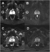
DWI of prostate performed using a turbo spin echo read-out (TSE) ((a), b-value of 0 s/mm2; (b), b-value of 500 s/mm2) and epi read-out ((c), b-value of 0 s/mm2; (d), b-value of 500 s/mm2). Due to rectal gas and resulting B0 inhomogeneities, images acquired using epi read-out are severely distorted (c, d). The Mullerian duct cyst ((b), white arrow) can be seen only on images acquired using TSE read out.
Fig. 8.
An example of four repeated DWI data acquisitions performed using an optimized b-value distribution for the biexponential function of one healthy volunteer with placement of ROIs (a–d). The DWI signal decay curve of mean signal intensity (SI) of ROI in red color is shown (e–h). Bi-exponential model has the smallest sum of squares of the vertical distance of the points from the curve (“fitting residuals”). Mono, monoexponential function; Stretched, stretched exponential function; Kurt, Kurtosis function; Biex, biexponential function; SI, signal intensity.
Fig. 9.
Axial T2W image ((a), arrow points to prostate cancer lesion) and trace DWI images of b = 0 (b), 200 (c), 400 (d), 600 (e), 800 (f), 1000 (g), 1200 (h), 1400 (i), 1600 (j), 1800 (k), and 2000 (l) s/mm2 of a patient with histologically confirmed Gleason score 4 + 3 prostate cancer demonstrate increasing contrast between the cancer lesion and benign tissue with increasing b-values. All trace DWI images have the same windowing setting. The MR acquisition protocol is provided in the Supporting Material (D_2000_b11.txt).
Fig. 10.
Axial T2W image ((a), arrow points to prostate cancer lesion) and trace DWI images of b = 0 (b), 300 (c), 600 (d), 900 (e), 1200 (f), 1500 (g), 1800 (h), 2100 (i), 2400 (j), 2700 (k), and 3000 (l) s/mm2 of a patient (the same patient as in Fig. 9) with histologically confirmed Gleason score 4 + 3 prostate cancer demonstrate increasing contrast between the cancer lesion and benign tissue with increasing b-values. All trace DWI images have the same windowing setting. The MR acquisition protocol is provided in the Supporting Material (D_3000_b11.txt).
Fig. 11.
An example of signal decay in prostate cancer and normal tissue as a function of b-values (x-axis) fitted using the monoexponential, stretched exponential, kurtosis, and biexponential functions. Mono, monoexponential function; Stretched, stretched exponential function; Kurt, Kurtosis function; Biex, biexponential function; SI, signal intensity.
Similar articles
- Diagnostic accuracy of biparametric vs multiparametric MRI in clinically significant prostate cancer: Comparison between readers with different experience.
Di Campli E, Delli Pizzi A, Seccia B, Cianci R, d'Annibale M, Colasante A, Cinalli S, Castellan P, Navarra R, Iantorno R, Gabrielli D, Buffone A, Caulo M, Basilico R. Di Campli E, et al. Eur J Radiol. 2018 Apr;101:17-23. doi: 10.1016/j.ejrad.2018.01.028. Epub 2018 Feb 1. Eur J Radiol. 2018. PMID: 29571792 - How to read biparametric MRI in men with a clinical suspicious of prostate cancer: Pictorial review for beginners with public access to imaging, clinical and histopathological database.
Jambor I, Martini A, Falagario UG, Ettala O, Taimen P, Knaapila J, Syvänen KT, Steiner A, Verho J, Perez IM, Merisaari H, Vainio P, Lamminen T, Saunavaara J, Carrieri G, Boström PJ, Aronen HJ. Jambor I, et al. Acta Radiol Open. 2021 Nov 30;10(11):20584601211060707. doi: 10.1177/20584601211060707. eCollection 2021 Nov. Acta Radiol Open. 2021. PMID: 34868663 Free PMC article. - DWI of the prostate: Comparison of a faster diagonal acquisition to standard three-scan trace acquisition.
Corcuera-Solano I, Wagner M, Hectors S, Lewis S, Titelbaum N, Stemmer A, Rastinehad A, Tewari A, Taouli B. Corcuera-Solano I, et al. J Magn Reson Imaging. 2017 Dec;46(6):1767-1775. doi: 10.1002/jmri.25705. Epub 2017 Mar 16. J Magn Reson Imaging. 2017. PMID: 28301097 - Prostate MRI technical parameters standardization: A systematic review on adherence to PI-RADSv2 acquisition protocol.
Cuocolo R, Stanzione A, Ponsiglione A, Verde F, Ventimiglia A, Romeo V, Petretta M, Imbriaco M. Cuocolo R, et al. Eur J Radiol. 2019 Nov;120:108662. doi: 10.1016/j.ejrad.2019.108662. Epub 2019 Sep 10. Eur J Radiol. 2019. PMID: 31539790 - Update on the ICUD-SIU consultation on multi-parametric magnetic resonance imaging in localised prostate cancer.
Barret E, Turkbey B, Puech P, Durand M, Panebianco V, Fütterer JJ, Renard-Penna R, Rouvière O. Barret E, et al. World J Urol. 2019 Mar;37(3):429-436. doi: 10.1007/s00345-018-2395-3. Epub 2018 Jul 12. World J Urol. 2019. PMID: 30003373 Review.
Cited by
- Understanding PI-QUAL for prostate MRI quality: a practical primer for radiologists.
Giganti F, Kirkham A, Kasivisvanathan V, Papoutsaki MV, Punwani S, Emberton M, Moore CM, Allen C. Giganti F, et al. Insights Imaging. 2021 May 1;12(1):59. doi: 10.1186/s13244-021-00996-6. Insights Imaging. 2021. PMID: 33932167 Free PMC article. Review. - Imaging quality and prostate MR: it is time to improve.
Giganti F, Allen C. Giganti F, et al. Br J Radiol. 2021 Feb 1;94(1118):20200934. doi: 10.1259/bjr.20200934. Epub 2020 Nov 11. Br J Radiol. 2021. PMID: 33002388 Free PMC article. Review. - Validation of IMPROD biparametric MRI in men with clinically suspected prostate cancer: A prospective multi-institutional trial.
Jambor I, Verho J, Ettala O, Knaapila J, Taimen P, Syvänen KT, Kiviniemi A, Kähkönen E, Perez IM, Seppänen M, Rannikko A, Oksanen O, Riikonen J, Vimpeli SM, Kauko T, Merisaari H, Kallajoki M, Mirtti T, Lamminen T, Saunavaara J, Aronen HJ, Boström PJ. Jambor I, et al. PLoS Med. 2019 Jun 3;16(6):e1002813. doi: 10.1371/journal.pmed.1002813. eCollection 2019 Jun. PLoS Med. 2019. PMID: 31158230 Free PMC article. Clinical Trial. - Long-Term Risk of Clinically Significant Prostate Cancer in Biopsy-Negative Patients With Baseline Biparametric Prostate MRI.
Parhiala L, Knaapila J, Jambor I, Verho J, Syvänen K, Aronen H, Boström P, Ettala O. Parhiala L, et al. J Magn Reson Imaging. 2025 Jun;61(6):2425-2432. doi: 10.1002/jmri.29668. Epub 2024 Nov 27. J Magn Reson Imaging. 2025. PMID: 39601084 Free PMC article. - Prostate magnetic resonance imaging (MRI) in patients with hip implants-presetting a protocol using a phantom.
Vasilev YA, Panina OY, Semenov DS, Akhmad ES, Sergunova KA, Kivasev SA, Petraikin AV. Vasilev YA, et al. Quant Imaging Med Surg. 2024 Oct 1;14(10):7128-7137. doi: 10.21037/qims-24-604. Epub 2024 Sep 26. Quant Imaging Med Surg. 2024. PMID: 39429618 Free PMC article.
References
- Siegel RL, Miller KD, Jemal A. Cancer statistics, 2016. CA Cancer J Clin 2016; 66: 7–30. - PubMed
- Torre LA, Bray F, Siegel RL, et al. Global cancer statistics, 2012. CA Cancer J Clin 2015; 65: 87–108. - PubMed
- Soher BJ, Dale BM, Merkle EM. A review of MR physics: 3T versus 1.5T. Magn Reson Imaging Clin N Am 2007; 15: 277–90. v. - PubMed
- Hardy CJ, Giaquinto RO, Piel JE, et al. 128-channel body MRI with a flexible high-density receiver-coil array. J Magn Reson Imaging 2008; 28: 1219–1225. - PubMed
Publication types
LinkOut - more resources
Full Text Sources
Other Literature Sources


