The Neuronal Gene Arc Encodes a Repurposed Retrotransposon Gag Protein that Mediates Intercellular RNA Transfer - PubMed (original) (raw)
. 2018 Jan 11;172(1-2):275-288.e18.
doi: 10.1016/j.cell.2017.12.024.
Cameron E Day 1, Rachel B Kearns 1, Madeleine Kyrke-Smith 1, Andrew V Taibi 1, John McCormick 2, Nathan Yoder 1, David M Belnap 3, Simon Erlendsson 4, Dustin R Morado 5, John A G Briggs 5, Cédric Feschotte 2, Jason D Shepherd 6
Affiliations
- PMID: 29328916
- PMCID: PMC5884693
- DOI: 10.1016/j.cell.2017.12.024
The Neuronal Gene Arc Encodes a Repurposed Retrotransposon Gag Protein that Mediates Intercellular RNA Transfer
Elissa D Pastuzyn et al. Cell. 2018.
Erratum in
- The Neuronal Gene Arc Encodes a Repurposed Retrotransposon Gag Protein that Mediates Intercellular RNA Transfer.
Pastuzyn ED, Day CE, Kearns RB, Kyrke-Smith M, Taibi AV, McCormick J, Yoder N, Belnap DM, Erlendsson S, Morado DR, Briggs JAG, Feschotte C, Shepherd JD. Pastuzyn ED, et al. Cell. 2018 Mar 22;173(1):275. doi: 10.1016/j.cell.2018.03.024. Cell. 2018. PMID: 29570995 Free PMC article. No abstract available.
Abstract
The neuronal gene Arc is essential for long-lasting information storage in the mammalian brain, mediates various forms of synaptic plasticity, and has been implicated in neurodevelopmental disorders. However, little is known about Arc's molecular function and evolutionary origins. Here, we show that Arc self-assembles into virus-like capsids that encapsulate RNA. Endogenous Arc protein is released from neurons in extracellular vesicles that mediate the transfer of Arc mRNA into new target cells, where it can undergo activity-dependent translation. Purified Arc capsids are endocytosed and are able to transfer Arc mRNA into the cytoplasm of neurons. These results show that Arc exhibits similar molecular properties to retroviral Gag proteins. Evolutionary analysis indicates that Arc is derived from a vertebrate lineage of Ty3/gypsy retrotransposons, which are also ancestors to retroviruses. These findings suggest that Gag retroelements have been repurposed during evolution to mediate intercellular communication in the nervous system.
Keywords: Arc; Gag; RNA trafficking; capsid; exosome; extracellular vesicle; retrotransposon; retrovirus; synaptic plasticity.
Copyright © 2017 Elsevier Inc. All rights reserved.
Conflict of interest statement
DECLARATION OF INTERESTS
The authors declare no competing interests.
Figures
Figure 1. Arc forms virus-like capsids via a conserved retroviral Gag CA domain
(A) Maximum likelihood phylogeny based on an amino acid alignment of tetrapod Arc, fly dArc1, and Gag sequences from related Ty3/gypsy retrotransposons. Schematics of Gag-only Arc genes and Ty3/gypsy elements are included to the right of the tree. In lineages without Arc genes, the most closely related sequences to Arc are Gag-pol poly-proteins flanked by long terminal repeats (LTRs) as expected in bona fide Ty3/gypsy retrotransposons. (B) (top) Representative negative stain EM images of full-length purified rat Arc (prArc) protein (1mg/mL, 42,000x). (i–iv) Magnified view of boxed particles. Scale bars=30nm. Representative cryo-EM images of prArc (2mg/mL, 62,000x). (v–vii) Magnified images of Arc capsids showing the double-layered capsid shell. Scale bars=30nm. (bottom) Dynamic light scattering analysis of prArc capsids. The weighted size distribution profile is represented as a histogram of the number of particles. (C) Schematic of Arc protein with the predicted matrix (MA) (orange), CA-NTD (green), and CA-CTD (blue) domains. Also depicted: ΔCTD deletion mutant and the CA domain constructs. Representative negative stain EM images of purified GST, prArc, the Drosophila Arc homologue dArc1, prArc-ΔCTD, and CA-prArc (all 1mg/mL, 20,000x). Inset scale bars=50nm. (bottom) Quantification of capsid formation. Fully formed capsids include spherical particles that are between 20–60nm and have clear double shells, while partially formed capsids do not have clear double shells (scale bars=100nm). Data is the average of 3 independent experiments±SEM using 3 different prArc preparations. ***p<0.001, two-way ANOVA with _post hoc t_-tests. (D) (top) To determine properties of Arc capsid stability, prArc was exchanged into buffers with increasing molar concentrations of salt and examined by negative stain EM. Arc capsids were counted manually and quantified in each buffer condition at a protein concentration of 1.5mg/mL. Data is the average of 3 independent experiments±SEM using different prArc preparations. **p<0.01, Student’s _t_-test. (bottom) Representative EM images of prArc under 0M NaCl and 0.5M NaPO4 conditions.
Figure 2. Arc protein interacts with mRNA
(A) (left) qRT-PCR of Arc mRNA and the bacterial mRNA asnA from prArc. (right) qRT-PCR of Arc and asnA mRNA from total bacteria lysate. Data presented as the mean±SEM normalized to the average of the asnA group (Student’s _t_-test, _n_=3 independent protein preparations, *p<0.05). (B) Protein preparations were treated with or without RNase A for 15 min and qRT-PCR was performed. RNase treatment did not affect Arc and asnA mRNA levels (paired _t_-test, _n_=5 independent protein samples), but significantly degraded exogenous/free GFP mRNA (paired _t_-test, _n_=3 independent samples, *p<0.05). Data presented as ±SEM normalized to the average of the untreated group. (C) (top) Representative Western blots of Arc protein that was immunoprecipitated (IP) from WT mouse cortical tissue using an Arc or IgG antibody. Input (I)=10% total lysate. (bottom, left) Quantification of Arc protein IP, showing significant enrichment of Arc protein using an Arc antibody. (bottom, right) qRT-PCR was performed on the eluted fractions from the IP. Arc mRNA was specifically pulled down in the IP (two-way ANOVA with repeated measures and Sidak’s multiple comparisons: Arc+Arc vs. Arc+IgG, _p_=0.01; Arc+Arc vs. GAPDH+Arc, _p_=0.013; Arc+Arc vs. GAPDH+IgG, _p_=0.011). Data presented as the mean±SEM normalized to the average of the IgG group. (D) qRT-PCR of Arc mRNA from prArc and prArc(RNA−). There was significantly less Arc mRNA in the prArc(RNA−) preparations. Presented as the mean±SEM normalized to the average of the prArc group (Student’s _t_-test, _n_=3 independent samples, *_p_=0.05). (E) (left) Representative negative stain EM images of prArc, prArc(RNA−), and prArc(RNA−) incubated with 7.3% (w/w) GFP mRNA at RT for 2h (0.25mg/mL, 15,000x). Fully formed capsids are indicated by red arrows (scale bars=100nm). (right) Capsids were quantified as in Figure 1C. Data is presented as the average of 6 images from each condition ±SEM. ***p<0.001, unpaired _t_-test.
Figure 3. Arc is released from cells in extracellular vesicles
(A) HEK cells in 10-cm dishes were transfected with full-length rat WT myc-Arc and media collected 24h later. Representative Western blots (_n_=3 independent experiments) show Arc protein in total cell lysates (cells) and the EV fraction purified from cell media in Arc transfected (+) and untransfected (−) cells. ALIX was used as an EV fraction marker. Ponceau stain was used to visualize the total amount of protein in each lane. (B) HEK293 cells were transfected with myc-Arc-WT or myc-Arc-ΔCTD and media collected 24h later. Representative Western blots (_n_=3 independent experiments) show Arc protein in total cell lysates (cells) and the EV fraction from cell media. Arc levels in the EV fraction were normalized to Arc protein levels in the cell lysate for each experiment and data is presented normalized to WT levels (_n_=3). *p<0.05, Student’s _t_-test. (C) HEK EV fractions were untreated (control) or treated with RNase (_n_=6 independent cultures) prior to RNA extraction. qRT-PCR was used to measure Arc mRNA levels and data is presented as the mean±SEM normalized to the average of the untreated group. Paired _t_-test. (D) Media was harvested from DIV15 cultured cortical neurons obtained from WT and Arc KO mice after 24h incubation and the EV fraction was purified from collected media. Blots indicate levels of Arc, ALIX, and actin from supernatant (S)/soluble fraction and pellet (P)/insoluble fraction for total cellular lysate (cells). (S)/last wash of the ultracentrifugation purification protocol and final pellet (P)/EV fraction for purified EV fraction (EVs). 2.5% of S and P were loaded for cellular lysates. 5% of S and P were loaded for the EV fraction. (E) RT-PCR using Arc and GAPDH primers was performed on WT or KO mouse cortical tissue, mouse cortical DIV15 WT or KO neurons (cells), and EVs purified from media collected from WT or KO cultured neurons. Arc mRNA was present in all three preparations, while GAPDH mRNA was absent from EVs. (F) (top) Immunogold labeling for Arc in EVs obtained from the same Arc KO or WT cultured neuronal media in (D). Red arrow indicates a 10nm immunogold particle (20,000x). (bottom) Quantification of EVs (vesicular structures <100nm) that were Arc-positive±SEM, using immunogold labeling (_n_=3 independent experiments/EV preparations). ***p<0.001, Student’s _t_-test.
Figure 4. Arc extracellular vesicles mediate intercellular transfer of protein and mRNA in HEK293 cells
(A) Donor HEK cells in 10-cm dishes were transfected with GFP-Arc, myc-Arc, or nuclear GFP (nucGFP) for 6h. Culture media containing plasmid DNA and transfection reagents was then removed and replaced with fresh culture media. 18h later, this media was removed and used to replace media on naïve recipient HEK cells on coverslips in 12-well plates. 24h later, these cells were fixed and combined FISH for Arc mRNA and ICC for Arc protein was performed. (left) Representative images of HEK cells grown on coverslips and transfected with the same protocol as in 10-cm dishes, showing Arc protein (ICC) and Arc mRNA (FISH). (Right) Representative images of recipient HEK cells showing Arc mRNA and protein were present in cells that received media from GFP-Arc- and myc-Arc-transfected cells, but not nucGFP-transfected cells. Scale bar=20μm. Representative of 7 independent experiments and cultures. (B) Donor HEK cells in 10-cm dishes were transfected as in (A) with membrane GFP (mGFP), myc-Arc, or both constructs together. The media was replaced after 6h, and 18h later, transferred to naïve recipient HEK cells in 12-well plates. 24h later, cells were fixed and combined FISH/ICC for GFP mRNA and Arc protein was performed. (left) Representative images of transfected HEK cells grown on coverslips, showing mGFP fluorescence, Arc protein and GFP mRNA. (right) Representative images of recipient HEK cells that show co-transfer of GFP protein and mRNA with Arc protein. No GFP transfer was observed in the mGFP only group. Scale bar=20μm. Representative of 3 independent experiments and cultures.
Figure 5. Arc capsids transfer Arc mRNA into neurons
(A) Representative images of Arc ICC from DIV15 cultured hippocampal Arc KO neurons treated for 1 or 4h with 4μg prArc or WT control neurons. prArc-treated neurons showed increased dendritic Arc levels in untreated KO neurons. (B) Neurons were treated as in (A); representative images of Arc mRNA (FISH) are shown. 4h of prArc treatment significantly increased dendritic Arc mRNA levels. (C) Representative images of Arc ICC from DIV15 cultured hippocampal KO neurons treated with 4μg prArc, prArc-ΔCTD, or CA-prArc for 4h. KO neurons treated with prArc-ΔCTD and CA-prArc showed lower levels of Arc protein than prArc. (D) Neurons were treated as in (C); representative images of Arc mRNA are shown. Neurons treated with prArc-ΔCTD and CA-prArc showed lower levels of Arc mRNA than prArc. Dendritic segments boxed in white are shown magnified beneath each corresponding image. 30-μm segments of two dendrites/neuron were analyzed for integrated density measurements in all groups (_n_=10 neurons). Arc mRNA and Arc protein levels were normalized to untreated KO neurons and displayed as fold-change±SEM. Student’s _t_-test: *p<0.05. **p<0.01. ***p<0.001. Scale bars=10μm. Images are false-colored with the Smart LUT from ImageJ. All data are representative of 3–7 independent experiments using different protein preparations and cultures.
Figure 6. Endogenous Arc transfers Arc mRNA into neurons via extracellular vesicles
(A) Representative images of Arc ICC from DIV15 cultured hippocampal Arc KO neurons treated for 1 or 4h with 10μg of the EV fraction prepared from 10-cm dishes of DIV15 high-density cortical WT or Arc KO neurons. 1 and 4h treatment with KO EVs did not increase dendritic Arc levels, whereas 1 and 4h of treatment with WT EVs significantly increased dendritic Arc protein levels. (B) Neurons were treated as in (A); representative images of Arc mRNA (FISH) are shown. 1 and 4h treatment with KO EV did not increase dendritic Arc mRNA levels. 1h of treatment with WT EV did not significantly increase dendritic Arc levels, while 4h increased dendritic Arc mRNA levels. 30-μm segments of two dendrites/neuron were analyzed for integrated density measurements in all groups (_n_=10 neurons). Arc mRNA and Arc protein levels were normalized to untreated KO neurons and displayed as fold-change±SEM. Student’s _t_-test: *p<0.05. **p<0.01. ***p<0.001. Scale bars=10μm. Representative of 6 independent experiments using different EV preparations and cultures.
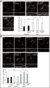
Figure 7. Arc capsid- and EV-transferred Arc mRNA is accessible for activity-dependent translation
(A) Representative images of Arc ICC from DIV15 cultured hippocampal Arc KO neurons treated for 4h with 4μg prArc. To induce translation of Arc mRNA, 30 min prior to fixation, neurons were treated with the mGluR1/5 agonist DHPG (100μM) for 5 min and then washed out. 1h prior to fixation, a subset of neurons was pretreated with cycloheximide (CHX; 180μM) to block protein translation. prArc significantly increased dendritic Arc expression in KO neurons and DHPG treatment further increased dendritic Arc levels, which was blocked by pretreatment with CHX. DHPG had no effect on untreated KO neurons. (B) Representative images of Arc ICC from DIV15 hippocampal Arc KO neurons treated for 4h with 10μg of the EV fraction prepared from 10-cm dishes of DIV15 high density cortical WT or Arc KO neurons. A subset of neurons was treated with DHPG and CHX as in (A). WT EVs significantly increased dendritic Arc expression in KO neurons, while KO EVs had no effect. DHPG treatment had no effect on dendritic Arc expression in untreated KO neurons or KO EV-treated KO neurons. However, DHPG treatment significantly increased dendritic Arc levels in WT EV-treated KO neurons, which was blocked by pretreatment with CHX. 30-μm segments of two dendrites/neuron were analyzed for integrated density measurements in all groups (_n_=10 neurons). Arc mRNA and Arc protein levels were normalized to untreated KO neurons and displayed as fold-change±SEM. Student’s _t_-test: *p<0.05. **p<0.01. ***p<0.001. Scale bars=10μm. Representative of 3 independent experiments using different EV/protein preparations and cultures.
Comment in
- Cell biology of the neuron: ARC goes viral.
Bray N. Bray N. Nat Rev Neurosci. 2018 Mar;19(3):120-121. doi: 10.1038/nrn.2018.9. Epub 2018 Feb 1. Nat Rev Neurosci. 2018. PMID: 29386612 No abstract available.
Similar articles
- Arc - An endogenous neuronal retrovirus?
Shepherd JD. Shepherd JD. Semin Cell Dev Biol. 2018 May;77:73-78. doi: 10.1016/j.semcdb.2017.09.029. Epub 2017 Sep 24. Semin Cell Dev Biol. 2018. PMID: 28941877 Free PMC article. Review. - Retrovirus-like Gag Protein Arc1 Binds RNA and Traffics across Synaptic Boutons.
Ashley J, Cordy B, Lucia D, Fradkin LG, Budnik V, Thomson T. Ashley J, et al. Cell. 2018 Jan 11;172(1-2):262-274.e11. doi: 10.1016/j.cell.2017.12.022. Cell. 2018. PMID: 29328915 Free PMC article. - mRNA Trafficking in the Nervous System: A Key Mechanism of the Involvement of Activity-Regulated Cytoskeleton-Associated Protein (Arc) in Synaptic Plasticity.
Fila M, Diaz L, Szczepanska J, Pawlowska E, Blasiak J. Fila M, et al. Neural Plast. 2021 Sep 23;2021:3468795. doi: 10.1155/2021/3468795. eCollection 2021. Neural Plast. 2021. PMID: 34603440 Free PMC article. Review. - Arc Oligomerization Is Regulated by CaMKII Phosphorylation of the GAG Domain: An Essential Mechanism for Plasticity and Memory Formation.
Zhang W, Chuang YA, Na Y, Ye Z, Yang L, Lin R, Zhou J, Wu J, Qiu J, Savonenko A, Leahy DJ, Huganir R, Linden DJ, Worley PF. Zhang W, et al. Mol Cell. 2019 Jul 11;75(1):13-25.e5. doi: 10.1016/j.molcel.2019.05.004. Epub 2019 May 28. Mol Cell. 2019. PMID: 31151856 Free PMC article. - Intercellular Communication in the Nervous System Goes Viral.
Hantak MP, Einstein J, Kearns RB, Shepherd JD. Hantak MP, et al. Trends Neurosci. 2021 Apr;44(4):248-259. doi: 10.1016/j.tins.2020.12.003. Epub 2021 Jan 21. Trends Neurosci. 2021. PMID: 33485691 Free PMC article. Review.
Cited by
- Unearthing LTR Retrotransposon gag Genes Co-opted in the Deep Evolution of Eukaryotes.
Wang J, Han GZ. Wang J, et al. Mol Biol Evol. 2021 Jul 29;38(8):3267-3278. doi: 10.1093/molbev/msab101. Mol Biol Evol. 2021. PMID: 33871607 Free PMC article. - PNMA2 forms non-enveloped virus-like capsids that trigger paraneoplastic neurological syndrome.
Xu J, Erlendsson S, Singh M, Regier M, Ibiricu I, Day GS, Piquet AL, Clardy SL, Feschotte C, Briggs JAG, Shepherd JD. Xu J, et al. bioRxiv [Preprint]. 2023 Feb 9:2023.02.09.527862. doi: 10.1101/2023.02.09.527862. bioRxiv. 2023. PMID: 36798413 Free PMC article. Updated. Preprint. - Plasticity of Spine Structure: Local Signaling, Translation and Cytoskeletal Reorganization.
Nakahata Y, Yasuda R. Nakahata Y, et al. Front Synaptic Neurosci. 2018 Aug 29;10:29. doi: 10.3389/fnsyn.2018.00029. eCollection 2018. Front Synaptic Neurosci. 2018. PMID: 30210329 Free PMC article. Review. - Extracellular Vesicles and Viruses: Two Intertwined Entities.
Moulin C, Crupi MJF, Ilkow CS, Bell JC, Boulton S. Moulin C, et al. Int J Mol Sci. 2023 Jan 5;24(2):1036. doi: 10.3390/ijms24021036. Int J Mol Sci. 2023. PMID: 36674550 Free PMC article. Review. - Intellectual disability and autism spectrum disorders 'on the fly': insights from Drosophila.
Coll-Tané M, Krebbers A, Castells-Nobau A, Zweier C, Schenck A. Coll-Tané M, et al. Dis Model Mech. 2019 May 13;12(5):dmm039180. doi: 10.1242/dmm.039180. Dis Model Mech. 2019. PMID: 31088981 Free PMC article. Review.
References
- Campillos M, Doerks T, Shah PK, Bork P. Computational characterization of multiple Gag-like human proteins. Trends Genet. 2006;22:585–589. - PubMed
Publication types
MeSH terms
Substances
Grants and funding
- P50 GM082545/GM/NIGMS NIH HHS/United States
- R01 GM112972/GM/NIGMS NIH HHS/United States
- R35 GM122550/GM/NIGMS NIH HHS/United States
- MC_UP_1201/16/MRC_/Medical Research Council/United Kingdom
- R00 NS076364/NS/NINDS NIH HHS/United States
- R01 MH112766/MH/NIMH NIH HHS/United States
- T32 HD007491/HD/NICHD NIH HHS/United States
- R01 GM077582/GM/NIGMS NIH HHS/United States
LinkOut - more resources
Full Text Sources
Other Literature Sources
Molecular Biology Databases


