Morphology and Phylogeny of a New Species of Anaerobic Ciliate, Trimyema finlayi n. sp., with Endosymbiotic Methanogens - PubMed (original) (raw)
Morphology and Phylogeny of a New Species of Anaerobic Ciliate, Trimyema finlayi n. sp., with Endosymbiotic Methanogens
William H Lewis et al. Front Microbiol. 2018.
Abstract
Many anaerobic ciliated protozoa contain organelles of mitochondrial ancestry called hydrogenosomes. These organelles generate molecular hydrogen that is consumed by methanogenic Archaea, living in endosymbiosis within many of these ciliates. Here we describe a new species of anaerobic ciliate, Trimyema finlayi n. sp., by using silver impregnation and microscopy to conduct a detailed morphometric analysis. Comparisons with previously published morphological data for this species, as well as the closely related species, Trimyema compressum, demonstrated that despite them being similar, both the mean cell size and the mean number of somatic kineties are lower for T. finlayi than for T. compressum, which suggests that they are distinct species. This was also supported by analysis of the 18S rRNA genes from these ciliates, the sequences of which are 97.5% identical (6 substitutions, 1479 compared bases), and in phylogenetic analyses these sequences grouped with other 18S rRNA genes sequenced from previous isolates of the same respective species. Together these data provide strong evidence that T. finlayi is a novel species of Trimyema, within the class Plagiopylea. Various microscopic techniques demonstrated that T. finlayi n. sp. contains polymorphic endosymbiotic methanogens, and analysis of the endosymbionts' 16S rRNA gene showed that they belong to the genus Methanocorpusculum, which was confirmed using fluorescence in situ hybridization with specific probes. Despite the degree of similarity and close relationship between these ciliates, T. compressum contains endosymbiotic methanogens from a different genus, Methanobrevibacter. In phylogenetic analyses of 16S rRNA genes, the Methanocorpusculum endosymbiont of T. finlayi n. sp. grouped with sequences from Methanomicrobia, including the endosymbiont of an earlier isolate of the same species, 'Trimyema sp.,' which was sampled approximately 22 years earlier, at a distant (∼400 km) geographical location. Identification of the same endosymbiont species in the two separate isolates of T. finlayi n. sp. provides evidence for spatial and temporal stability of the Methanocorpusculum-T. finlayi n. sp. endosymbiosis. T. finlayi n. sp. and T. compressum provide an example of two closely related anaerobic ciliates that have endosymbionts from different methanogen genera, suggesting that the endosymbionts have not co-speciated with their hosts.
Keywords: Methanocorpusculum; Trimyema; anaerobic; ciliate; endosymbiont; methanogen; phylogeny.
Figures
FIGURE 1
A schematic drawing of the (A) ventral and (B) dorsal sides of a Trimyema finlayi n. sp. cell. CCK, caudal cilium kinety; G1, first ciliary girdle; G2, second ciliary girdle; G3, third ciliary girdle; G4, fourth ciliary girdle; Ma, macronucleus; mn, micronucleus; OK, oral kineties.
FIGURE 2
Microscopic imaging of T. finlayi n. sp. whole cells. DIC images of silver carbonate impregnated cells, (A,B) and (C,D) show two sides of the same cells, (E) squashed cell showing oral kineties, (F) squashed cell showing cytoproct. (G,H) Maximum intensity projection of a Z-stack of confocal images across a single T. finlayi cell double-labeled with two FISH probes. (G) _Methanocorpusculum_-specific probe (SYM5) dual-labeled with 6-FAM. (H) Archaea-specific probe (ARCH915) dual-labeled with Cy3, white arrows indicate extracellular Archaea that were not labeled by the probe SYM5 (G). (I) F420 auto-fluorescence. (J–L) In vivo DIC images. CCK, caudal cilium kinety; CP, cytoproct; FV, food vacuole; G1, first ciliary girdle; G2, second ciliary girdle; G3, third ciliary girdle; G4, fourth ciliary girdle; MA, macronucleus; NK, N-kineties; OC, oral cavity; OK, oral kineties. Scale bars = 10 μm.
FIGURE 3
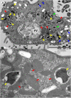
Transmission electron microscopy (TEM) images of T. finlayi n. sp. showing polymorphic methanogenic endosymbionts and hydrogenosomes (red arrowheads). Disc-shaped (blue arrowheads) and stellate form (yellow arrowheads) morphotypes are shown, as well as intermediate stages (green arrowheads). FV, food vacuole. Scale bars (A) = 5 μm, (B) = 1 μm.
FIGURE 4
Bayesian phylogeny inferred from 1640 nucleotide alignment of 18S rRNA genes of Plagiopylea species using the GTR+Γ+I model. Support values represent maximum likelihood bootstrap support/Bayesian posterior probabilities. Scale bar represents the number of substitutions per site.
FIGURE 5
Bayesian phylogeny inferred from a 1372 nucleotide alignment of methanogenic Archaea 16S rRNA genes using the GTR+Γ+I model. Support values represent maximum likelihood bootstrap support/Bayesian posterior probabilities. Scale bar represents the number of substitutions per site.
Similar articles
- Tripartite Symbiosis of an Anaerobic Scuticociliate with Two Hydrogenosome-Associated Endosymbionts, a _Holospora_-Related Alphaproteobacterium and a Methanogenic Archaeon.
Takeshita K, Yamada T, Kawahara Y, Narihiro T, Ito M, Kamagata Y, Shinzato N. Takeshita K, et al. Appl Environ Microbiol. 2019 Nov 27;85(24):e00854-19. doi: 10.1128/AEM.00854-19. Print 2019 Dec 15. Appl Environ Microbiol. 2019. PMID: 31585988 Free PMC article. - Systematic and morphological diversity of endosymbiotic methanogens in anaerobic ciliates.
Embley TM, Finlay BJ. Embley TM, et al. Antonie Van Leeuwenhoek. 1993-1994;64(3-4):261-71. doi: 10.1007/BF00873086. Antonie Van Leeuwenhoek. 1993. PMID: 8085789 - Phylogenetic analysis and fluorescence in situ hybridization detection of archaeal and bacterial endosymbionts in the anaerobic ciliate trimyema compressum.
Shinzato N, Watanabe I, Meng XY, Sekiguchi Y, Tamaki H, Matsui T, Kamagata Y. Shinzato N, et al. Microb Ecol. 2007 Nov;54(4):627-36. doi: 10.1007/s00248-007-9218-1. Epub 2007 Apr 29. Microb Ecol. 2007. PMID: 17468963 - Anaerobic ciliates as a model group for studying symbioses in oxygen-depleted environments.
Rotterová J, Edgcomb VP, Čepička I, Beinart R. Rotterová J, et al. J Eukaryot Microbiol. 2022 Sep;69(5):e12912. doi: 10.1111/jeu.12912. Epub 2022 May 3. J Eukaryot Microbiol. 2022. PMID: 35325496 Review. - Endosymbiotic interactions in anaerobic protozoa.
Hackstein JH, Vogels GD. Hackstein JH, et al. Antonie Van Leeuwenhoek. 1997 Feb;71(1-2):151-8. doi: 10.1023/a:1000154526395. Antonie Van Leeuwenhoek. 1997. PMID: 9049027 Review.
Cited by
- Anti-biofilm potential of human senescence marker protein 30 against Mycobacterium smegmatis.
Yadav P, Goel M, Gupta RD. Yadav P, et al. World J Microbiol Biotechnol. 2023 Dec 20;40(2):45. doi: 10.1007/s11274-023-03843-6. World J Microbiol Biotechnol. 2023. PMID: 38114754 - Genomes of two archaeal endosymbionts show convergent adaptations to an intracellular lifestyle.
Lind AE, Lewis WH, Spang A, Guy L, Embley TM, Ettema TJG. Lind AE, et al. ISME J. 2018 Nov;12(11):2655-2667. doi: 10.1038/s41396-018-0207-9. Epub 2018 Jul 10. ISME J. 2018. PMID: 29991760 Free PMC article. - Tripartite Symbiosis of an Anaerobic Scuticociliate with Two Hydrogenosome-Associated Endosymbionts, a _Holospora_-Related Alphaproteobacterium and a Methanogenic Archaeon.
Takeshita K, Yamada T, Kawahara Y, Narihiro T, Ito M, Kamagata Y, Shinzato N. Takeshita K, et al. Appl Environ Microbiol. 2019 Nov 27;85(24):e00854-19. doi: 10.1128/AEM.00854-19. Print 2019 Dec 15. Appl Environ Microbiol. 2019. PMID: 31585988 Free PMC article. - Some Aspects of the Physiology of the Nyctotherus velox, a Commensal Ciliated Protozoon Taken from the Hindgut of the Tropical Millipede Archispirostreptus gigas.
Kišidayová S, Scholcová N, Mihaliková K, Váradyová Z, Pristaš P, Weisskopf S, Chrudimský T, Chroňáková A, Šimek M, Šustr V. Kišidayová S, et al. Life (Basel). 2023 Apr 29;13(5):1110. doi: 10.3390/life13051110. Life (Basel). 2023. PMID: 37240755 Free PMC article.
References
- Augustin H., Foissner W., Adam H. (1987). Revision of the genera Acoineria, Trimyema and Trochiliopsis (Protozoa, Ciliophora). Bull. Br. Mus. Nat. Hist. 52 197–224.
- Baumgartner M., Stetter K. O., Foissner W. (2002). Morphological, small subunit rRNA, and physiological characterization of Trimyema minutum (Kahl, 1931), an anaerobic ciliate from submarine hydrothermal vents growing from 28°C to 52°C. J. Eukaryot. Microbiol. 49 227–238. 10.1111/j.1550-7408.2002.tb00527.x - DOI - PubMed
LinkOut - more resources
Full Text Sources
Other Literature Sources
Molecular Biology Databases


