Postcranial skeletal anatomy of the holotype and referred specimens of Buitreraptor gonzalezorum Makovicky, Apesteguía and Agnolín 2005 (Theropoda, Dromaeosauridae), from the Late Cretaceous of Patagonia - PubMed (original) (raw)
Postcranial skeletal anatomy of the holotype and referred specimens of Buitreraptor gonzalezorum Makovicky, Apesteguía and Agnolín 2005 (Theropoda, Dromaeosauridae), from the Late Cretaceous of Patagonia
Federico A Gianechini et al. PeerJ. 2018.
Abstract
Here we provide a detailed description of the postcranial skeleton of the holotype and referred specimens of Buitreraptor gonzalezorum. This taxon was recovered as an unenlagiine dromaeosaurid in several recent phylogenetic studies and is the best represented Gondwanan dromaeosaurid discovered to date. It was preliminarily described in a brief article, but a detailed account of its osteology is emerging in recent works. The holotype is the most complete specimen yet found, so an exhaustive description of it provides much valuable anatomical information. The holotype and referred specimens preserve the axial skeleton, pectoral and pelvic girdles, and both fore- and hindlimbs. Diagnostic postcranial characters of this taxon include: anterior cervical centra exceeding the posterior limit of neural arch; eighth and ninth cervical vertebral centra with lateroventral tubercles; pneumatic foramina only in anteriormost dorsals; middle and posterior caudal centra with a complex of shallow ridges on lateral surfaces; pneumatic furcula with two pneumatic foramina on the ventral surface; scapular blade transversely expanded at mid-length; well-projected flexor process on distal end of the humerus; dorsal rim of the ilium laterally everted; and concave dorsal rim of the postacetabular iliac blade. A paleohistological study of limb bones shows that the holotype represents an earlier ontogenetic stage than one of the referred specimens (MPCA 238), which correlates with the fusion of the last sacral vertebra to the rest of the sacrum in MPCA 238. A revised phylogenetic analysis recovered Buitreraptor as an unenlagiine dromaeosaurid, in agreement with previous works. The phylogenetic implications of the unenlagiine synapomorphies and other characters, such as the specialized pedal digit II and the distal ginglymus on metatarsal II, are discussed within the evolutionary framework of Paraves.
Keywords: Buitreraptor gonzalezorum; Dromaeosauridae; Late Cretaceous; Osteology; Paleohistology; Paraves; Patagonia; Unenlagiinae.
Conflict of interest statement
The authors declare that they have no competing interests.
Figures
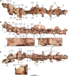
Figure 1. Anterior and mid cervical vertebrae of the holotype of Buitreraptor gonzalezorum (MPCA 245).
(A–C) Axis and third and fourth cervical vertebrae, in (A) dorsal, (B) left lateral and (C) ventral view. (D) Ventral view of the neural arch of the fourth vertebra, showing pneumatic foramina. (E–G) Fifth to seventh cervical vertebrae, in (E) dorsal, (F) left lateral and (G) ventral view. Scales: 2 cm for A–C and E–G, 1 cm for D. Ax, axis; cr, cervical rib; CV, cervical vertebra; ep, epipophysis; ns, neural spine; pf, pneumatic foramen; poz, postzygapophysis; pp, parapophysis; prez, prezygapophysis; tp, transverse process; vk, ventral keel.
Figure 2. Posterior cervical and anterior dorsal vertebrae of the holotype of Buitreraptor gonzalezorum (MPCA 245).
(A) Dorsal, (B) left lateral and (E) ventral view. (C) Detail of the box inset in B, showing pneumatic foramina on the first and second dorsal centra. (D) Detail of the centra of the last cervical and first dorsal vertebrae in right lateral view, showing pneumatic foramina. (F) Detail of the box inset in E, showing the posteroventral tubercles on the ninth cervical centrum. Scale: 2 cm for all elements, except for C, D and F. cp, carotid process; cr, cervical rib; CV, cervical vertebra; DV, dorsal vertebra; ep, epipophysis; ft, foramen transversarium; hp, hypapophysis; ns, neural spine; pf, pneumatic foramen; posf, post-spinal fossa; poz, postzygapophysis; pp, parapophysis; prez, prezygapophysis; pvt, posteroventral tubercle; tp, transverse process; vk, ventral keel.
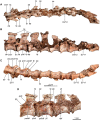
Figure 3. Mid and posterior dorsal vertebrae of the holotype of Buitreraptor gonzalezorum (MPCA 245).
(A) Dorsal, (B) left lateral and (C) ventral view. (D) Mid dorsal vertebrae, in lateroventral view. Scales: 2 cm for A–C, 1 cm for D. DV, dorsal vertebra; ns, neural spine; pacdf, parapophyseal centrodiapophyseal fossa; pcdl, posterior centrodiapoyseal lamina; pf, pneumatic foramen; pp, parapophysis; prdl, prezygodiapophyseal lamina; spf, septum between pneumatic foramina; SV, sacral vertebra; tp, transverse process; vk, ventral keel.
Figure 4. Sacral and caudal vertebrae and chevrons of the holotype of Buitreraptor gonzalezorum (MPCA 245).
(A–C) Sacrum and anterior caudal vertebrae, in (A) dorsal, (B) left lateral (C) and ventral view. (D) Detail of the box inset in C, showing the lateroventral tubercles of the last sacral vertebra. (E–G) 6th–10th caudal vertebrae, in (E) dorsal, (F) left lateral and (G) ventral view. (H–J) 10th and 11th caudal vertebrae, in (H) dorsal, (I) left lateral and (J) ventral view. (K) Distal caudal vertebrae, in left lateral view. (L) Distal chevrons, in dorsal view. Scale: 2 cm for all elements, except for D. aha, articular surfaces of the haemal arch; CaV, caudal vertebra; clr, convergent lateral ridges; ha, haemal arch; Ili, ilium; lSV, last sacral vertebra; lvts, lateroventral tubercle of the last sacral vertebra; ns, neural spine; poz, postzygapophysis; prez, prezygapophysis; prpol, “prezygopostzygapophyseal” lamina; tp, transverse process; vg, ventral groove.
Figure 5. Sacrum and pelvic girdle of the referred specimen of Buitreraptor gonzalezorum (MPCA 238).
(A–C) Sacrum and pelvic girdle, in (A) right lateral, (B) anterior and (C) left lateral view. (D–F) Detail of the sacrum and ilium, in (D) dorsal, (E) right lateral and (F) ventral view. (G) Detail of the box inset in F, showing the lateroventral tubercle of the last sacral vertebra. Scale: 2 cm for all elements, except for G. ac, acetabulum; ah, haemal arch; atr, antitrochanter; brf, brevis fossa; brsh, brevis shelf; edb, everted dorsal border of the ilium; Ili, ilium; ipil, ischiadic peduncle of the ilium; lvts, lateroventral tubercle of the last sacral vertebra; poaib, postacetabular iliac blade; ppil, pubic peduncle of the ilium; puap, pubic apron; Pub, pubis; sac, supracetabular crest; Sc, sacrum; tp, transverse process, vg, ventral groove.
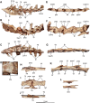
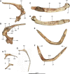
Figure 6. Pectoral girdle and dorsal ribs of the holotype of Buitreraptor gonzalezorum (MPCA 245).
(A, B) Left scapula and coracoid, in (A) medial and (B) lateral view. (C, D) Right scapula, in (C) dorsal and (D) ventral view. (E, F) Furcula, in (E) anterior and (F) posterior view. (G–I) Dorsal ribs, in anterior view. acr, acromion; cap, capitulum; cort, coracoid tuber; epi, epicleidum; gl, glenoid cavity; lgsc, lateral groove of the scapula; ltsc, lateral tubercle of the scapula; pf, pneumatic foramen; sb, scapular blade; sglf, subglenoid fossa; srib, shaft of the rib; tub, tuberculum; vbc, ventral blade of the coracoid; vhyp, vestigial hypocleidum.
Figure 7. Forelimb of the holotype of Buitreraptor gonzalezorum (MPCA 245).
(A, B) Right humerus, in (A) lateral and (B) medial view. (C–E) Right ulna and radius, in (C) anterior, (D) lateral (E) and medial view. (F) Detail of the proximal articular surface of the right ulna, in proximal view. Scales: 2 cm for A–E, 1 cm for F. ahum, articular surface for the humerus; bsc, bicipital scar; deltc, deltopectoral crest; deltf, flange on the lateral surface of the deltopectoral crest; dfpr, distal flexor process; dful, distal flange of the ulna; hh, humeral head; itub, internal tuberosity; ol, olecranon; pk, posterior keel; Ra, radius; radc, radial condyle; Ul, ulna.
Figure 8. Incomplete manual bones of the holotype (MPCA 245) and referred specimen (MPCA 471-C) of Buitreraptor gonzalezorum.
(A) Possible phalanges III-2 and III-3 or II-1 and II-2 of MPCA 245. (B) Possible metacarpal III or phalanx II-1 of MPCA 245, in lateral or medial view (indeterminate). (C–E) Possible metacarpal III or phalanx II-1 of MPCA 471-C, in (C) side, (D) dorsal and (E) ventral view. (F) Possible proximal end of phalanx III-2 or III-3 of MPCA 471-C, in side view. (G) Incomplete pre-ungual phalanx of MPCA 471-C, in side view. colf, fossa for the collateral ligament; extf, extensor fossa; flexf, flexor fossa; mph, manual phalanx; pvfp, proximoventral flexor process.
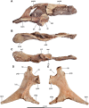
Figure 9. Pelvic bones of the holotype of Buitreraptor gonzalezorum (MPCA 245).
(A–C) Right ilium, in (A) lateral, (B) dorsal and (C) ventral views. (D, E) Right ischium, in (D) lateral and (E) medial view. ac, acetabulum; atr, antitrochanter; brf, brevis fossa; brsh, brevis shelf; dpis, distal process of the ischium; edb, everted dorsal border of the ilium; ilp, iliac peduncle of the ischium; ipil, ischiadic peduncle of the ilium; lcis, lateral crest of the ischium; obtp, obturator process; pdp, posterodorsal process; poaib, postacetabular iliac blade; ppis, pubic peduncle of the ischium; praib, preacetabular iliac blade; sac, supracetabular crest.
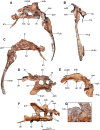
Figure 10. Femur of the holotype (MPCA 245) and referred specimen (MPCA 238) of Buitreraptor gonzalezorum.
(A–D) Right femur of MPCA 245, in (A) anterior, (B) lateral, (C) posterior (D) and medial view. (E) Detail of the proximal portion of the left femur of MPCA 245, in posterior view. (F–I) proximal portion of the right femur of MPCA 238, in (F) anterior, (G) lateral, (H) posterior and (I) medial view. Scales: 2 cm for A–D and F–I, 1 cm for E. bet, bioerosion trace fossils; ectt, ectocondylar tuberosity; fh, femur head; ftro, fourth trochanter; gtr, greater trochanter; ifint?, possible insertion point of the M. iliofemoralis internus; lcf, lateral condyle of the femur; lcfp, passage for the ligamentum capitis femoris; llf, lateral line of the femur; ltr, lesser trochanter; mcf, medial condyle of the femur; obcr, obturator crest; poplf, popliteal fossa; ptr, posterior trochanter; trsh, trochanteric shelf.
Figure 11. Right tibia and fibula of the holotype of Buitreraptor gonzalezorum (MPCA 245).
(A) Anterior, (B) lateral, (C) posterior and (D) medial view. (E) Distal portion of the right tibia, in anterior view. Scale: 2 cm for A–D, 1 cm for E. asts, articular surface for the ascendant process of the astragalus; cncr, cnemial crest; disf, distal part of the fibular shaft; fibc, fibular crest; ift, iliofibularis tubercle; inct, internal condyle of the tibia; prof, proximal part of the fibular shaft.
Figure 12. Incomplete right tibia articulated with the proximal tarsals of the referred specimen of Buitreraptor gonzalezorum (MPCA 238).
(A) Anterior, (B) lateral, (C) posterior and (D) medial view. As, astragalus; afib, articular surface for the fibula; asp, ascendant process of the astragalus; astf, anterior fossa of the astragalus; Ca, calcaneum; Fib, fibula.
Figure 13. Metatarsus and pedal phalanges of the holotype (MPCA 245) and referred specimen (MPCA 471-D) of Buitreraptor gonzalezorum.
(A, B) Articulated distal portions of the right metatarsals II, III and IV, and pedal phalanx II-1 of MPCA 245, in (A) anterior and (B) posterior view. (C) Transverse section of right metatarsal II, III and IV of MPCA 245, in proximal view. (D, E) Distal portion of the left metatarsal II of MPCA 245, in (D) anterior and (E) distal view. (F, G) Fragmentary articulated right metatarsals II, III and IV of MPCA 471-D, in (F) anterior and (G) posterior view. (H) Indeterminate pedal ungual phalanx of MPCA 471-D, in side view. Scales: 2 cm for A, B and D–H, 1 cm for C. extf, extensor fossa; ftub, flexor tubercle; icg, intercondylar groove; lgun, lateral groove of the ungual; MT, metatarsal; Ph, phalanx.
Figure 14. Metatarsus of the referred specimen of Buitreraptor gonzalezorum (MPCA 238).
(A–D) Articulated right metatarsals II, III and IV, in (A) anterior, (B) lateral, (C) posterior and (D) medial view. (E–I) Right metatarsal I, in (E) lateral, (F) posterior, (G) medial, (H) anterior and (I) distal view. Scales: 2 cm for A–D, 1 cm for E–I. asMT, articular surface for the metatarsal; colf, fossa for the collateral ligament; das, distal articular surface; MT, metatarsal; plf, posterolateral flange.
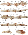
Figure 15. Metatarsal II and pedal phalanges of the referred specimen of Buitreraptor gonzalezorum (MPCA 478).
(A–D) Articulated distal portion of the right metatarsal II, and pedal phalanges II-1, II-2 and II-3, in (A) lateral, (B) dorsal, (C) medial and (D) ventral view. (E) Fragmentary articulated pedal phalanges, in side view. (F) Articulated distal portion of metatarsal III and proximal portion of pedal phalanx III-1, in side view. colf, fossa for the collateral ligament; extf, extensor fossa; ftub, flexor tubercle; lgun, lateral groove of the ungual; MT, metatarsal; Ph, phalanx; pvlr, posteroventral lateral ridge; pvmh, posteroventral medial heel; pvmr, posteroventral medial ridge.
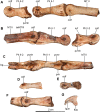
Figure 16. Metatarsal II and pedal phalanges of the holotype of Buitreraptor gonzalezorum (MPCA 245).
(A–C) Right metatarsal II and pedal phalanges II-1 and II-2, in (A) medial, (B) lateral and (C) ventral view (in C the phalanx II-2 is articulated with phalanx II-1). (D) Possible pedal pre-ungual phalanx of digit IV, in side view. (E) Indeterminate pedal phalanx, in side view. (F) Possible pedal phalanx IV-1 articulated with the proximal portion of the phalanx IV-2, in side view. (G) Ungual phalanx from pedal digit I, in side view. asMT, articular surface for the metatarsal; colf, fossa for the collateral ligament; ftub, flexor tubercle; icg, intercondylar groove; lgun, lateral groove of the ungual; MT, metatarsal; nvf, neurovascular foramen; Ph, phalanx; pvlr, posteroventral lateral ridge; pvmh, posteroventral medial heel; pvmr, posteroventral medial ridge.
Figure 17. Pedal phalanges of the referred specimen of Buitreraptor gonzalezorum (MPCA 238).
(A, B) Right pedal phalanx I-1, in (A) lateral and (B) medial view. (C, D) Distal portion of right pedal phalanx II-1, in (C) lateral and (D) medial view. (E–H) Right pedal phalanx II-2, in (E) lateral, (F) medial, (G) ventral and (H) proximal view. (I) Cast of the pedal ungual phalanx of digit II. colf, fossa for the collateral ligament; flexf, flexor fossa; icg, intercondylar groove; lc, lateral condyle; lct, lateral cotyle; lgun, lateral groove of the ungual; mc, medial condyle; mct, medial cotyle; pvmh, posteroventral medial heel.
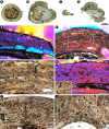
Figure 18. Long bone histology of Buitreraptor gonzalezorum.
(A–D) Complete cross section of selected bones corresponding to specimens MPCA 245 (A, B), MPCA 471-D (C) and MPCA 238 (D, E), including humerus (A), tibiae (B, D) and metatarsal (C); all the elements to the same scale. (E) Detail of the inner circumferential layer around the medullary cavity (small box inset in D). (F) General view of the compact bone of the humerus (box inset in A). Note the slight degree of birefringence of the compacta. (G) Detailed view of the cortical bone (box inset in F) showing the predominance of longitudinally oriented vascular spaces; a detailed view of the bone cell lacunae is showed in the upper right corner. (H) Cortical bone composed of fibrolamellar tissue (large box inset in D); a detailed view of the bone cell lacunae is showed in the lower left corner. (I) Enlarged view of the cortical bone showing two double LAGs; a detailed view of one of the double LAGs is showed in the lower left corner. (J) Inner cortex of the metatarsal (box inset in C) showing an ICL and secondary osteons; a detailed view of one secondary osteon is showed in the upper right corner. cl, cementing line; dLAG, double line of arrested growth; ICL, inner circumferential layer; mc, medullary cavity; po, primary osteon; rl, resorption line; so, secondary osteons.
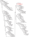
Figure 19. Reduced consensus tree of the MPTs obtained from the phylogenetic analysis, after the pruning of Kinnareemimus, Pamparaptor and Pyroraptor.
Numbers correspond to the different clades recognized: 1, Coelurosauria; 2, Tyrannosauroidea; 3, Compsognathidae; 4, Maniraptoriformes; 5, Ornithomimosauria; 6, Maniraptora; 7, Alvarezsauroidea; 8, Therizinosauria; 9, Oviraptorosauria; 10, Paraves; 11, Dromaeosauridae; 12, Unenlagiinae; 13, Troodontidae; 14, Avialae.
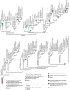
Figure 20. Optimization of the morphological characters discussed in the text.
Similar articles
- Diuqin lechiguanae gen. et sp. nov., a new unenlagiine (Theropoda: Paraves) from the Bajo de la Carpa Formation (Neuquén Group, Upper Cretaceous) of Neuquén Province, Patagonia, Argentina.
Porfiri JD, Baiano MA, Dos Santos DD, Gianechini FA, Pittman M, Lamanna MC. Porfiri JD, et al. BMC Ecol Evol. 2024 Jun 14;24(1):77. doi: 10.1186/s12862-024-02247-w. BMC Ecol Evol. 2024. PMID: 38872101 Free PMC article. - Osteology of the axial skeleton of Aucasaurus garridoi: phylogenetic and paleobiological inferences.
Baiano MA, Coria R, Chiappe LM, Zurriaguz V, Coria L. Baiano MA, et al. PeerJ. 2023 Nov 14;11:e16236. doi: 10.7717/peerj.16236. eCollection 2023. PeerJ. 2023. PMID: 38025666 Free PMC article. - Postcranial osteology of Beipiaosaurus inexpectus (Theropoda: Therizinosauria).
Liao CC, Zanno LE, Wang S, Xu X. Liao CC, et al. PLoS One. 2021 Sep 30;16(9):e0257913. doi: 10.1371/journal.pone.0257913. eCollection 2021. PLoS One. 2021. PMID: 34591927 Free PMC article. - Osteology and reassessment of Dineobellator notohesperus, a southern eudromaeosaur (Theropoda: Dromaeosauridae: Eudromaeosauria) from the latest Cretaceous of New Mexico.
Jasinski SE, Sullivan RM, Carter AM, Johnson EH, Dalman SG, Zariwala J, Currie PJ. Jasinski SE, et al. Anat Rec (Hoboken). 2023 Jul;306(7):1712-1756. doi: 10.1002/ar.25103. Epub 2022 Nov 7. Anat Rec (Hoboken). 2023. PMID: 36342817 - Unenlagiinae revisited: dromaeosaurid theropods from South America.
Gianechini FA, Apesteguia S. Gianechini FA, et al. An Acad Bras Cienc. 2011 Mar;83(1):163-95. doi: 10.1590/s0001-37652011000100009. An Acad Bras Cienc. 2011. PMID: 21437380 Review.
Cited by
- Functional constraints on the number and shape of flight feathers.
Kiat Y, O'Connor JK. Kiat Y, et al. Proc Natl Acad Sci U S A. 2024 Feb 20;121(8):e2306639121. doi: 10.1073/pnas.2306639121. Epub 2024 Feb 12. Proc Natl Acad Sci U S A. 2024. PMID: 38346196 Free PMC article. - Halszkaraptor escuilliei and the evolution of the paravian bauplan.
Brownstein CD. Brownstein CD. Sci Rep. 2019 Nov 11;9(1):16455. doi: 10.1038/s41598-019-52867-2. Sci Rep. 2019. PMID: 31712644 Free PMC article. - A new caudipterid from the Lower Cretaceous of China with information on the evolution of the manus of Oviraptorosauria.
Qiu R, Wang X, Wang Q, Li N, Zhang J, Ma Y. Qiu R, et al. Sci Rep. 2019 Apr 25;9(1):6431. doi: 10.1038/s41598-019-42547-6. Sci Rep. 2019. PMID: 31024012 Free PMC article. - Diuqin lechiguanae gen. et sp. nov., a new unenlagiine (Theropoda: Paraves) from the Bajo de la Carpa Formation (Neuquén Group, Upper Cretaceous) of Neuquén Province, Patagonia, Argentina.
Porfiri JD, Baiano MA, Dos Santos DD, Gianechini FA, Pittman M, Lamanna MC. Porfiri JD, et al. BMC Ecol Evol. 2024 Jun 14;24(1):77. doi: 10.1186/s12862-024-02247-w. BMC Ecol Evol. 2024. PMID: 38872101 Free PMC article.
References
- Agnolín FL, Novas FE. Avian ancestors: a review of the phylogenetic relationships of the theropods Unenlagiidae, Microraptoria, Anchiornis and Scansoriopterygidae. Dordrecht: Springer; 2013.
- Alifanov AR, Barsbold R. Ceratonykus oculatus gen. et sp. nov., a new dinosaur (?Theropoda, Alvarezsauria) from the Late Cretaceous of Mongolia. Paleontological Journal. 2009;43(1):94–106. doi: 10.1134/s0031030109010109. - DOI
- Alvarenga HMF, Bonaparte JF. A new flightless land bird from the cretaceous of Patagonia. Natural History Museum of Los Angeles County, Science Series. 1992;36:51–64.
Grants and funding
Fieldwork was supported by a NASA astrobiology grant to The Field Museum, and research was supported by funds of the Agencia Nacional de Promoción Científica y Tecnológica (PICT 2014-1449), The Jurassic Foundation and the American Museum of Natural History Collection Study Grant Program to Federico Gianechini, awards from the US National Science Foundation (EAR 0228693; ANT 1341475) to Peter J. Makovicky, and CONICET to Federico Gianechini, Sebastián Apesteguía and Ignacio Cerda. The funders had no role in study design, data collection and analysis, decision to publish, or preparation of the manuscript.
LinkOut - more resources
Full Text Sources
Other Literature Sources

