Pushing the resolution limit by correcting the Ewald sphere effect in single-particle Cryo-EM reconstructions - PubMed (original) (raw)
Pushing the resolution limit by correcting the Ewald sphere effect in single-particle Cryo-EM reconstructions
Dongjie Zhu et al. Nat Commun. 2018.
Abstract
The Ewald sphere effect is generally neglected when using the Central Projection Theorem for cryo electron microscopy single-particle reconstructions. This can reduce the resolution of a reconstruction. Here we estimate the attainable resolution and report a "block-based" reconstruction method for extending the resolution limit. We find the Ewald sphere effect limits the resolution of large objects, especially large viruses. After processing two real datasets of large viruses, we show that our procedure can extend the resolution for both datasets and can accommodate the flexibility associated with large protein complexes.
Conflict of interest statement
The authors declare no competing interests.
Figures
Fig. 1
The resolution limit caused by the effect of depth of field. a Positions marked with a cross (x) represent the calculated resolution limit of protein complexes of variable sizes by using the simulated 300 kV (black), 200 kV (red), and 120 kV (blue) cryo-EM data. The corresponding black, red, and blue lines are the resolution limits calculated using the modified empirical formula. Values for the dashed black line were obtained by applying the DeRoiser’s empirical formula that was used as a reference for comparison, . The black-edged square is the simulation result performed by Kenneth et al.. b Black and red circles with EMDB codes represent the resolutions of typical high-resolution protein complexes generated by cryo-EM SPA at 300 kV and 200 kV, respectively. Black squares represent the resolutions of PBCV-1 virus and HSV-2 virus that were reconstructed by using a block-based reconstruction method. Gray squares represent the resolution of these two viruses that were reconstructed by using a conventional method
Fig. 2
The block-based reconstruction, particle defocus, and local mean defoci of blocks. a To show how block-based reconstruction works, a density map (EMD-6775) was divided into four blocks, which are circled with red dashes. The distance between the center of mass of the model and the focal plane of objective lens along the Z axis is the particle defocus. Each block has its own local mean focus, which is the sum of the particle defocus values with the distance from the center of the block to the center of the model along the Z axis. b The Thon rings calculated from the whole virus (left) or after excluding the central part (right). The region of the virus in the 2D image (up right) corresponds to the part of the virus that is near the particle defocus plane in the 3D virus (down right). c The resolutions determined by gold standard FSC at threshold 0.143 of HSV-2 capsid reconstructions by a conventional reconstruction method (colored in black), by block-based reconstruction with local refinement of translational and rotational parameters of each block but without applying local mean defocus (colored in blue), and by block-based reconstruction with local refinement and local mean defocus being applied to overcome the Ewald sphere effect (colored in red). d The resolutions determined by gold standard FSC at threshold 0.143 of PBCV-1 virus reconstructions by a conventional reconstruction method (colored in black), by block-based reconstruction with local refinement of translational and rotational parameters of each block but without applying local mean defocus (colored in blue), and by block-based reconstruction with local refinement and local mean defocus being applied to overcome Ewald sphere effect (colored in red)
Fig. 3
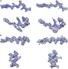
Comparison of cryo-EM densities from block-based and conventional reconstruction methods. a The densities belong to the major capsid protein in the 3.1 Å resolution HSV-2 reconstruction by block-based reconstruction method (left) and the densities that were from the same areas in the 4.0 Å resolution reconstruction by conventional method (right). The HSV-2 virus structure data are deposited in the Protein Databank with accession code 5ZAP (HSV-2 icosahedral reconstruction after block-based refinement) and in the Electron Microscopy Database with accession code EMD-6907. b The densities belong to the major capsid protein Vp54 in the 3.5 Å resolution PBCV-1 reconstruction by the block-based reconstruction method (left) and the densities that were from the same areas in the 4.2 Å resolution reconstruction by the conventional method (right)
Similar articles
- Correcting for the ewald sphere in high-resolution single-particle reconstructions.
Leong PA, Yu X, Zhou ZH, Jensen GJ. Leong PA, et al. Methods Enzymol. 2010;482:369-80. doi: 10.1016/S0076-6879(10)82015-4. Methods Enzymol. 2010. PMID: 20888969 Free PMC article. - Ewald sphere correction using a single side-band image processing algorithm.
Russo CJ, Henderson R. Russo CJ, et al. Ultramicroscopy. 2018 Apr;187:26-33. doi: 10.1016/j.ultramic.2017.11.001. Epub 2018 Jan 12. Ultramicroscopy. 2018. PMID: 29413409 Free PMC article. - The Ewald sphere/focus gradient does not limit the resolution of cryoEM reconstructions.
Heymann JB. Heymann JB. J Struct Biol X. 2022 Dec 30;7:100083. doi: 10.1016/j.yjsbx.2022.100083. eCollection 2023. J Struct Biol X. 2022. PMID: 36632443 Free PMC article. - Pushing the resolution limits in cryo electron tomography of biological structures.
Diebolder CA, Koster AJ, Koning RI. Diebolder CA, et al. J Microsc. 2012 Oct;248(1):1-5. doi: 10.1111/j.1365-2818.2012.03627.x. Epub 2012 Jun 7. J Microsc. 2012. PMID: 22670690 Review. - Determining the Crystal Structure of TRPV6.
Saotome K, Singh AK, Sobolevsky AI. Saotome K, et al. In: Kozak JA, Putney JW Jr, editors. Calcium Entry Channels in Non-Excitable Cells. Boca Raton (FL): CRC Press/Taylor & Francis; 2018. Chapter 14. In: Kozak JA, Putney JW Jr, editors. Calcium Entry Channels in Non-Excitable Cells. Boca Raton (FL): CRC Press/Taylor & Francis; 2018. Chapter 14. PMID: 30299652 Free Books & Documents. Review.
Cited by
- Structures of a large prolate virus capsid in unexpanded and expanded states generate insights into the icosahedral virus assembly.
Fang Q, Tang WC, Fokine A, Mahalingam M, Shao Q, Rossmann MG, Rao VB. Fang Q, et al. Proc Natl Acad Sci U S A. 2022 Oct 4;119(40):e2203272119. doi: 10.1073/pnas.2203272119. Epub 2022 Sep 26. Proc Natl Acad Sci U S A. 2022. PMID: 36161892 Free PMC article. - The receptor VLDLR binds Eastern Equine Encephalitis virus through multiple distinct modes.
Cao D, Ma B, Cao Z, Xu X, Zhang X, Xiang Y. Cao D, et al. Nat Commun. 2024 Aug 10;15(1):6866. doi: 10.1038/s41467-024-51293-x. Nat Commun. 2024. PMID: 39127734 Free PMC article. - Sub-2 Å Ewald curvature corrected structure of an AAV2 capsid variant.
Tan YZ, Aiyer S, Mietzsch M, Hull JA, McKenna R, Grieger J, Samulski RJ, Baker TS, Agbandje-McKenna M, Lyumkis D. Tan YZ, et al. Nat Commun. 2018 Sep 7;9(1):3628. doi: 10.1038/s41467-018-06076-6. Nat Commun. 2018. PMID: 30194371 Free PMC article. - Atomic structure of the human herpesvirus 6B capsid and capsid-associated tegument complexes.
Zhang Y, Liu W, Li Z, Kumar V, Alvarez-Cabrera AL, Leibovitch EC, Cui Y, Mei Y, Bi GQ, Jacobson S, Zhou ZH. Zhang Y, et al. Nat Commun. 2019 Nov 25;10(1):5346. doi: 10.1038/s41467-019-13064-x. Nat Commun. 2019. PMID: 31767868 Free PMC article. - Assembly of complex viruses exemplified by a halophilic euryarchaeal virus.
De Colibus L, Roine E, Walter TS, Ilca SL, Wang X, Wang N, Roseman AM, Bamford D, Huiskonen JT, Stuart DI. De Colibus L, et al. Nat Commun. 2019 Mar 29;10(1):1456. doi: 10.1038/s41467-019-09451-z. Nat Commun. 2019. PMID: 30926810 Free PMC article.
References
Publication types
LinkOut - more resources
Full Text Sources
Other Literature Sources


