The Parabrachial Nucleus: CGRP Neurons Function as a General Alarm - PubMed (original) (raw)
Review
The Parabrachial Nucleus: CGRP Neurons Function as a General Alarm
Richard D Palmiter. Trends Neurosci. 2018 May.
Abstract
The parabrachial nucleus (PBN), which is located in the pons and is dissected by one of the major cerebellar output tracks, is known to relay sensory information (visceral malaise, taste, temperature, pain, itch) to forebrain structures including the thalamus, hypothalamus, and extended amygdala. The availability of mouse lines expressing Cre recombinase selectively in subsets of PBN neurons and viruses for Cre-dependent gene expression is beginning to reveal the connectivity and functions of PBN component neurons. This review focuses on PBN neurons expressing calcitonin gene-related peptide (CGRPPBN) that play a major role in regulating appetite and transmitting real or potential threat signals to the extended amygdala. The functions of other specific PBN neuronal populations are also discussed. This review aims to encourage investigation of the numerous unanswered questions that are becoming accessible.
Keywords: anorexia; calcium imaging; fear and taste conditioning; threats.
Copyright © 2018 Elsevier Ltd. All rights reserved.
Conflict of interest statement
Conflict of interest: The author declares no conflicts of interest related to this review.
Figures
Figure 1
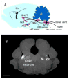
Location of parabrachial CGRP-expressing neurons that function as a general alarm. A. Cartoon sagittal section of mouse brain showing the approximate locations of CGRP neurons in the parabrachial nucleus (PBN), their inputs from the spinal cord and the nucleus tractus solitarius (NTS) where CCK- and DBH-expressing neurons reside, and their axonal projections to the central nucleus of the amygdala (CeA) and the oval region of the bed nucleus of the stria terminalis (BNST). All of these neurons are excitatory (glutamatergic, black) except for the inhibitory input from AgRP-expressing neurons in the arcuate nucleus (ARC) which are GABAergic (blue). The cerebellum (Cb) and cortex (Ctx) shown for reference. B. Coronal section of mouse brain showing expression of a fluorescent protein (white) in CGRP neurons (left side). The PBN has many subdivisions [4], the lateral region (L) that includes the external lateral subdivision where CGRP neurons reside, the medial region (M), the waist region that includes the superior cerebellar peduncle fibers (dark region between L and M), and adjacent lateral crest and Kölliker-Fuse regions (KF). Scale bar (bottom left): 1mm. Panel A drawn by the author; Panel B provided by Jane Chen.
Similar articles
- Activation of Parabrachial Tachykinin 1 Neurons Counteracts Some Behaviors Mediated by Parabrachial Calcitonin Gene-related Peptide Neurons.
Arthurs JW, Pauli JL, Palmiter RD. Arthurs JW, et al. Neuroscience. 2023 May 1;517:105-116. doi: 10.1016/j.neuroscience.2023.03.003. Epub 2023 Mar 9. Neuroscience. 2023. PMID: 36898496 Free PMC article. - Encoding of danger by parabrachial CGRP neurons.
Campos CA, Bowen AJ, Roman CW, Palmiter RD. Campos CA, et al. Nature. 2018 Mar 29;555(7698):617-622. doi: 10.1038/nature25511. Epub 2018 Mar 21. Nature. 2018. PMID: 29562230 Free PMC article. - Parabrachial CGRP neurons modulate active defensive behavior under a naturalistic threat.
Pyeon GH, Cho H, Chung BM, Choi JS, Jo YS. Pyeon GH, et al. Elife. 2025 Jan 10;14:e101523. doi: 10.7554/eLife.101523. Elife. 2025. PMID: 39791358 Free PMC article. - Parabrachial neurons promote nociplastic pain.
Palmiter RD. Palmiter RD. Trends Neurosci. 2024 Sep;47(9):722-735. doi: 10.1016/j.tins.2024.07.002. Epub 2024 Aug 14. Trends Neurosci. 2024. PMID: 39147688 Review. - Parabrachial Complex: A Hub for Pain and Aversion.
Chiang MC, Bowen A, Schier LA, Tupone D, Uddin O, Heinricher MM. Chiang MC, et al. J Neurosci. 2019 Oct 16;39(42):8225-8230. doi: 10.1523/JNEUROSCI.1162-19.2019. J Neurosci. 2019. PMID: 31619491 Free PMC article. Review.
Cited by
- Efferent projections of Vglut2, Foxp2, and Pdyn parabrachial neurons in mice.
Huang D, Grady FS, Peltekian L, Geerling JC. Huang D, et al. J Comp Neurol. 2021 Mar;529(4):657-693. doi: 10.1002/cne.24975. Epub 2020 Sep 21. J Comp Neurol. 2021. PMID: 32621762 Free PMC article. - The Parabrachial Nucleus as a Key Regulator of Neuropathic Pain.
Wang Z, Xu ZZ. Wang Z, et al. Neurosci Bull. 2021 Jul;37(7):1079-1081. doi: 10.1007/s12264-021-00676-x. Epub 2021 Apr 30. Neurosci Bull. 2021. PMID: 33929705 Free PMC article. No abstract available. - Autonomic dysfunction and treatment strategies in intracerebral hemorrhage.
Kang K, Shi K, Liu J, Li N, Wu J, Zhao X. Kang K, et al. CNS Neurosci Ther. 2024 Feb;30(2):e14544. doi: 10.1111/cns.14544. CNS Neurosci Ther. 2024. PMID: 38372446 Free PMC article. Review. - Conserved features of anterior cingulate networks support observational learning across species.
Burgos-Robles A, Gothard KM, Monfils MH, Morozov A, Vicentic A. Burgos-Robles A, et al. Neurosci Biobehav Rev. 2019 Dec;107:215-228. doi: 10.1016/j.neubiorev.2019.09.009. Epub 2019 Sep 8. Neurosci Biobehav Rev. 2019. PMID: 31509768 Free PMC article. Review. - Pain-related cortico-limbic plasticity and opioid signaling.
Neugebauer V, Presto P, Yakhnitsa V, Antenucci N, Mendoza B, Ji G. Neugebauer V, et al. Neuropharmacology. 2023 Jun 15;231:109510. doi: 10.1016/j.neuropharm.2023.109510. Epub 2023 Mar 20. Neuropharmacology. 2023. PMID: 36944393 Free PMC article. Review.
References
- Damasio A, Carvalho GB. The nature of feelings: evolutionary and neurobiological origins. Nat Rev Neurosci. 2013;14:143–152. - PubMed
- de Lacalle S, Saper CB. Calcitonin gene-related peptide-like immunoreactivity marks putative visceral sensory pathways in human brain. Neuroscience. 2000;100:115–130. - PubMed
- Hashimoto K, et al. Characterization of parabrachial subnuclei in mice with regard to salt tastants: possible independence of taste relay from visceral processing. Chem Senses. 34:253–267. - PubMed
- Ricardo JA, Koh ET. Anatomical evidence of direct projections from the nucleus of the solitary tract to hypothalamus, amygdala and other forebrain structures. Brain Res. 1978;153:1–26. - PubMed
Publication types
MeSH terms
Substances
LinkOut - more resources
Full Text Sources
Other Literature Sources
Research Materials
