Peripheral vascular disease assessment in the lower limb: a review of current and emerging non-invasive diagnostic methods - PubMed (original) (raw)
Review
Peripheral vascular disease assessment in the lower limb: a review of current and emerging non-invasive diagnostic methods
Elham Shabani Varaki et al. Biomed Eng Online. 2018.
Abstract
Background: Worldwide, at least 200 million people are affected by peripheral vascular diseases (PVDs), including peripheral arterial disease (PAD), chronic venous insufficiency (CVI) and deep vein thrombosis (DVT). The high prevalence and serious consequences of PVDs have led to the development of several diagnostic tools and clinical guidelines to assist timely diagnosis and patient management. Given the increasing number of diagnostic methods available, a comprehensive review of available technologies is timely in order to understand their limitations and direct future development effort.
Main body: This paper reviews the available diagnostic methods for PAD, CVI, and DVT with a focus on non-invasive modalities. Each method is critically evaluated in terms of sensitivity, specificity, accuracy, ease of use, procedure time duration, and training requirements where applicable.
Conclusion: This review emphasizes the limitations of existing methods, highlighting a latent need for the development of new non-invasive, efficient diagnostic methods. Some newly emerging technologies are identified, in particular wearable sensors, which demonstrate considerable potential to address the need for simple, cost-effective, accurate and timely diagnosis of PVDs.
Keywords: Ankle Brachial Index; Chronic venous insufficiency; Deep vein thrombosis; Doppler ultrasound; Peripheral arterial disease; Plethysmography.
Figures
Fig. 1
Examples of peripheral vascular function assessment in the lower limb using plethysmography techniques; a strain gauge plethysmography [37]; b photo plethysmography (PPG); c quantitative PPG/light reflection rheography (LRR); d Impedance Plethysmography, modified from [38]; e Air Plethysmography (APG) [39]
Fig. 2
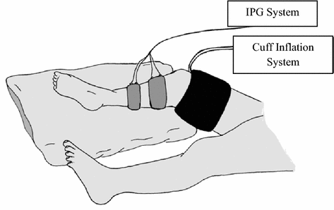
Schematic view of the use of impedance plethysmography for detection of DVT (adapted from [36])
Fig. 3
VOTI system and its sandal shaped measuring probe [165]
Fig. 4
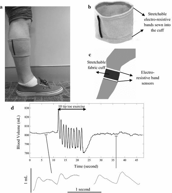
HeMo prototype; a a demonstration of HeMo worn on the calf [166]; b HeMo cuff (adopted from [166]); c diagram of HeMo worn on the calf (adopted from [166]); d blood flow variations recorded before, during and after tiptoe exercise by HeMo [167]
Fig. 5
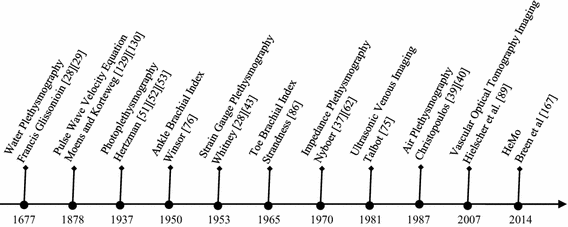
Advent of milestone technologies for noninvasive diagnosis of PVDs
Similar articles
- [Surgery for deep venous reflux in the lower limb].
Perrin M. Perrin M. J Mal Vasc. 2004 May;29(2):73-87. doi: 10.1016/s0398-0499(04)96718-2. J Mal Vasc. 2004. PMID: 15229402 Review. French. - Measurement of the clinical and cost-effectiveness of non-invasive diagnostic testing strategies for deep vein thrombosis.
Goodacre S, Sampson F, Stevenson M, Wailoo A, Sutton A, Thomas S, Locker T, Ryan A. Goodacre S, et al. Health Technol Assess. 2006 May;10(15):1-168, iii-iv. doi: 10.3310/hta10150. Health Technol Assess. 2006. PMID: 16707072 Review. - Stenting for peripheral artery disease of the lower extremities: an evidence-based analysis.
Medical Advisory Secretariat. Medical Advisory Secretariat. Ont Health Technol Assess Ser. 2010;10(18):1-88. Epub 2010 Sep 1. Ont Health Technol Assess Ser. 2010. PMID: 23074395 Free PMC article. - A systematic review of duplex ultrasound, magnetic resonance angiography and computed tomography angiography for the diagnosis and assessment of symptomatic, lower limb peripheral arterial disease.
Collins R, Cranny G, Burch J, Aguiar-Ibáñez R, Craig D, Wright K, Berry E, Gough M, Kleijnen J, Westwood M. Collins R, et al. Health Technol Assess. 2007 May;11(20):iii-iv, xi-xiii, 1-184. doi: 10.3310/hta11200. Health Technol Assess. 2007. PMID: 17462170 Review.
Cited by
- Understanding Gangrene in the Context of Peripheral Vascular Disease: Prevalence, Etiology, and Considerations for Amputation-Level Determination.
Bhargava A, Mahakalkar C, Kshirsagar S. Bhargava A, et al. Cureus. 2023 Nov 18;15(11):e49026. doi: 10.7759/cureus.49026. eCollection 2023 Nov. Cureus. 2023. PMID: 38116352 Free PMC article. Review. - The Effects of Spinal Cord Stimulators on End Organ Perfusion: A Literature Review.
Saini HS, Shnoda M, Saini I, Sayre M, Tariq S. Saini HS, et al. Cureus. 2020 Mar 12;12(3):e7253. doi: 10.7759/cureus.7253. Cureus. 2020. PMID: 32292667 Free PMC article. Review. - Evaluation of Short-Term Insulin Pump for Treatment of Patients with Type 2 Diabetes Mellitus Complicated with Lower Extremity Arterial Disease in Endocrinology by Ultrasonography.
Guo Q, Li Z, Chen H. Guo Q, et al. Comput Math Methods Med. 2022 May 27;2022:9128208. doi: 10.1155/2022/9128208. eCollection 2022. Comput Math Methods Med. 2022. PMID: 35669363 Free PMC article. Clinical Trial. - Concomitant chronic venous insufficiency in patients with peripheral artery disease: insights from MR angiography.
Ammermann F, Meinel FG, Beller E, Busse A, Streckenbach F, Teichert C, Weinrich M, Neumann A, Weber MA, Heller T. Ammermann F, et al. Eur Radiol. 2020 Jul;30(7):3908-3914. doi: 10.1007/s00330-020-06696-x. Epub 2020 Feb 25. Eur Radiol. 2020. PMID: 32100090 Free PMC article. - Enhancing Functional Efficiency and Quality of Life through Revascularization Surgery in Peripheral Arterial Disease: A Comparative Analysis of Objective and Subjective Indicators.
Nowaczyk A, Cwajda-Białasik J, Jawień A, Szewczyk MT. Nowaczyk A, et al. Med Sci Monit. 2023 Sep 18;29:e941673. doi: 10.12659/MSM.941673. Med Sci Monit. 2023. PMID: 37718505 Free PMC article.
References
- Artazcoz AV. Diagnosis of peripheral vascular disease: current perspectives. J Anesth Clin Res. 2015;6:1–7.
- Centre National Clinical Guideline. Lower limb peripheral arterial disease: diagnosis and management. London: Natl. Clin. Guidel. Cent, Royal College of Physicians; 2012. - PubMed
- Pierce GF, Mustoe TA. Pharmacologic enhancement of wound healing. Annu Rev Med. 1995;46:467–481. - PubMed
- Regensteiner JG, Hiatt WR. Treatment of peripheral arterial disease. Clin Cornerstone. 2002;4:26–37. - PubMed
- Bahr C. CVI and PAD: a review of venous and arterial disease. JAAPA. 2007;20:20–25. - PubMed
Publication types
MeSH terms
LinkOut - more resources
Full Text Sources
Other Literature Sources


