From the Guiana Highlands to the Brazilian Atlantic Rain Forest: four new species of Selaginella (Selaginellaceae - Lycopodiophyta: S. agioneuma, S. magnafornensis, S. ventricosa, and S. zartmanii) - PubMed (original) (raw)
From the Guiana Highlands to the Brazilian Atlantic Rain Forest: four new species of Selaginella (Selaginellaceae - Lycopodiophyta: S. agioneuma, S. magnafornensis, S. ventricosa, and S. zartmanii)
Iván A Valdespino et al. PeerJ. 2018.
Abstract
We describe four new species in the genus Selaginella (i.e., S. agioneuma, S. magnafornensis, S. ventricosa, and S. zartmanii) from Brazil, all presently classified in subg. Stachygynandrum. For each of the new taxa we discuss taxonomic affinities and provide information on habitat, distribution, and conservation status. In addition, line drawings and scanning electron microscope (SEM) images of stems sections, leaves, and spores (when present) are included. Selaginella agioneuma and S. magnafornensis are from the State of Espíritu Santo where they inhabit premontane to montane Atlantic rain forests in the Reserva Biológica Augusto Ruschi and Parque Estadual Forno Grande, respectively. Selaginella ventricosa was collected in upper montane forests at Parque Nacional Serra da Mocidade, State of Roraima and S. zartmanii in premontane Amazon rain forests on upper Rio Negro at Mpio. São Gabriel da Cachoeira, Amazonas State in both Serra Curicuriari and the Morro dos Seis Lagos Biological Reserve.
Keywords: Amazon; Atlantic forest; Idioblasts; Laminar flap; Lycophytes; Papillae; Prickles; Stomata; Tooth-like.
Conflict of interest statement
The authors declare there are no competing interests.
Figures
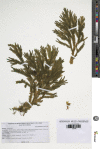
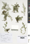
Figure 1. Scanned image of holotype of S. agioneuma.
Salino et al. 14088 (P). Image courtesy of Muséum national d’Histoire naturelle, Paris, France.
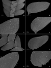
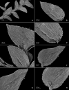
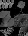
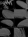
Figure 2. Scanning electron micrographs of branch sections and leaves of S. agioneuma.
(A) Section of upper surface of stem branch showing lateral and median leaves. (B) Close-up of lateral leaf from stem branch, upper surface. (C) Close-up distal portion of lateral leaf (same leaf shown in B); note, elongate and papillate idioblasts (a). (D) Close-up of proximal portion of lateral leaf (same leaf shown in B); note, elongate and papillate idioblasts (a). (E) Section of lower surface of stem branch showing lateral leaves. (F) Close-up of lateral leaf from stem branch, lower surface. (G) Close-up of distal portion of lateral leaf (same leaf shown in F); note, elongate and papillate idioblasts (a) and stomata along midrib (b) and submarginal and marginal (c) portions of lamina. (H) Close-up of proximal portion of lateral leaf (same leaf shown in F); note, elongate and papillate idioblasts (a) and stomata along midrib (b) and submarginal and marginal (c) portions of lamina. (A–H) taken from holotype, Salino et al. 14088 (P).
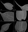
Figure 3. Scanning electron micrographs of median leaves of S. agioneuma.
(A) Close-up of median leaf from stem branch, upper surfaces. (B) Close-up of distal portion of median leaf (same leaf shown in A) showing aristate apex; note tooth-like projections on arista surface (a). (C) Close-up of distal portion of median leaf (same leaf shown in A) showing section of aristate apex; note, elongate and papillate idioblasts (a) and stomata along midrib (b) and submarginal and marginal portions of lamina (c). (D) Close-up of proximal portion of median leaf (same leaf shown in A); note, elongate and papillate idioblasts (a) and stomata along midrib (b) and submarginal and marginal (c) portions of lamina. (E) Close-up of median leaf from stem branch, lower surfaces. (F) Close-up of proximal portion of median leaf (same leaf shown in E); note, elongate idioblast-like cells on leaf lamina (a). (A–F) taken from holotype, Salino et al. 14088 (P).
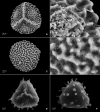
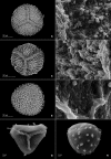
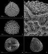
Figure 4. Scanning electron micrographs of mega- and microspores of S. agioneuma.
(A) Megaspore, proximal face. (B) Close-up of megaspore, proximal face. (C) Megaspore, distal face. (D) Close-up of megaspore, distal face. (E) Microspore, proximal face. (F) Microspore, distal face. (A–F) taken from holotype, Salino et al. 14088 (P).
Figure 5. Scanned image of holotype of S. magnafornensis.
Salino et al. 13704 (P). Image courtesy of Muséum national d’Histoire naturelle, Paris, France.
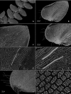
Figure 6. Scanning electron micrographs of branch section and leaves of S. magnafornensis.
(A) Section of lower surface of stem branch showing lateral and axillary leaves. (B) Close-up of lateral leaf from stem branch, lower surface; note elongate and interconnected, papillate idioblast (a) on both side of midrib and stomata (b) along midrib. (C) Close-up distal portion of lateral leaf (same leaf shown in B); note elongate and interconnected, papillate idioblast (a) on both side of midrib and stomata along midrib (b) and margin (c). (D) Close-up of proximal portion of lateral leaf (same leaf shown in B); note elongate and interconnected, papillate idioblast (a) on both side of midrib and stomata along midrib (b). (E) Close up of axillary leaf from stem branch, lower surface; note elongate and interconnected, papillate idioblast (a) on both side of midrib and stomata along midrib (b) and margins (c). (F) Close-up of lateral leaf from stem branch, upper surface; note papillate cells (a). (G) Close-up of distal portion of lateral leaf (same leaf shown in F); note papillate cells (a). (H) Close-up of proximal portion of lateral leaf (same leaf shown in F); note papillate cells (a). (A–H) taken from holotype, Salino et al. 13704 (P).
Figure 7. Scanning electron micrographs of branch section and leaves of S. magnafornensis.
(A) Section of upper surface of stem branch showing lateral and median leaves. (B) Close-up of median leaf from stem branch, upper surface. (C) Close-up distal portion of lateral leaf (same leaf shown in B) showing aristate apex; note papillate cells (a) and stomata (b) along the midrib. (D) Close-up of proximal portion of lateral leaf (same leaf shown in B) showing base; note papillate cells (a) and stomata along midrib (b) and submarginal near base (c). (A–D) taken from holotype, Salino et al. 13704 (P).
Figure 8. Scanning electron micrographs of mega- and microspores of S. magnafornensis.
(A) Megaspore, proximal face. (B) Close-up of megaspore (same megaspore shown in A), proximal face. (C) Megaspore (less mature), proximal face. (D) Close-up of megaspore (same megaspore shown in C), proximal face. (E) Megaspore distal face. (F) Close-up of megaspore (same megaspore shown in E), distal face. (G) Microspore, proximal face. (H) Microspore, distal face. (A–H) taken from holotype, Salino et al. 13704 (P).
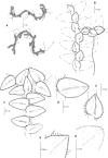
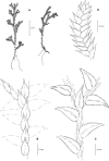
Figure 9. Line drawing of holotype of S. ventricosa.
(A) Habit. (B) Upper surface of stem showing median and outline of lateral leaves. (C) Median leaf, upper surface. (D) Distal portion of lateral leaf upper surface showing teeth-like or prickle projections near the apex. (E) Distal portion of median leaf upper surface showing teeth-like or prickle projections near the apex. (F) Lower surface of stem showing lateral leaves and axillary leaf. (G) Lateral leaf, lower surface. (A–G) from Terra-Araujo et al. 1320 (INPA). Illustration made by Rubén Lozano.
Figure 10. Scanning electron micrographs of branch sections and leaves of S. ventricosa.
(A) Section of upper surface of stem showing median and lateral leaves. (B) Close-up of median leaf, upper surface; note, papillate cells (a), stomata (b) along midrib, and hairs (c) on distal portion of lamina, and ciliate inner margin. (C) Close-up of mid-distal portion of median leaf (same leaf shown in B); note, elongate, idioblast-like marginal cells with jointed and line-like (a1) or individual (a2) rows of papillae, stomata (b) along midrib, and hairs (c) on distal portion of lamina, and ciliate inner margin. (D) Acroscopic and distal portion of the inner margins of median leaf (same leaf shown in B) showing cilia; note, many papillae on each lumen of roundish to quadrangular cells, elongate, idioblast-like marginal cells with jointed and line-like (a1) or one (a2) or two rows of individual (a3) papillae. (E) Close up of proximal portion of median leaf (same leaf shown in B); note, cell lumina without or with many papillae and stomata (a) along midrib. (F) Close-up of median leaf (same leaf shown in B); note, cell lumina with many papillae and stomata (a) along midrib. (G) Section of lower surface of stem showing lateral and axillary leaves; note, elongate idioblasts. (H) Close-up of median leaf, lower surface; note stomata (a) on margin and elongate, sinuate-walled, and entire or papillate cells. (A–H) taken from holotype, Terra-Araujo et al. 1320 (INPA).
Figure 11. Scanning electron micrographs of branch section and leaves of Selaginella ventricosa.
(A) Section of upper surface of stem showing lateral and median leaves. (B) Close-up of lateral leaf, upper surface. (C) Close-up of distal portion of lateral leaf (same leaf shown in B); note, many papillae on each cell lumen and hairs (a) on apical region. (D) Close-up of proximal portion of lateral leaf (same leaf shown in B); note, ciliate margin and roundish to quadrangular cells with or without many papillae on each cell lumen. (A–D) taken from holotype, Terra-Araujo et al. 1320 (INPA).
Figure 12. Scanning electron micrographs of branch section and leaves of S. ventricosa.
(A) Section of lower surface of stem showing lateral leaves and portions of median leaves; note idioblasts (a). (B) Close-up of lateral leaf, lower surface. (C) Close-up distal portion of lateral leaf (same leaf shown in B); note, idioblasts with a single elongate papilla (a), elongate cells with many papillae mostly in two rows (b), and stomata (c) along midrib. (D) Close-up of proximal portion of lateral leaf (same leaf shown in B); note, idioblasts with a single elongate papilla (a), elongate cells with many papillae mostly in two rows (b), and stomata (c) along midrib. (E) Close-up of same view as in (D); note, idioblasts with a single elongate papilla (a), elongate cells with many papillae mostly in two rows (b), and stomata. (F) Close-up of same view as in (D); note, idioblasts with a single elongate papilla (a) and elongate cells with many papillae mostly in two rows (b). (G) Close-up of proximal portion of lateral leaf, upper surface; note cells lumen mostly with papillae. (H) Close-up portion of lateral leaf, upper surface; note cells lumen with papillae. (A–H) taken from holotype, Terra-Araujo et al. 1320 (INPA).
Figure 13. Line drawing of holotype of S. zartmanii.
(A) Habit. (B) Upper surface of stem showing median leaves and outline of lateral leaves. (C) Lower surface of stem showing lateral leaves and axillary leaf. (D) Close-up of strobilus. (A–D) from Terra-Araujo et al. 1320 (INPA). Illustration made by Rubén Lozano.
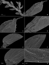
Figure 14. Scanning electron micrographs of branch section and leaves of S. zartmanii.
(A) Section of upper surface of stem showing median and lateral leaves. (B) Close-up of median leaf, upper surface; note, papillate cells (a) and stomata (b) along midrib. (C) Close-up of mid-distal portion of median leaf (same leaf shown in B); note, elongate, idioblast-like marginal cells with a single row of papillae, stomata (b) along midrib, and some cells with a single papilla (c) on cell lumen. (D) Close-up of mid-proximal portion of median leaf (same leaf shown in B); note, elongate, idioblast-like marginal cells with a single row of papillae stomata (b) along midrib, and some cells with a single papilla (c) on cell lumen. (E) Close up lateral leaf, upper surface. (F) Close-up of distal portion of lateral leaf (same leaf shown in E); note, some cell lumen with a single papilla. (G) Close-up of proximal portion of lateral leaf (same leaf shown in E). (H) Close-up of portion of basiscopic half of lateral leaf (same leaf shown in E); note, some cell lumen with a single papilla. (A–H) taken from holotype, Zartman 8586 (INPA).
Figure 15. Scanning electron micrographs of branch section and leaves of S. zartmanii.
(A) Section of lower surface of stem showing lateral leaves. (B) Close-up of lateral leaf, lower surface; note, revolute basiscopic margin. (C) Close-up of mid-distal portion of lateral leaf (same leaf shown in B); note, elongate and papillate, idioblast (a), stomata (b) along midrib and basiscopic margin revolute with marginal stomata (c) and tooth-like projections (d) on upper surface. (D) Close-up of mid-proximal portion of lateral leaf (same leaf shown in B); note, some elongate cells with papillae in a single row (a), stomata (b) along midrib, and marginal and submarginal stomata (c) on upper surface of basiscopic margin. (E) Close up lateral leaf, lower surface; note, elongate and papillate idioblasts (a) and stomata (b) along midrib. (F) Close-up of distal portion of lateral leaf (same leaf shown in E); note, elongate and papillate idioblasts (a), stomata (b) along midrib and marginal stomata (c) on upper surface of basiscopic margin. (G) Close-up of proximal portion of lateral leaf (same leaf shown in E); note, elongate and papillate idioblasts (a), stomata (b) along midrib and marginal stomata (c) on upper surface of basiscopic margin. (H) Close-up of portion of acroscopic base of lateral leaf; note, cilia (a). (A–H) taken from holotype, Zartman 8586 (INPA).
Figure 16. Scanning electron micrographs of mega- and microspores of S. zartmanii.
(A) Megaspore, proximal face. (B) Close-up of megaspore, proximal face. (C) Megaspore, distal face. (D) Close-up of megaspore, distal face. (E) Microspore, proximal face. (F) Microspore, distal face. (A–F) taken from holotype, Zartman 8586 (INPA).
Similar articles
- Novel fern- and centipede-like Selaginella (Selaginellaceae) species and a new combination from South America.
Valdespino IA. Valdespino IA. PhytoKeys. 2017 Nov 28;(91):13-38. doi: 10.3897/phytokeys.91.21417. eCollection 2017. PhytoKeys. 2017. PMID: 29308039 Free PMC article. - Novelties in Selaginella (Selaginellaceae - Lycopodiophyta), with emphasis on Brazilian species.
Valdespino IA. Valdespino IA. PhytoKeys. 2015 Dec 15;(57):93-133. doi: 10.3897/phytokeys.57.6489. eCollection 2015. PhytoKeys. 2015. PMID: 26752963 Free PMC article. - Rubiaceae in Brazilian Atlantic Forest remnants: floristic similarity and implications for conservation.
de Paiva AM, Barberena FFVA, Lopes RC. de Paiva AM, et al. Rev Biol Trop. 2016 Jun;64(2):655-65. doi: 10.15517/rbt.v64i2.19087. Rev Biol Trop. 2016. PMID: 29451761 - Seven new species of Selaginellasubg.Stachygynandrum (Selaginellaceae) from Brazil and new synonyms for the genus.
Valdespino IA, Heringer G, Salino A, Góes-Neto LA, Ceballos J. Valdespino IA, et al. PhytoKeys. 2015 Jun 16;(50):61-99. doi: 10.3897/phytokeys.50.4873. eCollection 2015. PhytoKeys. 2015. PMID: 26140021 Free PMC article. - Taxonomic innovations in South American Selaginella (Selaginellaceae, Lycopodiophyta): description of five new species and an additional range extension.
Valdespino IA. Valdespino IA. PhytoKeys. 2020 Sep 4;159:71-113. doi: 10.3897/phytokeys.159.55330. eCollection 2020. PhytoKeys. 2020. PMID: 32973390 Free PMC article.
Cited by
- Phylogeny, character evolution, and classification of Selaginellaceae (lycophytes).
Zhou XM, Zhang LB. Zhou XM, et al. Plant Divers. 2023 Jul 20;45(6):630-684. doi: 10.1016/j.pld.2023.07.003. eCollection 2023 Nov. Plant Divers. 2023. PMID: 38197007 Free PMC article.
References
- Alston AHG, Jermy AC, Rankin JM. The genus Selaginella in tropical South America. Bulletin of the British Museum (Natural History): Botany. 1981;9:233–330.
- Bastos CJP, Sierra AM, Zartman CE. Three new species of Cheilolejeunea (Spruce) Steph. (Marchantiophyta, Lejeuneaceae) from northern Brazil. Phytotaxa. 2016;277:36–46. doi: 10.11646/phytotaxa.277.1.3. - DOI
- Beauv P de AMFJ. Suite de l’Aethéogamie. Magasin Encyclopédique. 1804;9(5):472–483.
Grants and funding
This work was supported through a research stipend granted to Iván A. Valdespino by the Sistema Nacional de Investigación (SNI) of Panama. Instituto Nacional de Pesquisas da Amazônia (INPA), GRIFA Filmes, the Exército Brasileiro and Instituto Chico Mendes de Conservação da Biodiversidade (ICMBio) [Collection # 52268-1] supported the expedition in 2016 to Serra da Mocidade. There was no additional internal or external funding received for this study. The funders had no role in study design, data collection and analysis, decision to publish, or preparation of the manuscript.
LinkOut - more resources
Full Text Sources
Other Literature Sources
