Structure of the Human cGAS-DNA Complex Reveals Enhanced Control of Immune Surveillance - PubMed (original) (raw)
Structure of the Human cGAS-DNA Complex Reveals Enhanced Control of Immune Surveillance
Wen Zhou et al. Cell. 2018.
Abstract
Cyclic GMP-AMP synthase (cGAS) recognition of cytosolic DNA is critical for immune responses to pathogen replication, cellular stress, and cancer. Existing structures of the mouse cGAS-DNA complex provide a model for enzyme activation but do not explain why human cGAS exhibits severely reduced levels of cyclic GMP-AMP (cGAMP) synthesis compared to other mammals. Here, we discover that enhanced DNA-length specificity restrains human cGAS activation. Using reconstitution of cGAMP signaling in bacteria, we mapped the determinant of human cGAS regulation to two amino acid substitutions in the DNA-binding surface. Human-specific substitutions are necessary and sufficient to direct preferential detection of long DNA. Crystal structures reveal why removal of human substitutions relaxes DNA-length specificity and explain how human-specific DNA interactions favor cGAS oligomerization. These results define how DNA-sensing in humans adapted for enhanced specificity and provide a model of the active human cGAS-DNA complex to enable structure-guided design of cGAS therapeutics.
Keywords: STING; cGAS; innate immunity; structural biology.
Copyright © 2018 Elsevier Inc. All rights reserved.
Conflict of interest statement
Declaration of Interests
The Dana-Farber Cancer Institute and Harvard Medical School have patents pending for human cGAS technologies on which the authors are inventors.
Figures
Figure 1. A rapid cGAS activity assay in bacteria reveals the molecular determinants of human-specific cGAS regulation
(A) cGAS production of 2′3′ cGAMP in vitro with purified components. A concentration gradient of recombinant hcGAS and mcGAS was activated with 45 bp double-stranded DNA and 2′3′ cGAMP formation was monitored by incorporation of [α-32P] ATP. Reactions were visualized by treating with alkaline phosphatase and separating by thin-layer chromatography. (B) Schematic of a rapid, genetic cGAS activity assay. V. cholerae harboring an overexpression plasmid encoding an MBP fusion protein are inoculated onto chemotaxis agar. As bacteria grow and consume nutrients, they swim outward towards fresh medium (chemotaxis). cGAMP inhibits chemotaxis, which is visualized and quantified as the area of motile bacteria. (C) Chemotaxis of V. cholerae strains overexpressing indicated cGAMP synthases. Genes were codon optimized and expressed as N-terminal MBP fusion proteins. Below, expression of each fusion protein 1 h post induction at log phase, visualized by Western blot. Images are representative of at least three independent experiments. (D, E) Quantification of V. cholerae chemotaxis repression (see Star Methods) for strains overexpressing the synthases indicated. Dotted line represents mcGAS level of repression for reference. Data are represented as mean ± SEM for at least three independent experiments. Below, schematic of hcGAS Chimera 4.3 and alignment of the mcGAS N172–R181 region replacing hcGAS K187–R196. See also Figures S1, S2, and S3. (F) Analysis of hcGAS, mcGAS and hcGAS K187N/L195R enzyme kinetics. cGAS enzyme activity was measured as a function of varying ATP concentration, and 2′3′ cGAMP product formation was quantified and fit according to Michaelis-Menten kinetics accounting for substrate inhibition. Data are represented as mean ± SD of three independent experiments. See also Figures S1, S2, and S3.
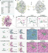
Figure 2. Structural basis of how K187 and L195 substitutions control hcGAS activity
(A) In vitro analysis of the role of hcGAS K187 and L195 variation in 2′3′ cGAMP synthesis regulation. Human and mouse amino acid sequences at 187 (human K187 and mouse-equivalent N187, denoted K or N) and 195 (denoted L or R) were analyzed in both hcGAS and mcGAS backgrounds. Enzymes were stimulated with 45 bp DNA, and 2′3′ cGAMP synthesis was measured as in Figure 1A and quantified relative to maximal activity observed with mcGAS. Data are represented as mean ± SD of three independent experiments. (B) Schematic and overview of the hcGAS–DNA complex. hcGAS forms a 2:2 complex with DNA where each cGAS monomer has two distinct DNA-binding surfaces (DNA A-Site and DNA B-Site). Stars in the schematic denote the enzyme metal-coordinating active-site residues, schematic not to scale. (C) Overview of a single 1:1 cGAS–DNA unit in the hcGAS–DNA–ATP complex. Zoom-in cutaways of the locations of K187 and L195 human-specific cGAS substitutions in the DNA A-site. The water molecule coordinated by the K187N mutation and Y215 is depicted as a grey sphere. See also Table S1 and Figure S4.
Figure 3. Mechanism of human-specific cGAS–DNA recognition
(A) Cladogram depicting evolution of hcGAS DNA A-site K187 and L195 positions in primates and relevant vertebrates. Human-specific substitutions are denoted in magenta, and the mouse cGAS sequence is denoted in blue for reference. (B) Electrophoretic mobility shift assay measurement of in vitro cGAS–DNA complex formation. hcGAS and mcGAS variants (labeled as in Figure 2A) were incubated with 45 bp DNA, assembled using gradient dialysis, and the resulting stable complexes were resolved on a 2% agarose gel. (C) Schematic map of protein–DNA contacts in the hcGAS–DNA complex. Human-specific contacts are highlighted in magenta, and black dots denote interactions bridged by water molecules. Black labels indicate contacts directly observed in the hcGAS–DNA complex, grey labels indicate water-mediated contacts potentially conserved with mcGAS but not observed in the hcGAS–DNA complex. See also Figure S4B. (D) Overview of the hcGAS–DNA complex highlighting the location of human-specific DNA A-site and B-site substitutions. A-site substitutions have a major role in enzyme regulation, and B-site substitutions play an additional minor role (See Figure S5). Human-specific substitutions are shown as sticks in magenta, and the mouse-like K187N and L195R DNA A-site mutations are denoted in blue. One cGAS protein monomer from the 2:2 complex is omitted for clarity. (E) Cartoon model of the hcGAS bound to short DNA (yellow) overlaid with the path of long DNA (orange) derived from the mcGAS-39 bp DNA structure (PDB 5N6I). Human-specific cGAS substitutions (magenta) weaken interactions with DNA in a portion of the DNA-binding surface that is not required for recognition of long DNA. Short and long DNA share identical interactions in the top conserved portion of the cGAS DNA A-site (blue), but assembly of an oligomerized cGAS–DNA complex causes long DNA to curve away and no longer make contacts with the bottom portion of the cGAS DNA A-site where the human-specific substitutions K187 and L195 are located. See also Figure S5.
Figure 4. Human cGAS adaptations re-shape DNA-length specificity
(A) In vitro analysis of cGAS DNA-length specificity. Purified hcGAS and mcGAS enzymes were stimulated with increasing concentration of 45 bp (top) or 17 bp (bottom) DNA. Enzyme activation was analyzed as in Figure 1A. Unlike mcGAS, hcGAS is only able to activate 2′3′ cGAMP synthesis in the presence of long 45 bp DNA. (B) Identical experiment as in A, using mcGAS with human-like K187 and L195 substitutions or hcGAS with mouse-like N187 and R195 substitutions. Human-specific K187 and L195 substitutions are necessary and sufficient for cGAS length-dependent DNA discrimination. (C) Quantification of cGAS DNA length-dependent activation experiments in A and B. 2′3′ cGAMP synthesis was quantified as in Figure 1A and graphed as total conversion of ATP to 2′3′ cGAMP. Data are represented as mean ± SD of three independent experiments. See also Figure S6.
Figure 5. The hcGAS–DNA structure provides insight into tumor-associated mutations and small-molecule inhibitor design
(A) Tumor-associated mutations in cGAS, and the structural role of each mutated residue predicted by the hcGAS–DNA–ATP ternary complex. (B) Highlights of the tumor-associated mutations in hcGAS on the hcGAS–DNA–ATP ternary complex. (C) Overview of a single 1:1 cGAS-unit in the hcGAS–DNA–ATP ternary complex with (D,E,F) zoom-in cutaways of the cGAS active-site showing protein residues in contact with ATP and small-molecule inhibitors. Nucleotide substrate and inhibiting compounds are shown in green, human-specific cGAS substitutions are in magenta, corresponding mcGAS residues are in blue, and conserved active-site amino acids are in grey. (G–J) Molecular docking analysis of the compatibility of existing cGAS inhibitors with the active hcGAS–DNA complex. All top docked inhibitor poses of the mcGAS inhibitor RU.521 (PDB 5XZG) and hcGAS inhibitor PF-06928215 (PDB 5V8N) shown in orange lines, are compatible with the mcGAS–DNA complex and apo hcGAS structure respectively, and agree with the experimentally derived crystallographic binding poses shown in green sticks for reference. In contrast, the docked inhibitor poses with the active hcGAS–DNA complex structure are distinct, further confirming that the hcGAS active site differs from previously observed structures.
Figure 6. The molecular basis of cGAS inhibitor specificity
(A) In vitro analysis of mcGAS and hcGAS K187N/L195R (hcGAS*) 2′3′ cGAMP synthesis with increasing concentration of the mcGAS inhibitor RU.521. Enzyme activation was analyzed as in Figure 1A. (B) Identical experiment as in A, using mcGAS with humanizing C419S/H467N mutations or hcGAS* with mouse-like S434C/N482H mutations in the inhibitor binding pocket. Mouse-specific C419 and H467 substitutions are necessary and sufficient for susceptibility to RU.521. (C) Quantification of cGAS inhibition by RU.521. 2′3′ cGAMP synthesis was quantified as in Figure 1A and was normalized to the DMSO control (set as 100%). Data are represented as mean ± SD of two independent experiments. See also Figure S7.
Figure 7. Determination of the hcGAS–DNA structure reveals that human-specific substitutions enhance regulation of cGAS activation
(A) Human-specific substitutions increase the DNA-length specificity of cGAS. Control of DNA sensing must be balanced to maintain sensitivity to pathogen-or stress-derived DNA, and allow accurate tolerance of self-DNA. Human substitutions in cGAS re-shape this balance, and reduce 2′3′ cGAMP synthesis in order to favor enhanced DNA selectivity. (B) Binding of DNA to cGAS induces a large conformational change, resulting in an “open” active-site conformation that is competent to associate with nucleotides for 2′3′ cGAMP synthesis. Structures of active human cGAS will be critical to guide drug development and analysis of human disease-related cGAS polymorphisms.
Comment in
- Human cGAS Has a Slightly Different Taste for dsDNA.
Decout A, Ablasser A. Decout A, et al. Immunity. 2018 Aug 21;49(2):206-208. doi: 10.1016/j.immuni.2018.08.005. Immunity. 2018. PMID: 30134199
Similar articles
- Cyclic GMP-AMP synthase is activated by double-stranded DNA-induced oligomerization.
Li X, Shu C, Yi G, Chaton CT, Shelton CL, Diao J, Zuo X, Kao CC, Herr AB, Li P. Li X, et al. Immunity. 2013 Dec 12;39(6):1019-31. doi: 10.1016/j.immuni.2013.10.019. Immunity. 2013. PMID: 24332030 Free PMC article. - The catalytic mechanism of cyclic GMP-AMP synthase (cGAS) and implications for innate immunity and inhibition.
Hall J, Ralph EC, Shanker S, Wang H, Byrnes LJ, Horst R, Wong J, Brault A, Dumlao D, Smith JF, Dakin LA, Schmitt DC, Trujillo J, Vincent F, Griffor M, Aulabaugh AE. Hall J, et al. Protein Sci. 2017 Dec;26(12):2367-2380. doi: 10.1002/pro.3304. Epub 2017 Oct 25. Protein Sci. 2017. PMID: 28940468 Free PMC article. - Structural basis for sequestration and autoinhibition of cGAS by chromatin.
Michalski S, de Oliveira Mann CC, Stafford CA, Witte G, Bartho J, Lammens K, Hornung V, Hopfner KP. Michalski S, et al. Nature. 2020 Nov;587(7835):678-682. doi: 10.1038/s41586-020-2748-0. Epub 2020 Sep 10. Nature. 2020. PMID: 32911480 - Cyclic Guanosine Monophosphate-Adenosine Monophosphate Synthase (cGAS), a Multifaceted Platform of Intracellular DNA Sensing.
Verrier ER, Langevin C. Verrier ER, et al. Front Immunol. 2021 Feb 23;12:637399. doi: 10.3389/fimmu.2021.637399. eCollection 2021. Front Immunol. 2021. PMID: 33708225 Free PMC article. Review. - Conserved strategies for pathogen evasion of cGAS-STING immunity.
Eaglesham JB, Kranzusch PJ. Eaglesham JB, et al. Curr Opin Immunol. 2020 Oct;66:27-34. doi: 10.1016/j.coi.2020.04.002. Epub 2020 Apr 15. Curr Opin Immunol. 2020. PMID: 32339908 Free PMC article. Review.
Cited by
- DNA-sensing pathways in health, autoinflammatory and autoimmune diseases.
Dong M, Fitzgerald KA. Dong M, et al. Nat Immunol. 2024 Nov;25(11):2001-2014. doi: 10.1038/s41590-024-01966-y. Epub 2024 Oct 4. Nat Immunol. 2024. PMID: 39367124 Review. - Cytosolic Sensors for Pathogenic Viral and Bacterial Nucleic Acids in Fish.
Mojzesz M, Rakus K, Chadzinska M, Nakagami K, Biswas G, Sakai M, Hikima JI. Mojzesz M, et al. Int J Mol Sci. 2020 Oct 2;21(19):7289. doi: 10.3390/ijms21197289. Int J Mol Sci. 2020. PMID: 33023222 Free PMC article. Review. - Supramolecular organizing centers at the interface of inflammation and neurodegeneration.
Sušjan-Leite P, Ramuta TŽ, Boršić E, Orehek S, Hafner-Bratkovič I. Sušjan-Leite P, et al. Front Immunol. 2022 Aug 1;13:940969. doi: 10.3389/fimmu.2022.940969. eCollection 2022. Front Immunol. 2022. PMID: 35979366 Free PMC article. Review. - Genotype-Phenotype Correlation and Functional Insights for Two Monoallelic TREX1 Missense Variants Affecting the Catalytic Core.
Amico G, Hemphill WO, Severino M, Moratti C, Pascarella R, Bertamino M, Napoli F, Volpi S, Rosamilia F, Signa S, Perrino F, Zedde M, Ceccherini I, On Behalf Of The Gaslini Stroke Study Group. Amico G, et al. Genes (Basel). 2022 Jun 30;13(7):1179. doi: 10.3390/genes13071179. Genes (Basel). 2022. PMID: 35885962 Free PMC article. - Acetylation Blocks cGAS Activity and Inhibits Self-DNA-Induced Autoimmunity.
Dai J, Huang YJ, He X, Zhao M, Wang X, Liu ZS, Xue W, Cai H, Zhan XY, Huang SY, He K, Wang H, Wang N, Sang Z, Li T, Han QY, Mao J, Diao X, Song N, Chen Y, Li WH, Man JH, Li AL, Zhou T, Liu ZG, Zhang XM, Li T. Dai J, et al. Cell. 2019 Mar 7;176(6):1447-1460.e14. doi: 10.1016/j.cell.2019.01.016. Epub 2019 Feb 21. Cell. 2019. PMID: 30799039 Free PMC article.
References
- Andreeva L, Hiller B, Kostrewa D, Lassig C, de Oliveira Mann CC, Jan Drexler D, Maiser A, Gaidt M, Leonhardt H, Hornung V, et al. cGAS senses long and HMGB/TFAM-bound U-turn DNA by forming protein-DNA ladders. Nature. 2017;549:394–398. - PubMed
Publication types
MeSH terms
Substances
Grants and funding
- S10 RR029205/RR/NCRR NIH HHS/United States
- P41 GM103403/GM/NIGMS NIH HHS/United States
- R01 AI018045/AI/NIAID NIH HHS/United States
- R01 CA214608/CA/NCI NIH HHS/United States
- T32 CA207021/CA/NCI NIH HHS/United States
LinkOut - more resources
Full Text Sources
Other Literature Sources
Molecular Biology Databases
Research Materials
Miscellaneous


