A new southern Laramidian ankylosaurid, Akainacephalus johnsoni gen. et sp. nov., from the upper Campanian Kaiparowits Formation of southern Utah, USA - PubMed (original) (raw)
A new southern Laramidian ankylosaurid, Akainacephalus johnsoni gen. et sp. nov., from the upper Campanian Kaiparowits Formation of southern Utah, USA
Jelle P Wiersma et al. PeerJ. 2018.
Abstract
A partial ankylosaurid skeleton from the upper Campanian Kaiparowits Formation of southern Utah is recognized as a new taxon, Akainacephalus johnsoni, gen. et sp. nov. The new taxon documents the first record of an associated ankylosaurid skull and postcranial skeleton from the Kaiparowits Formation. Preserved material includes a complete skull, much of the vertebral column, including a complete tail club, a nearly complete synsacrum, several fore- and hind limb elements, and a suite of postcranial osteoderms, making Akainacephalus johnsoni the most complete ankylosaurid from the Late Cretaceous of southern Laramidia. Arrangement and morphology of cranial ornamentation in Akainacephalus johnsoni is strikingly similar to Nodocephalosaurus kirtlandensis and some Asian ankylosaurids (e.g., Saichania chulsanensis, Pinacosaurus grangeri, and Minotaurasaurus ramachandrani); the cranium is densely ornamented with symmetrically arranged and distinctly raised ossified caputegulae which are predominantly distributed across the dorsal and dorsolateral regions of the nasals, frontals, and orbitals. Cranial caputegulae display smooth surface textures with minor pitting and possess a distinct conical to pyramidal morphology which terminates in a sharp apex. Character analysis suggests a close phylogenetic relationship with N. kirtlandensis, M. ramachandrani, Tarchia teresae, and S. chulsanensis, rather than with Late Cretaceous northern Laramidian ankylosaurids (e.g., Euoplocephalus tutus, Anodontosaurus lambei, and Ankylosaurus magniventris). These new data are consistent with evidence for distinct northern and southern biogeographic provinces in Laramidia during the late Campanian. The addition of this new ankylosaurid taxon from southern Utah enhances our understanding of ankylosaurid diversity and evolutionary relationships. Potential implications for the geographical distribution of Late Cretaceous ankylosaurid dinosaurs throughout the Western Interior suggest multiple time-transgressive biogeographic dispersal events from Asia into Laramidia.
Keywords: Biodiversity; Paleontology; Taxonomy.
Conflict of interest statement
The authors declare that they have no competing interests.
Figures
Figure 1. Distribution of Late Cretaceous Laramidian ankylosaurid dinosaurs.
Overview of stratigraphic, temporal, and biogeographic distribution of Late Cretaceous (upper Campanian–upper Maastrichtian) ankylosaurid dinosaurs, including Akainacephalus johnsoni, across northern Laramidian (Alberta, Montana, Wyoming), and southern Laramidian (Utah, New Mexico) basins. Noticeable is the higher number of taxa and more widespread distribution of ankylosaurids in northern Laramidia. Colored bars within stratigraphic formations represent ankylosaurid taxa and their respective temporal range. Paleogeographic map modified after Sampson et al. (2010). Stratigraphic intervals modified after Arbour & Currie (2013a); Roberts et al. (2013); Johnson, Nichols & Hartman (2002).
Figure 2. Location and stratigraphy of the Kaiparowits Formation.
Map of Grand Staircase-Escalante National Monument (GSENM), southern Utah (A), and generalized stratigraphic section of the upper Campanian Kaiprowits Formation (B). The approximate stratigraphic position of Akainacephalus johnsoni is located near the base of the middle unit within the Kaiparowits Formation. The map highlights the GSENM boundary (dashed line), showing the geological distribution and outcrops of the Cretaceous Kaiparowits, Wahweap, and Straight Cliffs formations. The red star indicates the Horse Mountain area, from which Akainacephalus johnsoni was recorded. Map and stratigraphic column modified from Roberts (2005). Radioisotopic dates used from Roberts, Deino & Chan (2005) and Roberts et al. (2013), respectively.
Figure 3. Skull of Akainacephalus johnsoni (UMNH VP 20202).
Photographs of the skull of Akainacephalus johnsoni in (A), left lateral; and (B), right lateral views. Line drawings in (C), left lateral; and (D), right lateral views highlight major anatomical features. Study sites: en, external naris; fca, frontal caputegulum; j, jugal; jca, jugal caputegulum; l, lacrimal; laca, lacrimal caputegulum; loca, loreal caputegulum; mx, maxilla; n, nasal; naca, nasal caputegulae; ns, nuchal shelf; orb, orbit; pmx, premaxilla; prfca, prefrontal caputegulum; snca, supranarial caputegulum; sob, supraorbital boss; q, quadrate; qjh, quadratojugal horn; sqh, squamosal horn.
Figure 4. Skull of Akainacephalus johnsoni (UMNH VP 20202).
Photographs of the skull of Akainacephalus johnsoni in (A), dorsal; and (B), ventral views. Line drawings in (C), dorsal; and (D), ventral views highlight major anatomical features. Study sites: bpt, basipterygoid; bs, basisphenoid; ch, choana; exo, exoccipital; fm, foramen magnum; fca, frontal caputegulum; ins, internarial septum; laca, lacrimal caputegulum; loca, loreal caputegulum; mx, maxilla; mxtr, maxillary tooth row; naca, nasal caputegulum; ns, nuchal shelf; oc, occipital condyle; pal, palatine; prfca, prefrontal caputegulum; pmx, premaxilla; pmxs, interpremaxillry suture with oblong depression; pop, paroccipital process; ptv, pterygoid vacuity; q, quadrate; qj, quadratojugal; qjh, quadratojuga horn; so, supra occipital; snca, supranarial caputegulum; sob, supraorbital boss; sqh, squamosal horn.
Figure 5. Skull of Akainacephalus johnsoni (UMNH VP 20202).
Photographs of the skull of Akainacephalus johnsoni in (A), posterior; and (B), anterior views. Line drawings in (C), posterior; and (D), anterior views highlight major anatomical features. Study sites: fm, foramen magnum; fca, frontal caputegulae; loca, loreal caputegulum; naca; nasal caputegulae; ns, nuchal shelf; oc, occipital condyle; pmx, premaxilla; pop, paroccipital process; prfca, prefrontal caputegulum; q, quadrate; qjh, quadratojugal horn; snca, supranarial caputegulum; so, supraoccipital; sob, supraorbital boss; sqh, squamosal horn.
Figure 6. Computed tomography (CT) scans of Akainacephalus johnsoni (UMNH VP 20202).
Transverse plane CT visualization of the skull of Akainacephalus johnsoni revealing important anatomical features, otherwise obscured by bone and/or matrix. Anatomical features in skull of A. johnsoni with horizontal lines indicating localities for the CT slices; (A), oblong-shaped olfactory lobes and outline of the endocranial cavity which provides space for the brain, including its olfactory lobes; (B), and (C), ocular cavities and their surrounding pre- and postocular walls. Presence of cranial nerves CN V, ?VII, IX, X, XI and the fenestra ovalis (FO) along the lateral surfaces of the endocranial cavity; (D), anatomical outline of a small portion of the complex nasal air passage system showing the rostral loop of the nasal vestibule; (E) outlines of the olfactory chambers which house the olfactory lobes. Images not to scale.
Figure 7. Variation in cranial ornamentation in selected Laramidian and Asian taxa, including Akainacephalus johnsoni.
Comparative line drawings highlighting major areas of cranial ornamentation in Akainacephalus johnsoni and closely related Laramidian and Asian taxa. Akainacephalus johnsoni (UMNH VP 20202) in (A), dorsal; (B), left lateral view compared to Nodocephalosaurus kirtlandensis (SMP VP-900) in (C), dorsal; and (D) left lateral view; Tarchia teresae (PIN 3142/250) in (E) dorsal; (F), left lateral view and Minotaurasaurus ramachandrani (INBR 21004) in (G), dorsal; and (H), left lateral view. Study sites: acc po, accessory postorbital ossification; asob, anterior supraorbital boss; frca, frontal caputegulum; laca, lacrimal caputegulum; loca, loreal caputegulum; mso, medial supraorbital; mx, maxilla; n, external naris; naca, nasal caputegulae; nuca, nuchal caputegulae; orb, orbital; pmx, premxilla; pnca, postnarial caputegulum; pos postocular ossicles; prfca, prefrontal caputegulum; psob, posterior supraorbital boss; pt, pterygoid; q, quadrate; qjh, quadratojugal horn; snca, supranarial caputegulum; sqh, squamosal horn. Color scheme after Arbour & Currie (2013a). Dorsal view of N. kirtlandensis modified after Arbour et al. (2014). T. teresea (=Saichania chulsanensis in Arbour, Currie & Badamgarav, 2014) and M. ramachandrani modified after Arbour, Currie & Badamgarav (2014).
Figure 8. Skull of Akainacephalus johnsoni compared with Nodocephalosaurus kirtlandensis.
Comparison of cranial features between closely related southern Laramidian taxa; (A), Akainacephalus johnsoni (UMNH VP 20202) from the Late Cretaceous Kaiparowits Formation of Utah; and (B), Nodocephalosaurus kirtlandensis (SMP VP-900) from the Late Cretaceous Kirtland Formation of New Mexico, in left lateral views. Various synapomorphies are shared with N. kirtlandensis (highlighted in black and white arrows) and includes “flaring nostrils”; enlarged, laterally projecting, loreal osteoderms that are situated directly dorsal to the external nares. Other synapomorphies include pyramid-shaped nasal and frontal osteoderms positioned on the dorsal regions of the skull. A number of significant differences have been observed between both specimens; in A. johnsoni, the anterior, and posterior supraorbital bosses form an enlarged element that is somewhat backswept, whereas in N. kirtlandensis, the posterior and anterior supraorbital bosses are clearly defined as individual osteoderms, and are much smaller in size. Additionally, the squamosal horn in Akainacephalus is very small but is prominent and tetrahedrally shaped in Nodocephalosaurus. The quadratojugal horn in Akainacephalus is massive, has a subtriangular morphology in lateral view and projects almost entirely ventral, whereas in Nodocephalosaurus, the quadratojugal horn is smaller and has a typical fin-shaped morphology. Study sites: asob, anterior supraorbital boss; ext naris, external naris; laca, lacrimal caputegulum; loca, loreal caputegulum; naca, nasal caputegulae; orb, orbit; psob, posterior supraorbital boss; qjh, quadratojugal horn; sqh, squamosal horn. Part B: Photograph of the holotype of Nodocephalosaurus kirtlandensis (SMP VP-900) is reproduced courtesy Robert M. Sullivan.
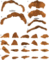
Figure 9. Mandibles and predentary.
Photographs of the mandibles and predentary of Akainacephalus johnsoni (UMNH VP 20202). Left mandible in (A), lateral; (B), medial; (C), ventral; and (D), dorsal views. Right mandible in (E), lateral; (F), medial; (G), ventral; and (H), dorsal view. Predentary in (I), anterior; (J), posterior; (K), dorsal; and (L), ventral views. Study sites: cp, coronoid process; cs, predentary cutting surface; mos, mandibular osteoderm; ms, mandibular symphysis; mtr, mandibular tooth row; pp, predentary protuberance.
Figure 10. Cervical vertebra.
Photographs of the cervical vertebra of Akainacephalus johnsoni (UMNH VP 20202) in (A), anterior; (B), posterior; (C) right lateral; (D), left lateral; (E), ventral; and (F), dorsal views. Study sites: c, centrum; na, neural arch; nc, neural canal; ns, neural spine; poz, postzygopophyses; prz, prezygopophyses.
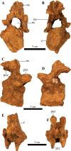
Figure 11. Free anterior dorsal vertebrae.
Photographs of the two anterior dorsal vertebrae of Akainacephalus johnsoni (UMNH VP 20202) in (A) and (F), anterior; (B) and (G), posterior; (C) and (H), right lateral; (D) and (I), left lateral; (E) and (J), dorsal views. Ribs are broken away in both vertebrae but articulation with the diapophyses and parapophyses are clearly visible. Study sites: c, centrum; diap, diapophysis; na, neural arch; nc, neural canal; ns, neural spine; parp, parapophysis; poz, postzygopophyses; prz, prezygopophyses; tp, transverse process.
Figure 12. Free mid-dorsal vertebrae.
Photographs of two mid-dorsal vertebrae with fused ribs of Akainacephalus johnsoni (UMNH VP 20202) in (A) and (D), anterior; (B) and (E), posterior; (C) and (F), dorsal views. Study sites: c, centrum; nc, neural canal; ns, neural spine; poz, postzygopophyses; prz, prezygopophyses.
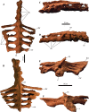
Figure 13. Sacrum, presacral, and caudosacral vertebrae.
Photographs of the sacrum, presacral rod, and caudosacral vertebrae of Akainacephalus johnsoni (UMNH VP 20202) in (A), ventral; (B), dorsal; (C), right lateral; (D), left lateral; (E), posterior; and (F), anterior views. Study sites: c, centrum; cs, caudosacral vertebra; csr, caudosacral ribs; ds, dorsosacral vertebrae; dsr, dorsosacral ribs; ns, neural spine; sr, sacral ribs.
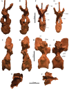
Figure 14. Anterior caudal vertebrae.
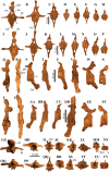
Photographs of eight anterior caudal vertebrae of Akainacephalus johnsoni (UMNH VP 20202) in (A–H), anterior; (I–P), posterior; (Q–X), left lateral; (Y–FF), right lateral; (GG–NN), dorsal; and (OO–VV), ventral views. Study sites: c, centrum; ha, haemal arch; hc, haemal canal; na, neural arch; nc, neural canal; ns, neural spine; poz, postzygopophyses; prz, prezygopophyses; tp, transverse process.
Figure 15. Fused caudal vertebrae (handle) and tail club knob.
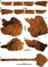
Photographs of distal caudal vertebrae (handle) and tail club of Akainacephalus johnsoni (UMNH VP 20202). Handle in (A), left lateral; and (B), right lateral view. Tail club in (C), dorsal; (D), ventral; (E), left lateral; and (F), right lateral views. Reconstructed handle and tail club in (G), dorsal view. Study sites: lo, lateral osteoderms; to, terminal osteoderms.
Figure 16. Ribs and rib fragments.
Photographs of ribs and rib fragments of Akainacephalus johnsoni (UMNH VP 20202) in (A), anterior; and (B), posterior views.
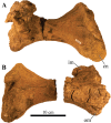
Figure 17. Left and right scapulae and right coracoid.
Photographs of the left and right scapulae and right coracoid of Akainacephalus johnsoni (UMNH VP 20202). Right scapula and coracoid in (A), medial; and (B), lateral views. Left scapula in (C), lateral; and (D), medial views. Study sites: acr, acromium; cor, coracoid; gl, glenoid; glf, glenoid fossa; mtlc, enthesis of M. triceps longus caudalis; scb, scapular blade; stp, sternal process.
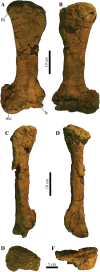
Figure 18. Right humerus.
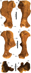
Photographs of the right humerus of Akainacephalus johnsoni (UMNH VP 20202) in (A), anterior; (B), posterior; (C), lateral; (D), medial; (E), dorsal; and (F), ventral views. Study sites: dpc, deltopectoral crest; hh, humeral head; it, internal tuberosity; mc, medial condyle; lc, lateral condyle.
Figure 19. Left ulna.
Photographs of a partial left ulna of Akainacephalus johnsoni (UMNH VP 20202) in (A), anterior; (B), posterior; (C), lateral; (D), medial; (E), distal; and (F), proximal views. Study sites: op, olecranon process.
Figure 20. Left ilium and right ischium.
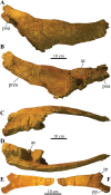
Photographs of the left ilium and right ischium of Akainacephalus johnsoni (UMNH VP 20202). Left ilium in (A), dorsal; (B), ventral; (C), lateral; and (D), medial views. Right ischium in (E), medial; and (F), lateral views. Study sites: ac, acetabulum; ip, iliac peduncle; poa, postacetabular process; pp, pubic peduncle; prea, preacetabular process.
Figure 21. Left femur.
Photographs of the left femur of Akainacephalus johnsoni (UMNH VP 20202) in (A), anterior; (B), posterior; (C), lateral; (D), medial; (E), dorsal; and (F), ventral views. Study sites: fh, femoral head; lc, lateral condyle; mc, medial condyle.
Figure 22. Right tibia.
Photographs of the right tibia of Akainacephalus johnsoni (UMNH VP 20202) in (A), anterior; and (B), posterior views. Study sites: cn, cnemial crest; im, inner malleolus; om, outer malleolus.
Figure 23. Left fibula.
Photographs of the left fibula of Akainacephalus johnsoni (UMNH VP 20202) in (A), posterior; and (B), anterior views.
Figure 24. Pedal phalanx.
Photographs of a single pedal phalanx of Akainacephalus johnsoni (UMNH VP 20202) in (A), anterior; (B), posterior; (C), dorsal; (D), ventral; (E), lateral; and (F), medial views.
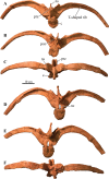
Figure 25. Cervical half rings and postcranial osteoderms.
Photographs of postcranial osteoderms of Akainacephalus johnsoni (UMNH VP 20202). Nearly complete cervical half ring in (A), anterior; and (B), posterior views. Right lateral portion of the partial half ring in (C), dorsal; (D), ventral; (E), posterior; and (F), anterior views. Various sharply keeled, type 1 (pup-tent), osteoderms associated with UMNH VP 20202. The osteoderms are displayed in (G), dorsal; (H), ventral; (I), medial; and (J), lateral views. No consensus currently exists regarding the exact position of type 1 osteoderms, but they appear to be positioned either laterally on the body along the sacral and proximal portion of the tail or dorsolaterally along the thoracic and pectoral regions as seen in Euoplocephalus and Scolosaurus (Maryańska, 1977; Ford, 2000).
Figure 26. Results of phylogenetic analyses for Akainacephalus johnsoni.
Phylogenetic position of Akainacephalus johnsoni in (A), a strict consensus of 21 equally most parsimonious trees, including the wildcard taxon Ahshislepelta minor, placing A. johnsoni within a large polytomy, consisting of crown group taxa that include Asian and all Laramidian ankylosaurids; and (B), the resulting strict consensus of six equally most phylogenetic trees, from which the wildcard taxon Ahshislepelta minor has been pruned. The crown group taxa are slightly better resolved in the pruned analysis, in which Akainacephalus johnsoni forms a clade with its sister taxon Nodocephalosaurus kirtlandensis, nested within the clade that also includes the Asian taxa Minotaurasaurus ramachandrani, Tarchia kilanae, and Shanxia tianzhenensis, suggesting a close taxonomic relationship with Nodocephalosaurus kirtlandensis and Asian taxa.
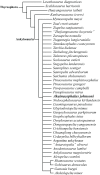
Figure 27. Phylogenetic results using character state codings from Arbour & Evans (2017).
Phylogenetic analysis resulting in a strict consensus of 1990 equally most parsimonious trees and the phylogenetic position of Akainacephalus johnsoni, implemented into the character matrix from Arbour & Evans (2017). The resulting consensus tree renders A. johnsoni poorly resolved, consistently placing it within a large polytomy.
Figure 28. Preserved elements and skeletal reconstructions of Akainacephalus johnsoni.
A composite showing all holotype skeletal material of _Akainacephalus johnson_i (UMNH VP 20202) anatomically arranged in dorsal view (A). Cartoon illustrating a full body reconstruction for A. johnsoni in (B), dorsal; and (C), left lateral view. Preserved material in the skeletal reconstructions is highlighted in orange.
Similar articles
- A new ankylosaurid dinosaur from the Upper Cretaceous (Kirtlandian) of New Mexico with implications for ankylosaurid diversity in the Upper Cretaceous of western North America.
Arbour VM, Burns ME, Sullivan RM, Lucas SG, Cantrell AK, Fry J, Suazo TL. Arbour VM, et al. PLoS One. 2014 Sep 24;9(9):e108804. doi: 10.1371/journal.pone.0108804. eCollection 2014. PLoS One. 2014. PMID: 25250819 Free PMC article. - New horned dinosaurs from Utah provide evidence for intracontinental dinosaur endemism.
Sampson SD, Loewen MA, Farke AA, Roberts EM, Forster CA, Smith JA, Titus AL. Sampson SD, et al. PLoS One. 2010 Sep 22;5(9):e12292. doi: 10.1371/journal.pone.0012292. PLoS One. 2010. PMID: 20877459 Free PMC article. - A remarkable short-snouted horned dinosaur from the Late Cretaceous (late Campanian) of southern Laramidia.
Sampson SD, Lund EK, Loewen MA, Farke AA, Clayton KE. Sampson SD, et al. Proc Biol Sci. 2013 Jul 17;280(1766):20131186. doi: 10.1098/rspb.2013.1186. Print 2013 Sep 7. Proc Biol Sci. 2013. PMID: 23864598 Free PMC article. - Euoplocephalus tutus and the diversity of ankylosaurid dinosaurs in the Late Cretaceous of Alberta, Canada, and Montana, USA.
Arbour VM, Currie PJ. Arbour VM, et al. PLoS One. 2013 May 8;8(5):e62421. doi: 10.1371/journal.pone.0062421. Print 2013. PLoS One. 2013. PMID: 23690940 Free PMC article. - A large non-marine turtle from the Upper Cretaceous of Alabama and a review of North American "Macrobaenids".
Gentry AD, Kiernan CR, Parham JF. Gentry AD, et al. Anat Rec (Hoboken). 2023 Jun;306(6):1411-1430. doi: 10.1002/ar.25054. Epub 2022 Aug 19. Anat Rec (Hoboken). 2023. PMID: 37158131 Review.
Cited by
- A brief review of non-avian dinosaur biogeography: state-of-the-art and prospectus.
Upchurch P, Chiarenza AA. Upchurch P, et al. Biol Lett. 2024 Oct;20(10):20240429. doi: 10.1098/rsbl.2024.0429. Epub 2024 Oct 30. Biol Lett. 2024. PMID: 39471833 Free PMC article. Review. - Lokiceratops rangiformis gen. et sp. nov. (Ceratopsidae: Centrosaurinae) from the Campanian Judith River Formation of Montana reveals rapid regional radiations and extreme endemism within centrosaurine dinosaurs.
Loewen MA, Sertich JJW, Sampson S, O'Connor JK, Carpenter S, Sisson B, Øhlenschlæger A, Farke AA, Makovicky PJ, Longrich N, Evans DC. Loewen MA, et al. PeerJ. 2024 Jun 20;12:e17224. doi: 10.7717/peerj.17224. eCollection 2024. PeerJ. 2024. PMID: 38912046 Free PMC article. - Calibrating the zenith of dinosaur diversity in the Campanian of the Western Interior Basin by CA-ID-TIMS U-Pb geochronology.
Ramezani J, Beveridge TL, Rogers RR, Eberth DA, Roberts EM. Ramezani J, et al. Sci Rep. 2022 Sep 26;12(1):16026. doi: 10.1038/s41598-022-19896-w. Sci Rep. 2022. PMID: 36163377 Free PMC article. - A new Cretaceous thyreophoran from Patagonia supports a South American lineage of armoured dinosaurs.
Riguetti FJ, Apesteguía S, Pereda-Suberbiola X. Riguetti FJ, et al. Sci Rep. 2022 Aug 11;12(1):11621. doi: 10.1038/s41598-022-15535-6. Sci Rep. 2022. PMID: 35953515 Free PMC article. - The phylogenetic nomenclature of ornithischian dinosaurs.
Madzia D, Arbour VM, Boyd CA, Farke AA, Cruzado-Caballero P, Evans DC. Madzia D, et al. PeerJ. 2021 Dec 9;9:e12362. doi: 10.7717/peerj.12362. eCollection 2021. PeerJ. 2021. PMID: 34966571 Free PMC article.
References
- Arbour VM, Burns ME, Sissons RL. A redescription of Dyoplosaurus acutosquameus Parks, 1924 (Ornithischia: Ankylosauria) and a revision of the genus. Journal of Vertebrate Paleontology. 2009;29:1117–1135.
- Arbour VM, Burns ME, Sullivan RM, Lucas SG, Cantrell AK, Fry J, Suazo TL. A new ankylosaurid dinosaur from the Upper Cretaceous (Kirtlandian) of New Mexico with implications for ankylosaurid diversity in the Upper Cretaceous of western North America. PLOS ONE. 2014;9(9):e108804. doi: 10.1371/journal.pone.0108804. - DOI - PMC - PubMed
- Arbour VM, Currie PJ. The taxonomic identity of a nearly complete ankylosaurid dinosaur skeleton from the Gobi Desert of Mongolia. Cretaceous Research. 2013b;46:24–30. doi: 10.1016/j.cretres.2013.08.008. - DOI
- Arbour VM, Currie PJ. Systematics, phylogeny, and palaeobiogeography of the ankylosaurid dinosaurs. Journal of Systematic Palaeontology. 2016;14(5):385–444. doi: 10.1080/14772019.2015.1059985. - DOI
Grants and funding
Research funding was provided by the Geological Society of America Graduate Student Research Grant (grant number 10631-14); the Bureau of Land Management National Landscape Conservation System Grant (grant numbers JSA071004 & L12AC20378); and the University of Utah, Department of Geology & Geophysics Graduate Student Travel Grant. The funders had no role in study design, data collection and analysis, decision to publish, or preparation of the manuscript.
LinkOut - more resources
Full Text Sources
Other Literature Sources
Miscellaneous


