Correlative Light-Electron Microscopy detects lipopolysaccharide and its association with fibrin fibres in Parkinson's Disease, Alzheimer's Disease and Type 2 Diabetes Mellitus - PubMed (original) (raw)
Correlative Light-Electron Microscopy detects lipopolysaccharide and its association with fibrin fibres in Parkinson's Disease, Alzheimer's Disease and Type 2 Diabetes Mellitus
Greta M de Waal et al. Sci Rep. 2018.
Abstract
Many chronic diseases, including those classified as cardiovascular, neurodegenerative, or autoimmune, are characterized by persistent inflammation. The origin of this inflammation is mostly unclear, but it is typically mediated by inflammatory biomarkers, such as cytokines, and affected by both environmental and genetic factors. Recently circulating bacterial inflammagens such as lipopolysaccharide (LPS) have been implicated. We used a highly selective mouse monoclonal antibody to detect bacterial LPS in whole blood and/or platelet poor plasma of individuals with Parkinson's Disease, Alzheimer's type dementia, or Type 2 Diabetes Mellitus. Our results showed that staining is significantly enhanced (P < 0.0001) compared to healthy controls. Aberrant blood clots in these patient groups are characterized by amyloid formation as shown by the amyloid-selective stains thioflavin T and Amytracker™ 480 or 680. Correlative Light-Electron Microscopy (CLEM) illustrated that the LPS antibody staining is located in the same places as where amyloid fibrils may be observed. These data are consistent with the Iron Dysregulation and Dormant Microbes (IDDM) hypothesis in which bacterial inflammagens such as LPS are responsible for anomalous blood clotting as part of the aetiology of these chronic inflammatory diseases.
Conflict of interest statement
The authors declare no competing interests.
Figures
Figure 1
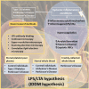
Overview of this paper, focusing on systemic inflammation in various inflammatory conditions, the presence of inflammagens such as LPS, and its contribution to hypercoagulation and amyloid formation, along with a list of novel research methods employed.
Figure 2
Overview of optimization of LPS antibody binding, using healthy platelet poor plasma (PPP) samples exposed to LPS.
Figure 3
Typical range of confocal micrographs of platelet poor plasma (PPP) with added thrombin, showing the fluorescence amyloid signal of (A) healthy individuals and (B) Parkinson’s Disease (PD) individuals. Platelet poor plasma (PPP) from each individual was incubated with three specific amyloidogenic fluorescent markers, thioflavin T and Amytracker™ 480 and 680.
Figure 4
Representative confocal micrographs of healthy platelet poor plasma (PPP) with added LPS to show optimization of detection of LPS, by using anti-E.coli LPS antibody and a secondary antibody (1:200 dilution for primary and secondary antibodies). We used five different LPS concentrations, and estimated that 0.5 **μ**gL−1 LPS is the lowest detectable concentration. For clarity, we inverted the micrographs, followed by applying the “find edges” function in ImageJ (FIJI), to show the decreasing fluorescence LPS signal with decreasing concentrations added.
Figure 5
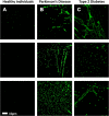
Representative platelet poor plasma (PPP) smears with added anti-E.coli LPS antibody and secondary antibody, from (A) healthy individuals, (B) Parkinson’s Disease (PD) individuals and (C) Type 2 Diabetes (T2D) individuals (1:200 dilution for primary and secondary antibodies).
Figure 6
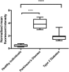
Boxplot of the distribution of the mean fluorescence intensity, normalised by dividing the mean fluorescence intensity values of the healthy individuals, Parkinson’s Disease (PD) individuals and Type 2 Diabetes (T2D) individuals, by the mean fluorescence intensity values of the corresponding secondary antibody control. In this way, we accounted for non-specific secondary antibody binding; ****P < 0.0001.
Figure 7
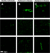
Representative whole blood (WB) smears with added anti-E.coli LPS antibody and secondary antibody, from (A) healthy individuals, (B) Parkinson’s Disease (PD) individuals and (C) Alzheimer’s Disease (AD) individuals (1:200 dilution for primary and secondary antibodies).
Figure 8
Representative CLEM and SEM micrographs of (A) stored platelet poor plasma (PPP) from a Type 2 Diabetes (T2D) individual, (B) freshly-collected whole blood (WB) from a Parkinson’s Disease (PD) individual and (C) stored whole blood (WB) from a Parkinson’s Disease (PD) individual (1:200 dilution for primary and secondary antibodies). The fluorescence microscopy modalities used were super-resolution (SR-SIM) for (B) and confocal for (A) and (C).
Figure 9
Representative (A) super-resolution (SR-SIM), (B) SEM and (C) CLEM micrographs of freshly-collected whole blood (WB) from a Parkinson’s Disease (PD) individual (1:200 dilution for primary and secondary antibodies). LPS antibody staining is closely associated with the fibre-like structures. Micrograph C colour was enhanced for publication clarity, by adjusting the vibrancy, brightness and contrast in Adobe Photoshop CS6.
Similar articles
- Lipopolysaccharide-binding protein (LBP) can reverse the amyloid state of fibrin seen or induced in Parkinson's disease.
Pretorius E, Page MJ, Mbotwe S, Kell DB. Pretorius E, et al. PLoS One. 2018 Mar 1;13(3):e0192121. doi: 10.1371/journal.pone.0192121. eCollection 2018. PLoS One. 2018. PMID: 29494603 Free PMC article. - Substantial fibrin amyloidogenesis in type 2 diabetes assessed using amyloid-selective fluorescent stains.
Pretorius E, Page MJ, Engelbrecht L, Ellis GC, Kell DB. Pretorius E, et al. Cardiovasc Diabetol. 2017 Nov 2;16(1):141. doi: 10.1186/s12933-017-0624-5. Cardiovasc Diabetol. 2017. PMID: 29096623 Free PMC article. - Acute induction of anomalous and amyloidogenic blood clotting by molecular amplification of highly substoichiometric levels of bacterial lipopolysaccharide.
Pretorius E, Mbotwe S, Bester J, Robinson CJ, Kell DB. Pretorius E, et al. J R Soc Interface. 2016 Sep;13(122):20160539. doi: 10.1098/rsif.2016.0539. J R Soc Interface. 2016. PMID: 27605168 Free PMC article. - Theoretical approaches to protein aggregation.
Gsponer J, Vendruscolo M. Gsponer J, et al. Protein Pept Lett. 2006;13(3):287-93. doi: 10.2174/092986606775338407. Protein Pept Lett. 2006. PMID: 16515457 Review. - Iron Dysregulation and Dormant Microbes as Causative Agents for Impaired Blood Rheology and Pathological Clotting in Alzheimer's Type Dementia.
Pretorius L, Kell DB, Pretorius E. Pretorius L, et al. Front Neurosci. 2018 Nov 16;12:851. doi: 10.3389/fnins.2018.00851. eCollection 2018. Front Neurosci. 2018. PMID: 30519157 Free PMC article. Review.
Cited by
- Intestinal Permeability, Dysbiosis, Inflammation and Enteric Glia Cells: The Intestinal Etiology of Parkinson's Disease.
Yang H, Li S, Le W. Yang H, et al. Aging Dis. 2022 Oct 1;13(5):1381-1390. doi: 10.14336/AD.2022.01281. eCollection 2022 Oct 1. Aging Dis. 2022. PMID: 36186124 Free PMC article. - A central role for amyloid fibrin microclots in long COVID/PASC: origins and therapeutic implications.
Kell DB, Laubscher GJ, Pretorius E. Kell DB, et al. Biochem J. 2022 Feb 17;479(4):537-559. doi: 10.1042/BCJ20220016. Biochem J. 2022. PMID: 35195253 Free PMC article. Review. - A Perspective on How Fibrinaloid Microclots and Platelet Pathology May be Applied in Clinical Investigations.
Pretorius E, Kell DB. Pretorius E, et al. Semin Thromb Hemost. 2024 Jun;50(4):537-551. doi: 10.1055/s-0043-1774796. Epub 2023 Sep 25. Semin Thromb Hemost. 2024. PMID: 37748515 Free PMC article. Review. - SARS-CoV-2 spike protein S1 induces fibrin(ogen) resistant to fibrinolysis: implications for microclot formation in COVID-19.
Grobbelaar LM, Venter C, Vlok M, Ngoepe M, Laubscher GJ, Lourens PJ, Steenkamp J, Kell DB, Pretorius E. Grobbelaar LM, et al. Biosci Rep. 2021 Aug 27;41(8):BSR20210611. doi: 10.1042/BSR20210611. Biosci Rep. 2021. PMID: 34328172 Free PMC article. - Iron Dysregulation and Inflammagens Related to Oral and Gut Health Are Central to the Development of Parkinson's Disease.
Vuuren MJV, Nell TA, Carr JA, Kell DB, Pretorius E. Vuuren MJV, et al. Biomolecules. 2020 Dec 29;11(1):30. doi: 10.3390/biom11010030. Biomolecules. 2020. PMID: 33383805 Free PMC article. Review.
References
- Marshall BJ. Helicobacter pylori. The American journal of gastroenterology. 1994;89:S116–128. - PubMed
Publication types
MeSH terms
Substances
LinkOut - more resources
Full Text Sources
Medical


