A new African Titanosaurian Sauropod Dinosaur from the middle Cretaceous Galula Formation (Mtuka Member), Rukwa Rift Basin, Southwestern Tanzania - PubMed (original) (raw)
A new African Titanosaurian Sauropod Dinosaur from the middle Cretaceous Galula Formation (Mtuka Member), Rukwa Rift Basin, Southwestern Tanzania
Eric Gorscak et al. PLoS One. 2019.
Abstract
The African terrestrial fossil record has been limited in its contribution to our understanding of both regional and global Cretaceous paleobiogeography, an interval of significant geologic and macroevolutionary change. A common component in Cretaceous African faunas, titanosaurian sauropods diversified into one of the most specious groups of dinosaurs worldwide. Here we describe the new titanosaurian Mnyamawamtuka moyowamkia gen. et sp. nov. from the Mtuka Member of the Galula Formation in southwest Tanzania. The new specimen preserves teeth, elements from all regions of the postcranial axial skeleton, parts of both appendicular girdles, and portions of both limbs including a complete metatarsus. Unique traits of M. moyowamkia include the lack of an interpostzygapophyseal lamina in posterior dorsal vertebrae, pronounced posterolateral expansion of middle caudal centra, and an unusually small sternal plate. Phylogenetic analyses consistently place M. moyowamkia as either a close relative to lithostrotian titanosaurians (e.g., parsimony, uncalibrated Bayesian analyses) or as a lithostrotian and sister taxon to Malawisaurus dixeyi from the nearby Aptian? Dinosaur Beds of Malawi (e.g., tip-dating Bayesian analyses). M. moyowamkia shares a few features with M. dixeyi, including semi-spatulate teeth and a median lamina between the neural canal and interpostzygapophyseal lamina in anterior dorsal vertebrae. Both comparative morphology and phylogenetic analyses support Mnyamawamtuka as a distinct and distant relative to Rukwatitan bisepultus and Shingopana songwensis from the younger Namba Member of the Galula Formation with these results largely congruent with newly constrained ages for the Mtuka Member (Aptian-Cenomanian) and Namba Member (Campanian). Coupled with recent discoveries from the Dahkla Oasis, Egypt (e.g., Mansourasaurus shahinae) and other parts of continental Afro-Arabia, the Tanzania titanosaurians refine perspectives on the development of African terrestrial faunas throughout the Cretaceous-a critical step in understanding non-marine paleobiogeographic patterns of Africa that have remained elusive until the past few years.
Conflict of interest statement
The authors have declared that no competing interests exist.
Figures
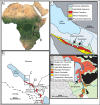
Fig 1. Map of research area.
Map of Africa, A, with expanded regional map of the Rukwa Rift Basin of Tanzania, B, with the type localities of Mnyamawamtuka moyowamkia and Rukwatitan bisepultus quarry near the Galula study area, C, and the Shingopana songwensis quarry near the Nsungwe study area, D. Malawi Dinosaur Beds (DB) marked in B to demonstrate the proximity of the deposits to the Galula Formation.
Fig 2. Quarry map.
Quarry map of the Mtuka bonebed locality RRBP 2004–06. Recovered elements of M. moyowamkia are color-coded and separated by dashed lines according to the year they were collected. The quarry map is represented as a four-by-six-meter grid. Unmarked elements on the map are either fragments or unidentified. Abbreviations: cac, caudal vertebral centrum; cana, caudal vertebral neural arch; cr, cervical rib; cvc, cervical vertebral centrum; dc, dorsal vertebral centrum; dic, distal caudal vertebra; dr, dorsal rib; dv, dorsal vertebra; fem, femur; fib, fibula; ha, haemal arch; hum, humerus; isc, ischium; mtc I, metacarpal I; sac, sacral centrum; scap, scapula; sp, sternal plate; sr, sacral rib; tib, tibia; ul, ulna; un, ungual.
Fig 3. Teeth associated with Mnyamawamtuka moyowamkia skeleton.
Teeth recovered from the Mnyamawamtuka moyowamkia quarry. A–D, tooth Morph A; E–F, tooth Morph B; and G–J, tooth Morph C. A, G, distal; B, E, H, labial; C, I, mesial; D, J, lingual; and F, occlusal views. Abbreviations: labwf, labial wear facet; linwf, lingual wear facet. Scale bar equals 1 cm.
Fig 4. Cervical vertebrae of Mnyamawamtuka moyowamkia.
A–D, anterior cervical neural arch; E–H, anterior cervical centrum; I–N, middle cervical centrum; and O–S, posterior cervical centrum. A, E, J, P, posterior; B, F, L, R, right lateral; C, G, I, O, anterior; D, M, S, ventral (anterior to the right); D, H, N, dorsal views (anterior to the right), and K, Q, left lateral views. Abbreviations: acdl, anterior centrodiapophyseal laminal; cpol, centropostzygapophyseal lamina; cprl, centroprezygapophyseal lamina; dia, diapophysis; epi, epipophysis; lsprl, lateral spinoprezygapophyseal ramus; msprl, medial spinoprezygapophyseal lamina ramus; nc, neural canal; ncs, neurocentral suture; pcdl, posterior centrodiapophyseal lamina; po, postzygapophysis; podl, postzygodiapophyseal; lamina; pp, parapophysis; prsl, prespinal lamina; sprl, spinoprezygapophyseal lamina. Scale bar equals 10 cm.
Fig 5. Example dorsal vertebra centrum of Mnyamawamtuka moyowamkia.
A, anterior; B, right lateral; C, dorsal, D, posterior; E, left lateral; and F, ventral views; anterior to the top of page in C and F. Abbreviations: ir, inner rim of pleurocoel; or, outer rim of pleurocoel; ped, pedicle; pf, pneumatic fossa. Scale bar equals 10 cm.
Fig 6. Anterior dorsal neural arch, D1, of Mnyamawamtuka moyowamkia.
A, anterior; B, left lateral; C, dorsal; D, posterior; E, right lateral; and F, ventral views; anterior to the top of page in C and F. Abbreviations: acdl, anterior centrodiapophyseal laminal; cpol, centropostzygapophyseal lamina; cprl, centroprezygapophyseal lamina; fos, fossa; medl, median lamina; pcdl, posterior centrodiapophyseal lamina; po, postzygapophysis; pr, prezygapophysis; prsl, prespinal lamina; spdl, spinodiapophyseal laminal; sprl, spinoprezygapophyseal lamina; tpol, interpostzygapophyseal lamina. Scale bar equals 10 cm.
Fig 7. Anterior dorsal vertebrae of Mnyamawamtuka moyowamkia.
A–F, anterior dorsal neural arch D2, and G–L, vertebra D3. A, G, anterior; B, H, left lateral; C, I, dorsal; D, J, posterior; E, K, right lateral; and F, L, ventral views. Anterior towards the top of the page in C, F, I, and L. Abbreviations: a+pcdl, merger of anterior and posterior centrodiapophyseal laminae; cf, circular foramen; cpol, centropostzygapophyseal lamina; dp, diapophysis; fos, fossa; medl, medial lamina; nc, neural canal; posl, postspinal lamina; pc, pleurocoel; pdf, paradiapophyseal lamina fossa; pocdf, postzygapophysis centrodiapophyseal fossa; pp, parapophysis; po, postzygapophysis; podl, postzygodiapophyseal lamina; prdl, prezygodiapophyseal laminal; prsl, prespinal lamina; spdl, spinodiapophyseal lamina; sprl, spinoprezygapophyseal lamina. Scale bar equals 10 cm.
Fig 8. Middle dorsal neural arch, D4, of Mnyamawamtuka moyowamkia.
A, anterior; B, left lateral; C, dorsal; D, posterior; and E, right lateral views. Anterior towards the top of the page in C. Abbreviations: a+pcdl, merger of anterior and posterior centrodiapophyseal laminae; acdl, anterior centrodiapophyseal lamina; cdf, centrodiapophyseal fossa; dp, diapophysis; pcdl, posterior centrodiapophyseal lamina; pp, parapophysis; ppdl, paradiapophyseal lamina; pr, prezygapophysis; spdl, spinodiapophyseal lamina; sprl, spinoprezygapophyseal lamina. Scale bar equals 10 cm.
Fig 9. Middle dorsal neural arch, D5, of Mnyamawamtuka moyowamkia.
A, anterior; B, left lateral; C, dorsal; D, posterior; E, right lateral; and F, ventral views. Anterior towards the top in C and F. Abbreviations: acdl, anterior centrodiapophyseal lamina; aspdl, accessary spinodiapophyseal lamina; cdf, centrodiapophyseal fossa; cpol, centropostzygapophyseal laminal; dp, diapophysis; fos, fossa; pcdl, posterior centrodiapophyseal lamina; pf, pneumatic fossa; po, postzygapophysis; podl, postzygadiapophyseal lamina; pp, parapophysis; ppdl, paradiapophyseal lamina; spdl, spinodiapophyseal lamina; spol, spinopostzygapophyseal laminal. Scale bar equals 10 cm.
Fig 10. Posterior dorsal neural arch, D6, of Mnyamawamtuka moyowamkia.
A, anterior; B, left lateral; C, dorsal; D, posterior; E, right lateral; and F, ventral views. Anterior towards the top of the page in C and F. Abbreviations: acdl, anterior centrodiapophyseal lamina; cpol, centropostzygapophyseal lamina; dp, diapophysis; medl, medial lamina; pcdl, posterior centrodiapophyseal lamina; po, postzygapophysis; podl, postzygodiapophyseal lamina; posl, postspinal lamina; pp, parapophysis; pr, prezygapophysis; spdl, spinodiapophyseal lamina; spol, spinopostzygapophyseal lamina; sprl, spinoprezygapophyseal lamina. Scale bar equals 10 cm.
Fig 11. Posterior dorsal neural arch, D7, of Mnyamawamtuka moyowamkia.
A, anterior; B, left lateral; C, dorsal; D, posterior; E, right lateral; and F, ventral views. Anterior towards the top of the page in C and F. Abbreviations: acpl, anterior centroparapophyseal lamina; al, accessary lamina; aml, anterior medial lamina; pcdl, posterior centrodiapophyseal lamina; cpol centropostzygapophyseal lamina; pcpl, posterior centroparapophyseal lamina; pml, posterior medial lamina; po, postzygapophysis; podl, postzygodiapophyseal lamina; posl, postspinal lamina; spdl, spinodiapophyseal lamina; spol, spinopostzygapophyseal lamina; sprl, spinoprezygapophyseal lamina. Scale bar equals 10 cm.
Fig 12. Sacral elements of Mnyamawamtuka moyowamkia.
A–E, anterior sacral centrum; F–J, middle sacral centrum; and K–N, right sacral rib. A, F, M, anterior; B, G, left lateral; C, H, K, posterior; D, I, dorsal; E, J, N, ventral; and L, medial views. Anterior towards the top of the page in D, E, I, J, and N. Abbreviations: cot, cotyle; ped, site for fusion with pedicle; pf, pneumatic fossa; srf, sacral rib facet. Scale bar equals 10 cm.
Fig 13. Anterior caudal vertebra of Mnyamawamtuka moyowamkia.
A, anterior; B, left lateral; C, posterior; D, right lateral; E, dorsal; and F, ventral views. Anterior towards the top of page in E and F. Abbreviations: fos, fossa anterior to postzygapophysis; ns, neural spine; po, postzygapophysis; posl, postspinal lamina; pr, prezygapophysis; prsl, prespinal lamina; ridge, ridge along pedicle; spol, spinopostzygapophyseal lamina; sprl, spinoprezygapophyseal lamina; tp, transverse process. Scale bar equals 10 cm.
Fig 14. Middle-posterior caudal vertebra of Mnyamawamtuka moyowamkia.
A, anterior; B, left lateral; C, posterior; D, right lateral; E, dorsal; and F, ventral views. Anterior towards the left in E and towards the right in F. Abbreviations: dle, dorsolateral expansion; fossa, unnamed neural arch fossa; po, postzygapophysis; pr, prezygapophysis. Scale bar equals 10 cm.
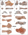
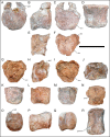
Fig 15. Middle and posterior caudal centra of Mnyamawamtuka moyowamkia.
A–D and E–F, middle caudal centra, G–J and K–N, middle–distal caudal centra, and O–R, posterior caudal centrum. A, G, K, O, anterior; B, E, H, L, P, right lateral; C, F, I, M, Q, posterior; D, J, R, ventral; and N, dorsal views. Anterior towards the top in D, J, N, and R. Abbreviations: dle, dorsolateral expansion; groove, ventral groove; ped, pedicle; vlm, ventrolateral margin. Scale bar equals 10 cm.
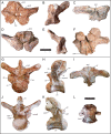
Fig 16. Caudal neural arches of Mnyamawamtuka moyowamkia.
A–D, First caudal neural arch, E–I, anterior caudal neural arch, J–N and O–S, middle caudal neural arches, and T–X, posterior caudal neural arch. A, E, J, O, T, anterior; B, F, K, P, U, left lateral; C, G, L, Q, V, posterior; D, H, M, R, W, right lateral; and I, N, S, X dorsal views. Anterior towards the top in D, I, N, S, X. Abbreviations: al, accessary lamina; cpol, centropostzygapophyseal lamina; ns, neural spine; po, postzygapophysis; posf, postspinal fossa; pr, prezygapophysis; prsl, prespinal lamina; spol, spinopostzygapophyseal laminal; sprl, spinoprezygapophyseal lamina. Scale bar equals 10 cm.
Fig 17. Comparison of caudal vertebrae of Mnyamawamtuka moyowamkia.
Mnyamawamtuka moyowamkia, A, Malawisaurus dixeyi MAL-197-8, B, and Lohuecotitan pandafilandi, C, in posterior view. Abbreviations: dle, dorsolateral expansion. Scale bar equals 10 cm.
Fig 18. Dorsal ribs of Mnyamawamtuka moyowamkia.
A–B, right anterior dorsal rib, and C–D, right posterior dorsal rib. A, C, anterior; and B, D, posterior views. Abbreviations: cap, capitulum; pf, pneumatic fossa; tub, tubercle; web, capitulotubercular webbing. Scale bar equals 10 cm.
Fig 19. Haemal arches of Mnyamawamtuka moyowamkia.
A–B, anterior haemal arch, and C–D, E–F, and G–H, middle haemal arches. A, C, E, G, anterior; and B, D, F, H, right lateral views. Scale bar equals 10 cm.
Fig 20. Pectoral elements of Mnyamawamtuka moyowamkia.
A–D, right scapula, and E, right sternal plate. A, lateral; B, medial; C, dorsal; D, ventral; and E, external/ventral views. Abbreviations: acro, acromion; ant, anterior; art, articular facet for coracoid; glen, glenoid fossa; lat, lateral; med, medial; pos, posterior; rid, dorsomedial ridge of the scapular blade. Scale bar equals 10 cm.
Fig 21. Forelimb elements of Mnyamawamtuka moyowamkia.
A–F, left humerus, and G–K, partial left ulna. A, J, anterior; B, lateral; C, I, posterior; D, H, medial; E, proximal; F, distal; G, anteromedial; and K, cross section views. Anterior towards the top of the page in E, F, and K. Abbreviations: alp, anterolateral process; amp, anteromedial process; dpc, deltopectoral crest; plb, posterolateral bulge; pp, posterior process; rc, radial condyle; scf, supracondylar fossa; uc, ulnar condyle. Scale bars equals 10 cm.
Fig 22. Metacarpals of Mnyamawamtuka moyowamkia.
A–F, left metacarpal I, and G–L, left metacarpal III. A, G, lateral; B, H, anterior; C, I, medial; D, J, posterior, E, K, proximal; and F, L, distal views. Anterior towards top of page in E, F, K, and L. Abbreviations: II, articular facets for metacarpal II; IV, articular facets for metacarpal IV. Scale bar equals 10 cm.
Fig 23. Pelvic elements of Mnyamawamtuka moyowamkia.
A–D, right pubis, and E–I, right ischium. A, G, lateral/external; B, F, anterior; C, E, medial/internal; D, H, posterior; and I, proximal views. Anterior towards top in I. Abbreviations: ace, acetabulum contribution; ilp, iliac peduncle; tub, tubercle. Scale bar equals 10cm.
Fig 24. Left femur of Mnyamawamtuka moyowamkia.
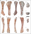
A, anterior; B, medial; C, posterior; D, lateral; E, distal views. Anterior towards top of page in E. Abbreviations: fibc, fibular condyle; ft, fourth trochanter; tibc, tibial condyle. Scale bar equals 10 cm.
Fig 25. Lower hind limb elements of Mnyamawamtuka moyowamkia.
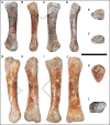
A–F, left tibia and G–L, left fibula. A, G, anterior; B, H, lateral; C, I, posterior; D, J, medial; E, K, proximal; and F, L, distal views. Anterior towards top of page in E, F, K, and L. Abbreviations: cc, cnemial crest; lt, lateral trochanter; ts, triangular scar area. Scale bar equals 10 cm.
Fig 26. Metatarsal elements of Mnyamawamtuka moyowamkia.
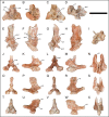
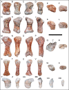
A–F, left metatarsal I; G–L, left metatarsal II; M–R, right metatarsal III; S–X, metatarsal IV; Y–DD. A, G, M, S, Y, dorsal; B, H, N, T, Z, lateral; C, I, O, U, AA, plantar; D, J, P, V, BB, medial; E, K, Q, W, CC, proximal; and F, L, R, X, DD, distal views. Anterior towards top of the page in E, F, K, L, Q, R, W, X, CC, and DD. Abbreviations: pdf, proximodorsal fossa; vr, ventral ridge. Scale bar equals 10 cm.
Fig 27. Pedal elements of Mnyamawamtuka moyowamkia.
A–F, left pedal phalanx, and G–L, and M–O, left pedal unguals. A, G, M, dorsal; B, H, medial; C, I, plantar; D, J, O, lateral; E, K, proximal; F, L, distal views. Anterior t owards top of page in E, F, K, and L. Abbreviations: tub, tubercle. Scale bar equals 5 cm.
Fig 28. Results of parsimony and uncalibrated Bayesian phylogenetic analyses.
Results of the majority-rule consensus tree of the parsimony phylogenetic analysis, A, and maximum clade credibility tree of the variable-rates Bayesian phylogenetic analysis, B. Numbers at the node represent frequency of recovered clades in A, and posterior probabilities in B.
Fig 29. Results of tip-dating phylogenetic analysis.
Maximum clade credibility tree for the variable-rates tip-dating Bayesian analysis. Numbers at the nodes represent posterior probabilities, blue bars at each node represent the 95% highest posterior density of each node age, and white bars at the tips represent the 95% highest posterior density of the sampled stratigraphic range of each terminal taxon. Abbreviations: MYA, millions of years ago.
Similar articles
- New Egyptian sauropod reveals Late Cretaceous dinosaur dispersal between Europe and Africa.
Sallam HM, Gorscak E, O'Connor PM, El-Dawoudi IA, El-Sayed S, Saber S, Kora MA, Sertich JJW, Seiffert ER, Lamanna MC. Sallam HM, et al. Nat Ecol Evol. 2018 Mar;2(3):445-451. doi: 10.1038/s41559-017-0455-5. Epub 2018 Jan 29. Nat Ecol Evol. 2018. PMID: 29379183 - A new titanosaurian sauropod from the Hekou Group (Lower Cretaceous) of the Lanzhou-Minhe Basin, Gansu Province, China.
Li LG, Li DQ, You HL, Dodson P. Li LG, et al. PLoS One. 2014 Jan 29;9(1):e85979. doi: 10.1371/journal.pone.0085979. eCollection 2014. PLoS One. 2014. PMID: 24489684 Free PMC article. - A Basal Lithostrotian Titanosaur (Dinosauria: Sauropoda) with a Complete Skull: Implications for the Evolution and Paleobiology of Titanosauria.
Martínez RD, Lamanna MC, Novas FE, Ridgely RC, Casal GA, Martínez JE, Vita JR, Witmer LM. Martínez RD, et al. PLoS One. 2016 Apr 26;11(4):e0151661. doi: 10.1371/journal.pone.0151661. eCollection 2016. PLoS One. 2016. PMID: 27115989 Free PMC article. - Biology of the sauropod dinosaurs: the evolution of gigantism.
Sander PM, Christian A, Clauss M, Fechner R, Gee CT, Griebeler EM, Gunga HC, Hummel J, Mallison H, Perry SF, Preuschoft H, Rauhut OW, Remes K, Tütken T, Wings O, Witzel U. Sander PM, et al. Biol Rev Camb Philos Soc. 2011 Feb;86(1):117-55. doi: 10.1111/j.1469-185X.2010.00137.x. Biol Rev Camb Philos Soc. 2011. PMID: 21251189 Free PMC article. Review. - Rethinking the nature of fibrolamellar bone: an integrative biological revision of sauropod plexiform bone formation.
Stein K, Prondvai E. Stein K, et al. Biol Rev Camb Philos Soc. 2014 Feb;89(1):24-47. doi: 10.1111/brv.12041. Epub 2013 May 6. Biol Rev Camb Philos Soc. 2014. PMID: 23647662 Review.
Cited by
- A nearly complete skull of the sauropod dinosaur Diamantinasaurus matildae from the Upper Cretaceous Winton Formation of Australia and implications for the early evolution of titanosaurs.
Poropat SF, Mannion PD, Rigby SL, Duncan RJ, Pentland AH, Bevitt JJ, Sloan T, Elliott DA. Poropat SF, et al. R Soc Open Sci. 2023 Apr 12;10(4):221618. doi: 10.1098/rsos.221618. eCollection 2023 Apr. R Soc Open Sci. 2023. PMID: 37063988 Free PMC article. - Deep reptilian evolutionary roots of a major avian respiratory adaptation.
Wang YY, Claessens LPAM, Sullivan C. Wang YY, et al. Commun Biol. 2023 Jan 17;6(1):3. doi: 10.1038/s42003-022-04301-z. Commun Biol. 2023. PMID: 36650231 Free PMC article. - New specimens of Baurutitan britoi and a taxonomic reassessment of the titanosaur dinosaur fauna (Sauropoda) from the Serra da Galga Formation (Late Cretaceous) of Brazil.
Silva Junior JCG, Martinelli AG, Marinho TS, da Silva JI, Langer MC. Silva Junior JCG, et al. PeerJ. 2022 Nov 15;10:e14333. doi: 10.7717/peerj.14333. eCollection 2022. PeerJ. 2022. PMID: 36405026 Free PMC article. - Sauropod dinosaur teeth from the lower Upper Cretaceous Winton Formation of Queensland, Australia and the global record of early titanosauriforms.
Poropat SF, Frauenfelder TG, Mannion PD, Rigby SL, Pentland AH, Sloan T, Elliott DA. Poropat SF, et al. R Soc Open Sci. 2022 Jul 13;9(7):220381. doi: 10.1098/rsos.220381. eCollection 2022 Jul. R Soc Open Sci. 2022. PMID: 35845848 Free PMC article. - First titanosaur dinosaur nesting site from the Late Cretaceous of Brazil.
Fiorelli LE, Martinelli AG, da Silva JI, Hechenleitner EM, Soares MVT, Silva Junior JCG, da Silva JC, Borges ÉMR, Ribeiro LCB, Marconato A, Basilici G, da Silva Marinho T. Fiorelli LE, et al. Sci Rep. 2022 Mar 24;12(1):5091. doi: 10.1038/s41598-022-09125-9. Sci Rep. 2022. PMID: 35332244 Free PMC article.
References
- Jacobs LL, Winkler DA, Gomani EM. Cretaceous dinosaurs of Africa: Examples from Cameroon and Malawi. Mem Queensl Mus. 1996;39: 595–610.
- Gomani EM. Sauropod dinosaurs from the Early Cretaceous of Malawi, Africa. Palaeontol Electronica 2005;8: 27A.
- O'Connor PM, Gottfried MD, Stevens NJ, Roberts EM, Ngasala S, Kapilima S, et al. A new vertebrate fauna from the Cretaceous Red Sandstone Group, Rukwa Rift Basin, Southwestern Tanzania. J Afr Earth Sci. 2006;44: 277–288.
Publication types
MeSH terms
Grants and funding
This research was supported by the U.S. National Science Foundation (NSF EAR_0617561, EAR_085421, EAR_1349825; P.M.O.), the National Geographic Society (Committee for Research and Exploration Grant) (P.M.O.), the Ohio University Heritage College of Osteopathic Medicine, Ohio University Office of Research and Sponsored Programs (P.M.O.), Ohio University African Studies Program, Ohio University Student Enhancement Award and Original Work Grant (E.G.), the Jurassic Foundation (E. G.), and the Paleontological Society (E.G.). The funders had no role in study design, data collection and analysis, decision to publish, or preparation of the manuscript.
LinkOut - more resources
Full Text Sources
Miscellaneous
