Stem cells from human exfoliated deciduous teeth ameliorate type II diabetic mellitus in Goto-Kakizaki rats - PubMed (original) (raw)
Stem cells from human exfoliated deciduous teeth ameliorate type II diabetic mellitus in Goto-Kakizaki rats
Nanquan Rao et al. Diabetol Metab Syndr. 2019.
Abstract
Background: By 2030, diabetes mellitus (DM) will be the 7th leading cause of death worldwide. Type 2 DM (T2DM) is the most common type of DM and is characterized by insulin resistance and defective β-cell secretory function. Stem cells from human exfoliated deciduous teeth (SHED) are favorable seed cells for mesenchymal stem cells (MSCs)-based therapy due to their higher proliferation rates and easier access to retrieval. Currently, no study has revealed the therapeutic efficiency of MSCs for T2DM in Goto-Kakizaki (GK) rats. Hence, we aimed to explore the effect of SHED on T2DM in GK rats.
Materials and methods: We investigated the effects of SHED on the progression of T2DM in GK rats. SHED and bone marrow mesenchymal stem cells (BMSCs) were injected via the tail vein. Body weight, fasting blood glucose and non-fasting blood glucose were measured before and after administration. At 8 weeks after injection, intraperitoneal insulin tolerance tests (IPITTs) and insulin release tests (IRTs) were performed. Additionally, hematoxylin-eosin (HE) staining, periodic acid-Schiff (PAS) staining and double-label immunofluorescence staining were used to explore the pathological changes in pancreatic islets and the liver. Immunohistochemistry (IHC) was employed to detect SHED engraftment in the liver. Additionally, real-time PCR and western blotting were used to explore glycogen synthesis, glycolysis and gluconeogenesis in the liver.
Results: At 8 weeks after SHED injection, T2DM was dramatically attenuated, including hyperglycemia, IPGTT and IRT. Additionally, histological analysis showed that SHED injection improved pancreatic islet and liver damage. Real-time PCR analysis showed that SHED significantly reversed the diabetic-induced increase of G-6-Pase, Pck1 and PK; and significantly reversed the diabetic-induced decrease of GSK3β, GLUT2, and PFKL. In addition, western blotting demonstrated that SHED significantly reversed the diabetic-induced increase of G-6-Pase and reversed the diabetic-induced decrease of GLUT2, GSK3β and PFKM.
Conclusion: Stem cells from human exfoliated deciduous teeth offers a potentially effective therapeutic modality for ameliorating T2DM, including hyperglycemia, insulin resistance, pancreatic islets and liver damage, and decreased glycogen synthesis, inhibited glycolysis and increased gluconeogenesis in the liver.
Keywords: Goto-Kakizaki (GK) rats; Insulin resistance; Stem cells from human exfoliated deciduous teeth (SHED); Type 2 diabetes mellitus.
Figures
Fig. 1
Characterization of SHED and BMSCs. a Characterization of SHED. SHED showed a fibroblast-like morphology. Multilineage differentiation potency including osteogenesis as identified by Alizarin Red staining; adipogenesis as identified by Oil Red O staining. b BMSCs showed a fibroblast-like morphology. c Flow cytometry of SHED. SHED expressed low levels of CD34 (0.4%) and CD45 (1.2%) and expressed high levels of CD73 (97.7%), CD90 (98.1%), CD105 (70.2%), and CD146 (63.5%)
Fig. 2
Effects of SHED on physical and biochemical parameters. a Body weight. b Fasting blood glucose. c Non-fasting blood glucose of rats in different groups over 8 weeks. d IPITTs, by injecting 2 g glucose/kg body weight. e HOMA-β of each group, HOMA-β (HBCI) = (20 * FINS [in units/L])/(FBG [in mmol/L] − 3.5). f IR index of each group, HOMA-IR index = (FBG [in mmol/L] * FINS [in units/L])/22.5. Values of a–f are mean ± SD. n = 8 rats per group. *P < 0.05 vs. normal group, *#P < 0.05 vs. both normal and PBS group
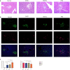
Fig. 3
Infusion of SHED promotes restoration of pancreatic islets in T2DM rats. a Pancreas histology was studied via HE staining, observed under light microscopy and focused on islet structures. b Pancreatic islets were characterized by immunofluorescence according to the presence and distribution of insulin-producing (green) and glucagon-producing (red) cells in four groups of rats at 8 weeks after infusion. Images were composite overlays of the individually stained nuclei, insulin and glucagon. SHED and BMSCs injection significantly improved abnormalities in pancreatic islets. c The proportion of the irregular islets and the proportion of β-cells in 4 group. ***P<0.001
Fig. 4
Effects of SHED on liver histology and SHED engraftment in the liver. a PAS staining showed the storage of glycogen in the liver. SHED and BMSC injection significantly improved glycogen storage in T2DM. b Immunohistochemical representative micrograph of liver tissue from the SHED group and BMSCs group. Red arrow indicated SHED and BMSCs engraftment
Fig. 5
Quantitative real-time PCR analysis and western blotting of the liver. a Effect of SHED on G-6-Pase, Pck1, GSK3b, GLUT2, PFKL and PK mRNA expression by real-time PCR. *P < 0.05, **P < 0.01, ***P < 0.001, ****P < 0.0001. b Effect of MSCs on GSK3β, G-6-Pase, GLUT2 and PFKM expression by western blotting
Similar articles
- Stem Cells from Human Exfoliated Deciduous Teeth Ameliorate Diabetic Nephropathy In Vivo and In Vitro by Inhibiting Advanced Glycation End Product-Activated Epithelial-Mesenchymal Transition.
Rao N, Wang X, Xie J, Li J, Zhai Y, Li X, Fang T, Wang Y, Zhao Y, Ge L. Rao N, et al. Stem Cells Int. 2019 Dec 1;2019:2751475. doi: 10.1155/2019/2751475. eCollection 2019. Stem Cells Int. 2019. PMID: 31871464 Free PMC article. - Effects and mechanism of stem cells from human exfoliated deciduous teeth combined with hyperbaric oxygen therapy in type 2 diabetic rats.
Xu Y, Chen J, Zhou H, Wang J, Song J, Xie J, Guo Q, Wang C, Huang Q. Xu Y, et al. Clinics (Sao Paulo). 2020 Jun 3;75:e1656. doi: 10.6061/clinics/2020/e1656. eCollection 2020. Clinics (Sao Paulo). 2020. PMID: 32520222 Free PMC article. - Enhanced Glucose Tolerance and Pancreatic Beta Cell Function by Low Dose Aspirin in Hyperglycemic Insulin-Resistant Type 2 Diabetic Goto-Kakizaki (GK) Rats.
Amiri L, John A, Shafarin J, Adeghate E, Jayaprakash P, Yasin J, Howarth FC, Raza H. Amiri L, et al. Cell Physiol Biochem. 2015;36(5):1939-50. doi: 10.1159/000430162. Epub 2015 Jul 17. Cell Physiol Biochem. 2015. PMID: 26202354 - Therapeutic effects of stem cells from human exfoliated deciduous teeth on diabetic peripheral neuropathy.
Xie J, Rao N, Zhai Y, Li J, Zhao Y, Ge L, Wang Y. Xie J, et al. Diabetol Metab Syndr. 2019 May 17;11:38. doi: 10.1186/s13098-019-0433-y. eCollection 2019. Diabetol Metab Syndr. 2019. PMID: 31131042 Free PMC article. - Type 2 diabetes mitigation in the diabetic Goto-Kakizaki rat by elevated bile acids following a common-bile-duct surgery.
Gao F, Zhang X, Zhou L, Zhou S, Zheng Y, Yu J, Fan W, Zhu Y, Han X. Gao F, et al. Metabolism. 2016 Feb;65(2):78-88. doi: 10.1016/j.metabol.2015.09.014. Epub 2015 Sep 26. Metabolism. 2016. PMID: 26773931
Cited by
- Human mesenchymal stem/stromal cell based-therapy in diabetes mellitus: experimental and clinical perspectives.
Zeinhom A, Fadallah SA, Mahmoud M. Zeinhom A, et al. Stem Cell Res Ther. 2024 Oct 29;15(1):384. doi: 10.1186/s13287-024-03974-z. Stem Cell Res Ther. 2024. PMID: 39468609 Free PMC article. Review. - Bone marrow mesenchymal stem cells improve thymus and spleen function of aging rats through affecting P21/PCNA and suppressing oxidative stress.
Wang Z, Lin Y, Jin S, Wei T, Zheng Z, Chen W. Wang Z, et al. Aging (Albany NY). 2020 Jun 19;12(12):11386-11397. doi: 10.18632/aging.103186. Epub 2020 Jun 19. Aging (Albany NY). 2020. PMID: 32561691 Free PMC article. - Therapeutic potential of stem cells from human exfoliated deciduous teeth infusion into patients with type 2 diabetes depends on basal lipid levels and islet function.
Li W, Jiao X, Song J, Sui B, Guo Z, Zhao Y, Li J, Shi S, Huang Q. Li W, et al. Stem Cells Transl Med. 2021 Jul;10(7):956-967. doi: 10.1002/sctm.20-0303. Epub 2021 Mar 4. Stem Cells Transl Med. 2021. PMID: 33660433 Free PMC article. - Sinking Our Teeth in Getting Dental Stem Cells to Clinics for Bone Regeneration.
Shoushrah SH, Transfeld JL, Tonk CH, Büchner D, Witzleben S, Sieber MA, Schulze M, Tobiasch E. Shoushrah SH, et al. Int J Mol Sci. 2021 Jun 15;22(12):6387. doi: 10.3390/ijms22126387. Int J Mol Sci. 2021. PMID: 34203719 Free PMC article. Review.
References
- World Health Organization. Global report on diabetes; 2006. http://www.who.int/diabetes/global-report/en/.
- Association AD. Diagnosis and classification of diabetes mellitus. Diabetes Care. 2012;31(Suppl. 1):S55–S60. - PubMed
LinkOut - more resources
Full Text Sources
Miscellaneous


