Association between endoscopic findings of eosinophilic esophagitis and responsiveness to proton pump inhibitors - PubMed (original) (raw)
Association between endoscopic findings of eosinophilic esophagitis and responsiveness to proton pump inhibitors
Akinari Sawada et al. Endosc Int Open. 2019 Apr.
Abstract
Background and study aims Endoscopic findings of esophageal eosinophilia sometimes localize to small areas of the esophagus. A previous study suggested that pathogenesis of localized-type eosinophilic esophagitis (LEoE) was associated with acid reflux. However, LEoE treatment outcomes have not been studied. We aimed to analyze the clinical and histologic significance of LEoE in comparison with diffuse-type eosinophilic esophagitis (DEoE). Patients and methods This study included 106 patients with esophageal eosinophilia. Esophageal eosinophilia was defined as a condition where the maximum number of intraepithelial eosinophils was ≥ 15 per high-power field. LEoE was defined as an endoscopic lesion confined to one-third of the esophagus: upper, middle, or lower. Esophageal eosinophilia encompassing more than two-thirds of the esophagus was defined as DEoE. We retrospectively compared LEoE and DEoE in terms of clinical characteristics, histologic findings, and proportion of proton pump inhibitor (PPI) responders. Results Of 106 patients, 12 were classified as having LEoE and 94 were classified as having DEoE. The proportion of asymptomatic patients was significantly higher in the LEoE group than the DEoE group (42 % vs 7 %, P < 0.01). In the LEoE group, 10 patients (84 %) had endoscopic lesions in the lower esophagus. The maximum number of eosinophils did not differ between the groups (54 [24 - 71] for LEoE, 40 [20 - 75] for DEoE, P = 0.65). The prevalence of PPI responders was significantly higher in the LEoE group than the DEoE group (100 % vs 63 %, P = 0.01). Conclusion LEoE can be a sign of good responsiveness to PPI therapy.
Conflict of interest statement
Competing interests None
Figures
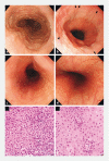
Fig. 1
Endoscopic and histologic findings of eosinophilic esophagitis.aEndoscopic findings of DEoE. Rings, white exudates, and linear furrows were observed in the etire esophagus.bCase 1 of LEoE. White turbidity was observed along the entire circumference just above the EGJ. In addition, linear furrows (black arrows) and white exudates (arrowhead) were distributed on white turbidity.cCase 2 of LEoE. White exudates and subtle rings were located along only half of the circumference of the upper esophagus.dNormal esophageal mucosa was found from the EGJ to middle esophagus.e, fHistologic findings in Case 2.eEsophageal biopsy samples taken from the mucosa with endoscopic findings around imagecshowed > 15 eosinophils/hpf, other inflammatory cells, and dilated intracellular space in the esophageal epithelium.fIn contrast, a sample taken from the mucosa without endoscopic findings around imagedshowed no intraepithelial eosinophils. LEoE, localized-type eosinophilic esophagitis; DEoE, diffuse-type eosinophilic esophagitis; EGJ, esophagogastric junction.
Fig. 2
Number of eosinophils in each esophageal biopsy sample with or without endoscopic findings in localized-type eosinophilic esophagitis. *P < 0.01.
Fig. 3
Response rate to PPI therapy of each type of eosinophilic esophagitis. LEoE, localized-type eosinophilic esophagitis; DEoE, diffuse-type eosinophilic esophagitis. *P < 0.05.
Similar articles
- Similarities and differences among eosinophilic esophagitis, proton-pump inhibitor-responsive esophageal eosinophilia, and reflux esophagitis: comparisons of clinical, endoscopic, and histopathological findings in Japanese patients.
Jiao D, Ishimura N, Maruyama R, Ishikawa N, Nagase M, Oshima N, Aimi M, Okimoto E, Mikami H, Izumi D, Okada M, Ishihara S, Kinoshita Y. Jiao D, et al. J Gastroenterol. 2017 Feb;52(2):203-210. doi: 10.1007/s00535-016-1213-1. Epub 2016 Apr 23. J Gastroenterol. 2017. PMID: 27108416 - Optimal Biopsy Protocol to Evaluate Histological Effectiveness of Proton Pump Inhibitor Therapy in Patients with Eosinophilic Esophagitis.
Fujiwara Y, Hashimoto A, Uemura R, Sawada A, Otani K, Tanaka F, Yamagami H, Tanigawa T, Watanabe T, Kabata D, Kuwae Y, Shintani A, Ohsawa M. Fujiwara Y, et al. Digestion. 2019;100(1):64-71. doi: 10.1159/000494253. Epub 2018 Nov 8. Digestion. 2019. PMID: 30408792 - [Proton Pump Inhibitor-responsive Esophageal Eosinophilia: An Overview of Cases from One University Hospital Center].
Ahn B, Lee DH, Lee CM, Hwang JJ, Yoon H, Shin CM, Park YS, Kim N. Ahn B, et al. Korean J Gastroenterol. 2016 Apr 25;67(4):178-82. doi: 10.4166/kjg.2016.67.4.178. Korean J Gastroenterol. 2016. PMID: 27112243 Korean. - Eosinophilic esophagitis: Update in diagnosis and management. Position paper by the Italian Society of Gastroenterology and Gastrointestinal Endoscopy (SIGE).
de Bortoli N, Penagini R, Savarino E, Marchi S. de Bortoli N, et al. Dig Liver Dis. 2017 Mar;49(3):254-260. doi: 10.1016/j.dld.2016.11.012. Epub 2016 Dec 2. Dig Liver Dis. 2017. PMID: 27979389 Review. - The role of proton pump inhibitor therapy in the management of eosinophilic esophagitis.
Molina-Infante J, Prados-Manzano R, Gonzalez-Cordero PL. Molina-Infante J, et al. Expert Rev Clin Immunol. 2016 Sep;12(9):945-52. doi: 10.1080/1744666X.2016.1178574. Epub 2016 Apr 28. Expert Rev Clin Immunol. 2016. PMID: 27097787 Review.
Cited by
- Long-term course of untreated asymptomatic esophageal eosinophilia and minimally symptomatic eosinophilic esophagitis.
Abe Y, Kikuchi R, Sasaki Y, Mizumoto N, Yagi M, Onozato Y, Watabe T, Goto H, Miura T, Sato R, Ito M, Tsuchiya H, Ueno Y. Abe Y, et al. Endosc Int Open. 2024 Apr 15;12(4):E545-E553. doi: 10.1055/a-2280-8277. eCollection 2024 Apr. Endosc Int Open. 2024. PMID: 38628394 Free PMC article. - Endoscopic Diagnosis of Eosinophilic Esophagitis: Basics and Recent Advances.
Abe Y, Sasaki Y, Yagi M, Mizumoto N, Onozato Y, Umehara M, Ueno Y. Abe Y, et al. Diagnostics (Basel). 2022 Dec 16;12(12):3202. doi: 10.3390/diagnostics12123202. Diagnostics (Basel). 2022. PMID: 36553209 Free PMC article. Review. - Extent of eosinophilic esophagitis predicts response to treatment.
Ghoz H, Stancampiano FF, Valery JR, Nordelo K, Malviya B, Lacy BE, Francis D, DeVault K, Bouras E, Krishna M, Palmer WC. Ghoz H, et al. Endosc Int Open. 2021 Aug;9(8):E1234-E1242. doi: 10.1055/a-1492-2650. Epub 2021 Jul 16. Endosc Int Open. 2021. PMID: 34447870 Free PMC article. - Clinical Features of Esophageal Eosinophilia According to Endoscopic Phenotypes.
Kon T, Abe Y, Sasaki Y, Kikuchi R, Uchiyama S, Kusaka G, Yaoita T, Yagi M, Shoji M, Onozato Y, Mizumoto N, Ueno Y. Kon T, et al. Intern Med. 2020 Dec 1;59(23):2971-2979. doi: 10.2169/internalmedicine.4447-20. Epub 2020 Aug 4. Intern Med. 2020. PMID: 32759578 Free PMC article. - Symptom-based diagnostic approach for eosinophilic esophagitis.
Fujiwara Y. Fujiwara Y. J Gastroenterol. 2020 Sep;55(9):833-845. doi: 10.1007/s00535-020-01701-y. Epub 2020 Jul 27. J Gastroenterol. 2020. PMID: 32720208 Free PMC article. Review.
References
- Arias Á, Pérez-Martínez I, Tenías J M et al.Systematic review with meta-analysis: the incidence and prevalence of eosinophilic oesophagitis in children and adults in population-based studies. Aliment Pharmacol Ther. 2016;43:3–15. - PubMed
- Lucendo A J, Arias Á, Molina-Infante J. Efficacy of proton pump inhibitor drugs for inducing clinical and histologic remission in patients with symptomatic esophageal eosinophilia: a systematic review and meta-analysis. Clin Gastroenterol Hepatol. 2016;14:13–2.2E12. - PubMed
LinkOut - more resources
Full Text Sources


