RNA m6A methylation regulates the epithelial mesenchymal transition of cancer cells and translation of Snail - PubMed (original) (raw)
doi: 10.1038/s41467-019-09865-9.
Guoshi Chai 2, Yingmin Wu 1, Jiexin Li 1, Feng Chen 1, Jianzhao Liu 3 4, Guanzheng Luo 2 3, Jordi Tauler 3, Jun Du 1, Shuibin Lin 5, Chuan He 3, Hongsheng Wang 6
Affiliations
- PMID: 31061416
- PMCID: PMC6502834
- DOI: 10.1038/s41467-019-09865-9
RNA m6A methylation regulates the epithelial mesenchymal transition of cancer cells and translation of Snail
Xinyao Lin et al. Nat Commun. 2019.
Retraction in
- Retraction Note: RNA m6A methylation regulates the epithelial mesenchymal transition of cancer cells and translation of Snail.
Lin X, Chai G, Wu Y, Li J, Chen F, Liu J, Luo G, Tauler J, Du J, Lin S, He C, Wang H. Lin X, et al. Nat Commun. 2023 Nov 16;14(1):7424. doi: 10.1038/s41467-023-43307-x. Nat Commun. 2023. PMID: 37973993 Free PMC article. No abstract available.
Abstract
N6-Methyladenosine (m6A) modification has been implicated in the progression of several cancers. We reveal that during epithelial-mesenchymal transition (EMT), one important step for cancer cell metastasis, m6A modification of mRNAs increases in cancer cells. Deletion of methyltransferase-like 3 (METTL3) down-regulates m6A, impairs the migration, invasion and EMT of cancer cells both in vitro and in vivo. m6A-sequencing and functional studies confirm that Snail, a key transcription factor of EMT, is involved in m6A-regulated EMT. m6A in Snail CDS, but not 3'UTR, triggers polysome-mediated translation of Snail mRNA in cancer cells. Loss and gain functional studies confirm that YTHDF1 mediates m6A-increased translation of Snail mRNA. Moreover, the upregulation of METTL3 and YTHDF1 act as adverse prognosis factors for overall survival (OS) rate of liver cancer patients. Our study highlights the critical roles of m6A on regulation of EMT in cancer cells and translation of Snail during this process.
Conflict of interest statement
The authors declare no conflict of interest.
Figures
Fig. 1
EMT in cancer cells is regulated by m6A levels of mRNAs. a HeLa and HepG2 cells were treated with or without 10 ng/ml TGF-β for 3 days, the m6A/A ratio of the total mRNA were determined by LC–MS/MS. b Wound healing of wild-type (control) or Mettl3 Mut/− cells was recorded (left) and quantitatively analyzed (right). c Wild-type or Mettl3 Mut/− cells were allowed to invade for 24 h and tested by CytoSelect™ 24-well Cell Invasion assay kits (8 µm, colorimetric format); d, e mRNA (d) and protein (e) expressions of MMP2, FN, and E-Cad in wild-type and Mettl3 Mut/− HeLa cells were measured by qRT-PCR and western blot analysis, respectively. f HeLa cells were transfected with pcDNA/ALKBH5 or a vector control for 48 h, protein expression was determined by western blot analysis (left) and quantitatively analyzed (right). g Wild-type or Mettl3 Mut/− cells were treated with or without 10 ng/ml TGF-β for 3 days, protein expression was determined by western blot analysis (left) and quantitatively analyzed (right). h The expression of METTL3 in liver cancer and its matched adjacent normal tissues of 50 patients from TCGA database. i Correlation between METTL3 and CDH1 in liver cancer patients (n = 364) from TCGA database. j HeLa cells were pretreated with or without Smad2/3 inhibitor SB431542 (10 μM) and then further treated with 10 ng/ml TGF-β for 3 days, the m6A/A ratio of the total mRNA were determined by LC–MS/MS. Data are presented as means ± SD from three independent experiments. *p < 0.05, **p < 0.01, NS, no significant, by Student’s t test. Red bar = 200 μm
Fig. 2
Variations of m6A-regulated genes in cells undergoing EMT. a Predominant consensus motif GGAC was detected in both the control and EMT cells in m6A-seq. b Density distribution of m6A peaks across mRNA transcripts. Regions of the 5′ untranslated region (5′UTR), coding region (CDS), and 3′ untranslated region (3′UTR) were split into 100 segments, then percentages of m6A peaks that fall within each segment were determined. c Proportion of m6A peak distribution in the 5′UTR, start codon, CDS, stop codon or 3′UTR region across the entire set of mRNA transcripts. d A cluster profiler identified the enriched gene ontology processes of 128 genes, which showed 1.5-fold m6A expression upregulation in EMT cells compared with control cells
Fig. 3
Snail is involved in m6A-regulated EMT in cancer cells. a Overlapping of 2.0-fold m6A expression changes in EMT cells and EMT-related functional genes. b m6A peaks were enriched in CDS and 3′UTRs of SNAI1 genes from m6A RIP-seq data. Squares marked increases of m6A peaks in cancer cells undergoing EMT; c m6A RIP-qPCR analysis of SNAI1 mRNA in the control and EMT undergoing HeLa cells. d Protein expression of Snail in Mettl3 Mut/− or AKLBH5 transfected (24 h) HeLa cells and the control. e The wound healing of wild-type or Mettl3 Mut/− HeLa cells transfected with or without pcDNA/Snail for 48 h were recorded (left) and quantitatively analyzed (right). f Wild-type or Mettl3 Mut/− HeLa cells were transfected with or without pcDNA/Snail for 48 h, expression of Snail, FN and E-Cad were measured by western blot analysis. Data are presented as means ± SD from three independent experiments. *p < 0.05, NS, no significant, by Student’s t test. Red bar = 200μm
Fig. 4
m6A triggers translation of Snail mRNA in cancer cells. a Precursor and mature mRNA of SNAI1 in wild-type (WT) and Mettl3 Mut/− cells. b The relative levels of the nuclear versus cytoplasmic SNAI1 mRNA in wild-type (WT) or Mettl3 Mut/− cells. c, d Wild-type (WT) or Mettl3 Mut/− cells were pretreated with Act-D for 90 min, then precursor (c) or mature (d) SNAI1 mRNA were analyzed at indicated times. e Wild-type (WT) or Mettl3 Mut/− cells were treated with CHX for the indicated times, and protein expression of Snail was analyzed by western blot analysis (left) and quantitatively analyzed (right); f Mettl3 Mut/− cells were pretreated with CHX or MG-132 for 6 h and then further treated with or without 10 ng/ml TGF-β for 48 h, the expression of Snail was detected by western blot analysis (left) and quantitatively analyzed (right). g Wild-type (WT) or Mettl3 Mut/− cells were transfected with pmirGLO-Snail reporter for 24 h, and the translation efficiency of Snail is defined as the quotient of reporter protein production (F-luc/R-luc) divided by mRNA abundanceh Polysome profiling of wild-type (WT) or Mettl3 Mut/− HeLa cells were analyzed. i Analysis of Snail mRNA in non-ribosome portion (< 40S), 40S, 60S, 80S, and polysome for the Mettl3 Mut/− cells compared to control cells. j Analysis of Snail mRNA in non-ribosome portion (< 40S), 40S, 60S, 80S, and polysome for EMT undergoing HeLa cells compared with control cells. Data are presented as means ± SD from three independent experiments. *p < 0.05, NS, no significant, by Student’s t test
Fig. 5
m6A methylated CDS regulates translation of Snail. a Schematic representation of positions of m6A motifs within Snail mRNA. b Schematic representation of mutated (GG
A
C to GG
C
C) 3’UTR of pmirGLO vector to investigate the m6A roles on Snail expression. c pmirGLO-3′UTR or pmirGLO-3′UTR-Mut1/2 reporter was transfected into wild-type or Mettl3 Mut/− HeLa cells for 24 h. d pcDNA-Snail-CDS-WT, pcDNA-Snail-mut1/2, or pcDNA/ZEB1 were transfected into HeLa cells for 24 h. Protein expression was measured by western blot analysis (left) and quantitatively analyzed (right). e Schematic representation of mutation in CDS to investigate the m6A roles on Snail expression. f After transfected with pcDNA-Snail-CDS-WT or pcDNA-Snail-mut1/2, HeLa cells were further treated with or without 10 ng/ml TGF-β for 48 h. The expression of Snail was measured by western blot analysis (left) and quantitatively analyzed (right). g YTHDF1 RIP-qPCR analysis of SNAI1 mRNA in wild-type or Mettl3 Mut/− HeLa cells. h Binding of YTHDF1 with the CDS or 3’UTR in wild-type or Mettl3 Mut/− cells were analyzed by YTHDF1 RIP-qPCR using fragmented RNA. i After pre-transfected with siNC or si-YTHDF1 for 12 h, HeLa cells were further treated with or without 10 ng/ml TGF-β for 48 h. Protein expression was measured by western blot analysis (left) and quantitatively analyzed (right). j eEF-1 and eEF-2 RIP-qPCR analysis of SNAI1 mRNA in wild-type or Mettl3 Mut/− HeLa cells. k Binding between YTHDF1 with eEF-1 or eEF-2 in wild-type or Mettl3 Mut/− HeLa cells was checked by immunoprecipitation (left) and quantitatively analyzed (right). Data are presented as means ± SD from three independent experiments. *p < 0.05, NS, no significant, by Student’s t test
Fig. 6
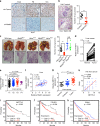
m6A methylation regulates in vivo cancer progression. a IHC (Snail, FN, and Vim)-stained paraffin-embedded sections obtained from sh-control and sh-Mettl3 Huh7 xenografts when the tumor volumes were about 100 mm3 for each group. b The sh-control and sh-Mettl3 Huh7 cells were injected into the nude mice by tail vein injection. Representative images of metastatic lung tumors and the H&E staining results were shown (left), and the number of lung tumors was quantitatively analyzed (right). c HeLa WT, Mettl3 Mut/− HeLa, HeLa_Snail_, and Mettl3 Mut/− HeLa_Snail_ stable cells were injected into the nude mice by tail vein injection. Representative images of metastatic lung tumors and the H&E staining results were shown (left), and the number of lung tumors was quantitatively analyzed (right). d Expression of YTHDF1 in paired human liver (n = 26) cancer tissues and adjacent normal mucosa tissues from TCGA database. e YTHDF1 expression in liver cancers of T1 (n = 153), T2 (n = 77), T3 (n = 65), and T4 (n = 13) stages from TCGA database. f Correlation between YTHDF1 and SNAI1 in liver cancer patients (n = 80) from TCGA database. g METTL3 expression in HCC tumor tissues (n = 35), liver cirrhosis tissues (n = 13), and normal liver tissues (n = 10) from Oncomine database (Wurmbach liver cancers). h Correlation between Mettl3 and Snail protein in liver cancer patients (100) by use of tissue microarray (CS03–01–008, Alenabio, Xian, Shanxi, China). i, j, k Kaplan–Meier survival curves of OS based on METTL3 (i), YTHDF1 (j), or SNAI1 (k) mRNA expression in HCC patients. The log-rank test was used to compare differences between two groups. Data are presented as means ± SD from three independent experiments. *p < 0.05, **p < 0.01, NS, no significant, by one-way ANOVA with Bonferroni test. Red bar = 200 μm
Similar articles
- N6-Methyladenosine Regulates the Expression and Secretion of TGFβ1 to Affect the Epithelial-Mesenchymal Transition of Cancer Cells.
Li J, Chen F, Peng Y, Lv Z, Lin X, Chen Z, Wang H. Li J, et al. Cells. 2020 Jan 25;9(2):296. doi: 10.3390/cells9020296. Cells. 2020. PMID: 31991845 Free PMC article. - Mouse double minute 2 (MDM2) upregulates Snail expression and induces epithelial-to-mesenchymal transition in breast cancer cells in vitro and in vivo.
Lu X, Yan C, Huang Y, Shi D, Fu Z, Qiu J, Yin Y. Lu X, et al. Oncotarget. 2016 Jun 14;7(24):37177-37191. doi: 10.18632/oncotarget.9287. Oncotarget. 2016. PMID: 27184007 Free PMC article. - Mechanism of methyltransferase like 3 in epithelial-mesenchymal transition process, invasion, and metastasis in esophageal cancer.
Liang X, Zhang Z, Wang L, Zhang S, Ren L, Li S, Xu J, Lv S. Liang X, et al. Bioengineered. 2021 Dec;12(2):10023-10036. doi: 10.1080/21655979.2021.1994721. Bioengineered. 2021. PMID: 34666602 Free PMC article. - N6-methyladenosine RNA modification in stomach carcinoma: Novel insights into mechanisms and implications for diagnosis and treatment.
Lu Z, Lyu Z, Dong P, Liu Y, Huang L. Lu Z, et al. Biochim Biophys Acta Mol Basis Dis. 2025 Jun;1871(5):167793. doi: 10.1016/j.bbadis.2025.167793. Epub 2025 Mar 14. Biochim Biophys Acta Mol Basis Dis. 2025. PMID: 40088577 Review. - [Research progress of m6A methylation modification in renal cell carcinoma].
Zhang Y, Cao ZF, Zhang YS. Zhang Y, et al. Zhonghua Bing Li Xue Za Zhi. 2024 Aug 8;53(8):876-882. doi: 10.3760/cma.j.cn112151-20231215-00417. Zhonghua Bing Li Xue Za Zhi. 2024. PMID: 39103278 Review. Chinese.
Cited by
- Trends and frontiers of RNA methylation in cancer over the past 10 years: a bibliometric and visual analysis.
Liu BN, Gao XL, Piao Y. Liu BN, et al. Front Genet. 2024 Oct 14;15:1461386. doi: 10.3389/fgene.2024.1461386. eCollection 2024. Front Genet. 2024. PMID: 39473440 Free PMC article. - RNA m6A modification in liver biology and its implication in hepatic diseases and carcinogenesis.
Ma W, Wu T. Ma W, et al. Am J Physiol Cell Physiol. 2022 Oct 1;323(4):C1190-C1205. doi: 10.1152/ajpcell.00214.2022. Epub 2022 Aug 29. Am J Physiol Cell Physiol. 2022. PMID: 36036444 Free PMC article. Review. - The Role of RNA Methyltransferase METTL3 in Hepatocellular Carcinoma: Results and Perspectives.
Pan F, Lin XR, Hao LP, Chu XY, Wan HJ, Wang R. Pan F, et al. Front Cell Dev Biol. 2021 May 11;9:674919. doi: 10.3389/fcell.2021.674919. eCollection 2021. Front Cell Dev Biol. 2021. PMID: 34046411 Free PMC article. Review. - Transcriptome and N6-Methyladenosine RNA Methylome Analyses in Aortic Dissection and Normal Human Aorta.
Zhou X, Chen Z, Zhou J, Liu Y, Fan R, Sun T. Zhou X, et al. Front Cardiovasc Med. 2021 May 28;8:627380. doi: 10.3389/fcvm.2021.627380. eCollection 2021. Front Cardiovasc Med. 2021. PMID: 34124185 Free PMC article. - So close, no matter how far: multiple paths connecting transcription to mRNA translation in eukaryotes.
Slobodin B, Dikstein R. Slobodin B, et al. EMBO Rep. 2020 Sep 3;21(9):e50799. doi: 10.15252/embr.202050799. Epub 2020 Aug 16. EMBO Rep. 2020. PMID: 32803873 Free PMC article. Review.
References
- Perry JK, Kelley DE. Existence of methylated messenger RNA in mouse L cells. Cell. 1974;1:37–42. doi: 10.1016/0092-8674(74)90153-6. - DOI
Publication types
MeSH terms
Substances
LinkOut - more resources
Full Text Sources
Other Literature Sources
Medical
Molecular Biology Databases
Research Materials


