What are the most accurate categories for mammal tarsus arrangement? A review with attention to South American Notoungulata and Litopterna - PubMed (original) (raw)
Review
. 2019 Dec;235(6):1024-1035.
doi: 10.1111/joa.13065. Epub 2019 Aug 2.
Affiliations
- PMID: 31373392
- PMCID: PMC6875937
- DOI: 10.1111/joa.13065
Review
What are the most accurate categories for mammal tarsus arrangement? A review with attention to South American Notoungulata and Litopterna
Malena Lorente. J Anat. 2019 Dec.
Abstract
The arrangement of the tarsus has been used to differentiate afrotherian and laurasiatherian ungulates for more than a century, and it is often present in morphological matrices that include appendicular features. Traditionally, it has two states: (i) an alternating tarsus, where proximal elements are interlocked with central and distal elements positioned like the bricks of a wall; and (ii) a serial tarsus, where elements are not interlocked. Over the years, these states became synonymous with the presence or absence of an astragalocuboid contact. Within the South American order Notoungulata, a third disposition was recognized: the reversed alternating tarsus, associated with a calcaneonavicular contact. This state was considered to be a synapomorphy of 'advanced' Toxodontia families (Notohippidae, Leontiniidae and Toxodontidae), but a further inspection of its distribution shows that it occurs throughout Mammalia. Additionally, it overlaps the serial tarsus condition as originally defined, and it probably has no functional or phylogenetic significance. Calcaneonavicular and astragalocuboid contacts are non-exclusive, and their presence within a species, genus or family is not constant. Serial and alternating imply movements of the articulations of the mid-tarsus in the transverse axis, while reverse alternating refers to a small calcaneonavicular contact that sometimes occurs in a serial condition or to a significant displacement of the tarsal articulations in a different (proximodistal) axis. The proximodistal arrangement of the joints could be functionally significant. Two new states are observed and defined: (i) 'flipped serial', present in Macropodidae, in which the calcaneocuboid articulation is medially displaced and significantly larger than the astragalonavicular contact, but the relationships between proximal and central elements are one to one; and (ii) 'distal cuboid', an extreme proximodistal displacement of the astragalonavicular joint. Serial and alternating, as originally defined (i.e. without any reference to which bone contacts which), seem to be the best states for classifying tarsal arrangement though as the disposition of distal or central bones in relationship to proximal bones.
Keywords: Mammalia; Meridiungulata; morphology; phylogeny; tarsus.
© 2019 Anatomical Society.
Figures
Figure 1
Outline of serial, alternating tarsal and previously proposed reverse alternating tarsal arrangements.
Figure 2
(A) Plantar view of right astragalus of Tremarctos ornatus (
MLP
1.I.03.62); shading in the surface of contact of cuboid bone. (B) Plantar view of left astragalus of Notostylops sp. (
LIEB
‐
PV
4016); shading in the space between navicular and sustentacular facets. (C) Plantar view of left astragalus of: (1) a juvenile of perissodactyl Tapirus terrestris (
MLP
1); (2) an adult Tapirus terretris (
MLP
1070). In yellow shading, ectal facet; in green shading, sustentacular facet; in pink shading, anterior astragalocalcaneal facet. Observe the disconnection between sustentacular and anterior astragalocalcaneal facet in the adult. (D) Left astragalus of Lagostomus maximus (
MLP
1683). Plantar view, slightly oblique to better observe the facets. Facets as previous image; in soft dark blue shading, the facet for the sesamoid. Scale bar: 10 mm.
Figure 3
Proximal view of the left navicular of proterotheriid Eoauchenia (
MLP
48‐
XII
‐16‐1). In light blue shading, sesamoid facet; in green shading, calcaneal facet; in red shading, astragalar facet. Scale bar: 10 mm.
Figure 4
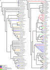
Presence of facets mapped over: (A) strict consensus cladogram from the analysis of Billet (2011, fig. 9); and (B) Bayesian consensus tree of
COL
1 protein sequence data, with chicken (Gallus) as outgroup from the analysis of Welker et al. (2015).
Figure 5
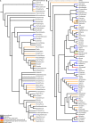
Displacement of joints in the proximodistal axis mapped over: (A) strict consensus cladogram from the analysis of Billet (2011, fig. 9); and (B) Bayesian consensus tree of
COL
1 protein sequence data, with chicken (Gallus) as outgroup from the analysis of Welker et al. (2015).
Figure 6
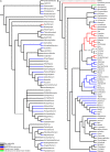
Displacement of joints in the transversal axis mapped over: (A) strict consensus cladogram from the analysis of Billet (2011, fig. 9); and (B) Bayesian consensus tree of
COL
1 protein sequence data, with chicken (Gallus) as outgroup from the analysis of Welker et al. (2015).
Figure 7
(A) Dorsal view of right tarsus of Eutypotherium lehmannnistchei (
MLP
12‐1701). (B) Dorsal view of right tarsus of Theosodon sp. (
MLP
700). In blue shading, astragalus; in green shading, navicular; in red shading, calcaneus; in yellow shading, cuboid. Scale bar: 10 mm.
Figure 8
(A) Dorsal view of right tarsus of Macropodidae indet. (
MLP
951). (B) Dorsal view of right tarsus of Loxodonta africana (
MLP
1123; specimen in exposition). In blue shading, astragalus; in green shading, navicular; in red shading, calcaneus; in orange shading, cuboid.
Similar articles
- Taxeopody in the carpus and tarsus of Oligocene Pliohyracidae (Mammalia: Hyracoidea) and the phyletic position of hyraxes.
Rasmussen DT, Gagnon M, Simons EL. Rasmussen DT, et al. Proc Natl Acad Sci U S A. 1990 Jun;87(12):4688-91. doi: 10.1073/pnas.87.12.4688. Proc Natl Acad Sci U S A. 1990. PMID: 2352942 Free PMC article. - [Anatomy and kinematics of the human ankle joint].
Pretterklieber ML. Pretterklieber ML. Radiologe. 1999 Jan;39(1):1-7. doi: 10.1007/s001170050469. Radiologe. 1999. PMID: 10065468 German. - Morphology and ontogeny of carpus and tarsus in stereospondylomorph temnospondyls.
Witzmann F, Fröbisch N. Witzmann F, et al. PeerJ. 2023 Oct 26;11:e16182. doi: 10.7717/peerj.16182. eCollection 2023. PeerJ. 2023. PMID: 37904842 Free PMC article. - Biometry of the calcaneocuboid joint: biomechanical implications.
Bonnel F, Teissier P, Colombier JA, Toullec E, Assi C. Bonnel F, et al. Foot Ankle Surg. 2013 Jun;19(2):70-5. doi: 10.1016/j.fas.2012.12.001. Epub 2012 Dec 29. Foot Ankle Surg. 2013. PMID: 23548445 Review. - [Functional anatomy of the foot].
Putz R, Müller-Gerbl M. Putz R, et al. Orthopade. 1991 Mar;20(1):2-10. Orthopade. 1991. PMID: 2034442 Review. German.
References
- Bergqvist LP (1996) Reassociação do pós‐cránio às espécies de ungulados da Bacia de S. J. de Itaboraí (Paleoceno), estado do Rio de Janeiro, e filogenia dos ‘condylarthra’ e ungulados sul‐americanos com base no póscrânio. Doctoral dissertation, Universidade Federal do Rio Grande do Sul.
- Billet G (2011) Phylogeny of the Notoungulata (Mammalia) based on cranial and dental characters. J Syst Palaeontol 9, 481–497.
- Bond M (1986) Los ungulados fósiles de Argentina: evolución y paleoambientes. Actas, IV Congreso Argentino de Paleontología y Bioestratigrafía, Mendoza 2, 173–185.
- Chaffee R (1952) The Deseadan vertebrate fauna of the Scarritt Pocket, Patagonia. Bull Am Mus Nat Hist 98, 503–562.
Publication types
MeSH terms
LinkOut - more resources
Full Text Sources


