Reconstitution and Substrate Specificity of the Thioether-Forming Radical S-Adenosylmethionine Enzyme in Freyrasin Biosynthesis - PubMed (original) (raw)
Reconstitution and Substrate Specificity of the Thioether-Forming Radical _S_-Adenosylmethionine Enzyme in Freyrasin Biosynthesis
Timothy W Precord et al. ACS Chem Biol. 2019.
Erratum in
- Correction to "Reconstitution and Substrate Specificity of the Thioether-Forming Radical _S_-Adenosylmethionine Enzyme in Freyrasin Biosynthesis".
Precord TW, Mahanta N, Mitchell DA. Precord TW, et al. ACS Chem Biol. 2021 Sep 17;16(9):1792. doi: 10.1021/acschembio.1c00536. Epub 2021 Aug 24. ACS Chem Biol. 2021. PMID: 34428027 No abstract available.
Abstract
The radical non-α-carbon thioether peptides (ranthipeptides) are a newly described class of ribosomally synthesized and post-translationally modified peptide (RiPP). Ranthipeptide biosynthetic gene clusters are characterized by a Cys-rich precursor peptide and a radical _S_-adenosylmethionine (rSAM)-dependent enzyme that forms a thioether linkage between a Cys donor and an acceptor residue. Unlike the sulfur-to-α-carbon linked thioether peptides (sactipeptides), known ranthipeptides contain thioethers to either the β- or γ-carbon (i.e., non-α-carbon) of an acceptor residue. Recently, we reported the discovery of freyrasin, a ranthipeptide from Paenibacillus polymyxa, which contains six thioethers from Cys-X3-Asp motifs present in the precursor peptide (PapA). The linkages are exclusively to the β-carbon of Asp (S-Cβ). In this report, we performed mutational analysis of PapA and the cognate thioether-forming rSAM enzyme (PapB) to define the substrate scope. Using a mass spectrometry-based activity assay, our data show that PapB is intolerant toward Ala and Asn in the acceptor position but tolerates Glu-containing variants. NMR spectroscopic data of a Glu variant demonstrated that the thioether linkage was to the 4-position of Glu (S-Cγ). Furthermore, we demonstrate that PapB is intolerant to expansion and contraction of the thioether motifs (Cys-X_n_-Asp, n = 2 or 4), although a minimal substrate featuring only one Cys-X3-Asp motif was competent for thioether formation. Akin to the sactipeptides, PapB was dependent on a RiPP recognition element (RRE) to bind the cognate precursor peptide, with deletion resulting in loss-of-function in vivo. The activity of PapB could be restored in vivo by supplying the excised RRE in trans. Finally, we reconstituted the activity of PapB in vitro, which led to modification of all six Cys residues in PapA. These studies provide insights into ranthipeptide biosynthesis and expand our understanding of rSAM enzyme chemistry in natural product biosynthesis.
Figures
Figure 1.. Ranthipeptide biosynthesis and freyrasin.
(A) Overview of ranthipeptide biosynthesis. Known thioether linkages (red) catalyzed by ranthipeptide rSAM enzymes are depicted. (B) Freyrasin BGC diagram. (C) Primary sequence of PapA with thioether linkages indicated by blue lines and expanded structure of freyrasin with Cys (red) and Asp (blue) colored. While all thioether linkages in freyrasin are known to be S-Cβ, the location of the leader peptide cleavage site (here shown between Tyr and Gly) is speculative. Negative numbering is used for the tentative leader region of PapA. CteA: thermocellin precursor; PapA: freyrasin precursor; QhpC: quinohemoprotein amine dehydrogenase small subunit precursor.
Figure 2.. Determination of PapB substrate tolerance.
(A) Schematic overview for obtaining freyrasin variants. The primary sequence of WT PapA is shown for reference along with the WT thioether linkage installed by PapB. (B) MALDI-TOF-MS results of PapB reactions on PapA Asp to Ala scan with predicted substructures shown. (C) Same as (B), but with Asp to Asn variants. (D) Asp to Glu variants. (E) Cys to Ala variants.
Figure 3.. Multidimensional NMR analysis of PapA D6E variant.
(A) TOCSY of PapA-D6E modified by PapB. The chemical shifts in the spin system are most consistent with a γ-linkage. The Cys spin system is not shown here for clarity (see Figure S25) (B) Indicated NOE contacts for the crosslink-site proton in the 1H-1H NOESY. (C) NOE contacts visible for the crosslink (γ) proton signal. All observed crosslinks support a thioether linkage at the γ position on Glu6. (D) NOE contacts for Glu6 β methylene protons. A strong NOE contact to both the α and γ positions supports its identity as a β proton.
Figure 4.. Thioether installation by PapB is leader-dependent.
(A) Leader peptide sequence logo generated for all 17 identified Cys-X3-Asp-like ranthipeptide precursors. (B) Mass of core peptide after co-expression with PapB as determined by MALDI-TOF-MS. Region of leader peptide subjected to Ala scan is indicated in red. These data support the hypothesis that PapB activity is hindered by the loss of Asn(−13) in vivo, but not abolished.
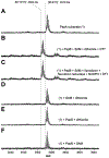
Figure 5:. MALDI-TOF-MS analysis of PapA peptide treated with purified PapB in vitro.
(A) Mass spectrum of the PapA peptide, m/z 3570 Da. Mass loss of 6 Da is consistent with three disulfide bond formation as reported previously. (B) Mass spectrum of the peptide after reaction with the full array of reactants, m/z 3564 Da. This is consistent with the formation of six thioether bonds (loss of 12 Da from the unmodified peptide), similar to what is obtained from in vivo co-expression experiments. (C) Identical to B except flavodoxin, flavodoxin reductase and NADPH was used as the reductant. (D) No PapB control reaction. (E) No SAM control reaction. (F) No dithionite control reaction.
Similar articles
- Bioinformatic Mapping of Radical S-Adenosylmethionine-Dependent Ribosomally Synthesized and Post-Translationally Modified Peptides Identifies New Cα, Cβ, and Cγ-Linked Thioether-Containing Peptides.
Hudson GA, Burkhart BJ, DiCaprio AJ, Schwalen CJ, Kille B, Pogorelov TV, Mitchell DA. Hudson GA, et al. J Am Chem Soc. 2019 May 22;141(20):8228-8238. doi: 10.1021/jacs.9b01519. Epub 2019 May 13. J Am Chem Soc. 2019. PMID: 31059252 Free PMC article. - Leveraging Substrate Promiscuity of a Radical _S_-Adenosyl-L-methionine RiPP Maturase toward Intramolecular Peptide Cross-Linking Applications.
Eastman KAS, Kincannon WM, Bandarian V. Eastman KAS, et al. ACS Cent Sci. 2022 Aug 24;8(8):1209-1217. doi: 10.1021/acscentsci.2c00501. Epub 2022 Aug 1. ACS Cent Sci. 2022. PMID: 36032765 Free PMC article. - A Promiscuous rSAM Enzyme Enables Diverse Peptide Cross-linking.
Eastman KAS, Mifflin MC, Oblad PF, Roberts AG, Bandarian V. Eastman KAS, et al. ACS Bio Med Chem Au. 2023 Aug 15;3(6):480-493. doi: 10.1021/acsbiomedchemau.3c00043. eCollection 2023 Dec 20. ACS Bio Med Chem Au. 2023. PMID: 38144258 Free PMC article. - Current Advancements in Sactipeptide Natural Products.
Chen Y, Wang J, Li G, Yang Y, Ding W. Chen Y, et al. Front Chem. 2021 May 20;9:595991. doi: 10.3389/fchem.2021.595991. eCollection 2021. Front Chem. 2021. PMID: 34095082 Free PMC article. Review. - Radical S-adenosylmethionine enzyme catalyzed thioether bond formation in sactipeptide biosynthesis.
Flühe L, Marahiel MA. Flühe L, et al. Curr Opin Chem Biol. 2013 Aug;17(4):605-12. doi: 10.1016/j.cbpa.2013.06.031. Epub 2013 Jul 24. Curr Opin Chem Biol. 2013. PMID: 23891473 Review.
Cited by
- Biosynthetic potential of the gut microbiome in longevous populations.
Liu S, Zhang Z, Wang X, Ma Y, Ruan H, Wu X, Li B, Mou X, Chen T, Lu Z, Zhao W. Liu S, et al. Gut Microbes. 2024 Jan-Dec;16(1):2426623. doi: 10.1080/19490976.2024.2426623. Epub 2024 Nov 11. Gut Microbes. 2024. PMID: 39529240 Free PMC article. - Advancements in the Application of Ribosomally Synthesized and Post-Translationally Modified Peptides (RiPPs).
Han SW, Won HS. Han SW, et al. Biomolecules. 2024 Apr 15;14(4):479. doi: 10.3390/biom14040479. Biomolecules. 2024. PMID: 38672495 Free PMC article. Review. - Genome Mining for New Enzyme Chemistry.
Nguyen DT, Mitchell DA, van der Donk WA. Nguyen DT, et al. ACS Catal. 2024 Mar 12;14(7):4536-4553. doi: 10.1021/acscatal.3c06322. eCollection 2024 Apr 5. ACS Catal. 2024. PMID: 38601780 Free PMC article. Review. - Discovery and engineering of ribosomally synthesized and post-translationally modified peptide (RiPP) natural products.
Li H, Ding W, Zhang Q. Li H, et al. RSC Chem Biol. 2023 Nov 21;5(2):90-108. doi: 10.1039/d3cb00172e. eCollection 2024 Feb 7. RSC Chem Biol. 2023. PMID: 38333193 Free PMC article. Review. - Peptide Selenocysteine Substitutions Reveal Direct Substrate-Enzyme Interactions at Auxiliary Clusters in Radical _S_-Adenosyl-l-methionine Maturases.
Rush KW, Eastman KAS, Kincannon WM, Blackburn NJ, Bandarian V. Rush KW, et al. J Am Chem Soc. 2023 May 10;145(18):10167-10177. doi: 10.1021/jacs.3c00831. Epub 2023 Apr 27. J Am Chem Soc. 2023. PMID: 37104670 Free PMC article.
References
- Arnison PG; Bibb MJ; Bierbaum G; Bowers AA; Bugni TS; Bulaj G; Camarero JA; Campopiano DJ; Challis GL; Clardy J; Cotter PD; Craik DJ; Dawson M; Dittmann E; Donadio S; Dorrestein PC; Entian KD; Fischbach MA; Garavelli JS; Göransson U; Gruber CW; Haft DH; Hemscheidt TK; Hertweck C; Hill C; Horswill AR; Jaspars M; Kelly WL; Klinman JP; Kuipers OP; Link AJ; Liu W; Marahiel MA; Mitchell DA; Moll GN; Moore BS; Müller R; Nair SK; Nes IF; Norris GE; Olivera BM; Onaka H; Patchett ML; Piel J; Reaney MJ; Rebuffat S; Ross RP; Sahl HG; Schmidt EW; Selsted ME; Severinov K; Shen B; Sivonen K; Smith L; Stein T; Süssmuth RD; Tagg JR; Tang GL; Truman AW; Vederas JC; Walsh CT; Walton JD; Wenzel SC; Willey JM; van der Donk WA Ribosomally Synthesized and Post-Translationally Modified Peptide Natural Products: Overview and Recommendations for a Universal Nomenclature. Nat. Prod. Rep 2013, 30, 108–160. - PMC - PubMed
Publication types
MeSH terms
Substances
LinkOut - more resources
Full Text Sources
Other Literature Sources


