Single-cell connectomic analysis of adult mammalian lungs - PubMed (original) (raw)
. 2019 Dec 4;5(12):eaaw3851.
doi: 10.1126/sciadv.aaw3851. eCollection 2019 Dec.
Micha Sam Brickman Raredon 1 2 3, Yasir Suhail 5, Jonas Christian Schupp 4, Sergio Poli 6, Nir Neumark 4 7, Katherine L Leiby 1 2 3, Allison Marie Greaney 1 2, Yifan Yuan 8, Corey Horien 3 9, George Linderman 3 10, Alexander J Engler 1 2, Daniel J Boffa 11, Yuval Kluger 7 10 12, Ivan O Rosas 6, Andre Levchenko 1 5, Naftali Kaminski 4, Laura E Niklason 1 2 8
Affiliations
- PMID: 31840053
- PMCID: PMC6892628
- DOI: 10.1126/sciadv.aaw3851
Single-cell connectomic analysis of adult mammalian lungs
Micha Sam Brickman Raredon et al. Sci Adv. 2019.
Abstract
Efforts to decipher chronic lung disease and to reconstitute functional lung tissue through regenerative medicine have been hampered by an incomplete understanding of cell-cell interactions governing tissue homeostasis. Because the structure of mammalian lungs is highly conserved at the histologic level, we hypothesized that there are evolutionarily conserved homeostatic mechanisms that keep the fine architecture of the lung in balance. We have leveraged single-cell RNA sequencing techniques to identify conserved patterns of cell-cell cross-talk in adult mammalian lungs, analyzing mouse, rat, pig, and human pulmonary tissues. Specific stereotyped functional roles for each cell type in the distal lung are observed, with alveolar type I cells having a major role in the regulation of tissue homeostasis. This paper provides a systems-level portrait of signaling between alveolar cell populations. These methods may be applicable to other organs, providing a roadmap for identifying key pathways governing pathophysiology and informing regenerative efforts.
Copyright © 2019 The Authors, some rights reserved; exclusive licensee American Association for the Advancement of Science. No claim to original U.S. Government Works. Distributed under a Creative Commons Attribution NonCommercial License 4.0 (CC BY-NC).
Figures
Fig. 1. Cross-species pulmonary scRNAseq dataset.
(A) Lungs from mice, rats, pigs, and human donors were dissociated, barcoded using 10× genomics, and sequenced on Illumina sequencers. Cells were clustered following genome alignment and dimensionality reduction. Markers were identified via differential expression and correlated to literature and the Human Protein Atlas (24) and in-house immunostaining. Comparative ligand-receptor analysis reveals stereotyped cellular roles in tissue homeostasis. (B) A comparable, heterogeneous community of cells was profiled in all four species. Species-conserved cell types include type I, type II, ciliated, and secretory epithelium; capillary, vascular, and lymphatic endothelium; B cells, T cells, and natural killer (NK) cells; SMCs; Col13+ and Col14+ fibroblasts; and two distinct subsets of macrophages.
Fig. 2. Cross-species cell atlas representation.
(A) The species-split DotPlot shows cross-species comparison of selected top markers for all cell populations profiled across all species. Color saturation indicates the strength of expression in positive cells, while dot size reflects the percentage of each cell cluster expressing the gene. Each color represents one species. Certain cell types were only profiled in certain species, such as basal cells marked by KRT5 (pig and human) and Sox9+ epithelium (rat). ITGA8/Itga8 reliably marks the Col13a1+ fibroblast population across species. Histology for highly expressed specific markers of conserved cell types, from the Human Protein Atlas [immunoperoxidase images (D)] and from in-house immunostaining [immunofluorescent images (B and C)], shows positive physical locations of identified cell type–specific markers. Markers shown are those that were present in the human dataset having homologs in the nonhuman species. Scale bars, 50 μm (D) and 62 μm (B and C).
Fig. 3. Global network comparison reveals conserved signaling patterns underlying adult lung homeostasis.
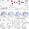
(A) Schematic of data processing. Gene expression was averaged by cluster and mapped against the FANTOM5 ligand-receptor database. When two nodes expressed a cognate ligand-receptor pair, a weighted edge was created (see Supplementary Text). (B) Promiscuous expression across cell types (ICAM1) causes a low distribution of edge weights. (C) Cell type–specific expression (VEGFA) causes a higher edge weight distribution (black arrows). (D) Global connectomes across the four species, showing the sum of edge weights between conserved nodes. Vertex (colored cell node) size is proportional to the Kleinberg hub score, while the thickness of the edges is proportional to the sum of the weights between two nodes. Similarities in overall signaling structure between the cell nodes are readily observable. (E) Comparison of degree rankings and the effect of thresholding. Stromal cells have the highest degrees, while lymphoid cells have the lowest. The x axis (“percent_exp”) is percent of cluster expressing the marker, and the y axis is the degree of each node plotted on a logarithmic scale. (F) Quantitative cross-species correlations. Plots of Spearman correlation coefficients (“ρ”), with human as a reference, of the rankings of nodal centrality metrics over increasing thresholds of the fraction of cells in a node that expresses the ligand and receptor. Note that the correlation coefficients are relatively high (>0.75) and remain so up to ~40% expression, meaning that 40% of the cells in the node express either the ligand or the receptor. This demonstrates that node-node relationships are robust to thresholding. This suggests a high degree of evolutionary conservation in pulmonary connectomic structure, because all four species are very highly correlated.
Fig. 4. Niche character visualizations from species-conserved signaling.
(A) Cell classes in close physical proximity in alveolar lung. Downstream analysis was limited to nine cell types spatially registering to the alveolar septal wall. All mapped ligand-receptor pairs were classified into 1 of 38 signaling families (see legend). Hive plot illustrations of niche character are presented here for (B) alveolar macrophages and (C) capillary endothelium (see fig. S7 for all other niche visualizations). Alveolar macrophages display high connectivity and express distinctly high levels of cell-cell adhesion molecules (*). Capillary endothelium, in contrast, expresses numerous receptors for VEGF family signaling (**).
Fig. 5. ATI cells dominate VEGF and SEMA family niche networks in all species.
(A) Capillary endothelia and ATI cells are in extremely close proximity in the pulmonary alveolus, often separated by less than 100 nm of shared basement membrane. (B) ATI cells, marked by Aqp5 in green, express high levels of Sema3e and Vegfa at the protein level, correlating with scRNAseq findings. (C) Ligand-receptor mapping shows that endothelial populations express high levels of receptors for both Vegfa and Sema3e, allowing receipt of spatial and growth cue information from ATI cells. (D) Network centrality analysis shows that ATI cells have dominant hub scores and top cumulative outgoing edge weight, while capillary endothelia have dominant authority scores and top cumulative incoming edge weight, in both the VEGF and SEMA family signaling networks.
Fig. 6. Network centrality analysis categorizes each signaling network by dominant producers and receivers.
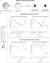
(A) The species-conserved connectome is first subset to a single signaling family. Outgoing centrality, defined as the cumulative outgoing edge weight and the Kleinberg network hub score, and incoming centrality, defined as the cumulative incoming edge weight and the Kleinberg network authority score, are then calculated for each node. (B) Identification of signaling family networks dominated by epithelial cells (top) and macrophage populations (bottom). The left side of each panel shows outgoing centrality metrics (cumulative outgoing edge weight for each cell, Kleinberg hub scores), while the right side shows incoming centrality metrics (cumulative incoming edge weight for each cell, Kleinberg authority scores). These trends suggest species-conserved cellular roles in pulmonary tissue and allow identification of dominant producers and receivers of each class of signal in homeostatic lung tissue. Additional results detailing endothelia- and mesenchyme-dominated networks are shown in fig. S9. Note here the ATI dominance of cell-cell adhesion networks acting predominantly on alveolar macrophages versus ATII dominance of CSF networks acting predominantly on interstitial macrophages. Macrophages predictably dominate networks based on immunoregulatory cues such as CCL and interleukin family ligand-receptor pairs.
Similar articles
- A mammalian Wnt5a-Ror2-Vangl2 axis controls the cytoskeleton and confers cellular properties required for alveologenesis.
Zhang K, Yao E, Lin C, Chou YT, Wong J, Li J, Wolters PJ, Chuang PT. Zhang K, et al. Elife. 2020 May 12;9:e53688. doi: 10.7554/eLife.53688. Elife. 2020. PMID: 32394892 Free PMC article. - Defining the role of pulmonary endothelial cell heterogeneity in the response to acute lung injury.
Niethamer TK, Stabler CT, Leach JP, Zepp JA, Morley MP, Babu A, Zhou S, Morrisey EE. Niethamer TK, et al. Elife. 2020 Feb 24;9:e53072. doi: 10.7554/eLife.53072. Elife. 2020. PMID: 32091393 Free PMC article. - Integrated Single-Cell Atlas of Endothelial Cells of the Human Lung.
Schupp JC, Adams TS, Cosme C Jr, Raredon MSB, Yuan Y, Omote N, Poli S, Chioccioli M, Rose KA, Manning EP, Sauler M, DeIuliis G, Ahangari F, Neumark N, Habermann AC, Gutierrez AJ, Bui LT, Lafyatis R, Pierce RW, Meyer KB, Nawijn MC, Teichmann SA, Banovich NE, Kropski JA, Niklason LE, Pe'er D, Yan X, Homer RJ, Rosas IO, Kaminski N. Schupp JC, et al. Circulation. 2021 Jul 27;144(4):286-302. doi: 10.1161/CIRCULATIONAHA.120.052318. Epub 2021 May 25. Circulation. 2021. PMID: 34030460 Free PMC article. - P2 Purinergic Signaling in the Distal Lung in Health and Disease.
Wirsching E, Fauler M, Fois G, Frick M. Wirsching E, et al. Int J Mol Sci. 2020 Jul 14;21(14):4973. doi: 10.3390/ijms21144973. Int J Mol Sci. 2020. PMID: 32674494 Free PMC article. Review. - Semaphorin regulation of cellular morphology.
Tran TS, Kolodkin AL, Bharadwaj R. Tran TS, et al. Annu Rev Cell Dev Biol. 2007;23:263-92. doi: 10.1146/annurev.cellbio.22.010605.093554. Annu Rev Cell Dev Biol. 2007. PMID: 17539753 Review.
Cited by
- Recent Advances in Molecular Diagnosis of Pulmonary Fibrosis for Precision Medicine.
Jeong MH, Han H, Lagares D, Im H. Jeong MH, et al. ACS Pharmacol Transl Sci. 2022 Jul 20;5(8):520-538. doi: 10.1021/acsptsci.2c00028. eCollection 2022 Aug 12. ACS Pharmacol Transl Sci. 2022. PMID: 35983278 Free PMC article. Review. - A census of the lung: CellCards from LungMAP.
Sun X, Perl AK, Li R, Bell SM, Sajti E, Kalinichenko VV, Kalin TV, Misra RS, Deshmukh H, Clair G, Kyle J, Crotty Alexander LE, Masso-Silva JA, Kitzmiller JA, Wikenheiser-Brokamp KA, Deutsch G, Guo M, Du Y, Morley MP, Valdez MJ, Yu HV, Jin K, Bardes EE, Zepp JA, Neithamer T, Basil MC, Zacharias WJ, Verheyden J, Young R, Bandyopadhyay G, Lin S, Ansong C, Adkins J, Salomonis N, Aronow BJ, Xu Y, Pryhuber G, Whitsett J, Morrisey EE; NHLBI LungMAP Consortium. Sun X, et al. Dev Cell. 2022 Jan 10;57(1):112-145.e2. doi: 10.1016/j.devcel.2021.11.007. Epub 2021 Dec 21. Dev Cell. 2022. PMID: 34936882 Free PMC article. - The lung microenvironment shapes a dysfunctional response of alveolar macrophages in aging.
McQuattie-Pimentel AC, Ren Z, Joshi N, Watanabe S, Stoeger T, Chi M, Lu Z, Sichizya L, Aillon RP, Chen CI, Soberanes S, Chen Z, Reyfman PA, Walter JM, Anekalla KR, Davis JM, Helmin KA, Runyan CE, Abdala-Valencia H, Nam K, Meliton AY, Winter DR, Morimoto RI, Mutlu GM, Bharat A, Perlman H, Gottardi CJ, Ridge KM, Chandel NS, Sznajder JI, Balch WE, Singer BD, Misharin AV, Budinger GRS. McQuattie-Pimentel AC, et al. J Clin Invest. 2021 Feb 15;131(4):e140299. doi: 10.1172/JCI140299. J Clin Invest. 2021. PMID: 33586677 Free PMC article. - Deciphering the Complexities of Pulmonary Hypertension: The Emergent Role of Single-Cell Omics.
Rafikov R, de Jesus Perez V, Dekan A, Kudryashova TV, Rafikova O. Rafikov R, et al. Am J Respir Cell Mol Biol. 2024 Aug 14;72(1):32-40. doi: 10.1165/rcmb.2024-0145PS. Online ahead of print. Am J Respir Cell Mol Biol. 2024. PMID: 39141563 - Reading the palimpsest of cell interactions: What questions may we ask of the data?
Pavlicev M, Wagner GP. Pavlicev M, et al. iScience. 2024 Apr 5;27(5):109670. doi: 10.1016/j.isci.2024.109670. eCollection 2024 May 17. iScience. 2024. PMID: 38665209 Free PMC article. Review.
References
- Amit I., Winter D. R., Jung S., The role of the local environment and epigenetics in shaping macrophage identity and their effect on tissue homeostasis. Nat. Immunol. 17, 18–25 (2016). - PubMed
Publication types
MeSH terms
Substances
Grants and funding
- R01 HL141852/HL/NHLBI NIH HHS/United States
- UL1 TR001863/TR/NCATS NIH HHS/United States
- R01 HG008383/HG/NHGRI NIH HHS/United States
- F30 HL143906/HL/NHLBI NIH HHS/United States
- R21 EB024889/EB/NIBIB NIH HHS/United States
- F30 HL143880/HL/NHLBI NIH HHS/United States
- T32 GM007205/GM/NIGMS NIH HHS/United States
- R01 HL138540/HL/NHLBI NIH HHS/United States
- U01 HL122626/HL/NHLBI NIH HHS/United States
- U01 HL145567/HL/NHLBI NIH HHS/United States
- R01 GM131642/GM/NIGMS NIH HHS/United States
- U01 HL145560/HL/NHLBI NIH HHS/United States
- U54 CA209992/CA/NCI NIH HHS/United States
- R01 HL127349/HL/NHLBI NIH HHS/United States
LinkOut - more resources
Full Text Sources
Other Literature Sources
Molecular Biology Databases


