CT Imaging Features of 2019 Novel Coronavirus (2019-nCoV) - PubMed (original) (raw)
. 2020 Apr;295(1):202-207.
doi: 10.1148/radiol.2020200230. Epub 2020 Feb 4.
Adam Bernheim 1, Xueyan Mei 1, Ning Zhang 1, Mingqian Huang 1, Xianjun Zeng 1, Jiufa Cui 1, Wenjian Xu 1, Yang Yang 1, Zahi A Fayad 1, Adam Jacobi 1, Kunwei Li 1, Shaolin Li 1, Hong Shan 1
Affiliations
- PMID: 32017661
- PMCID: PMC7194022
- DOI: 10.1148/radiol.2020200230
CT Imaging Features of 2019 Novel Coronavirus (2019-nCoV)
Michael Chung et al. Radiology. 2020 Apr.
Abstract
In this retrospective case series, chest CT scans of 21 symptomatic patients from China infected with the 2019 novel coronavirus (2019-nCoV) were reviewed, with emphasis on identifying and characterizing the most common findings. Typical CT findings included bilateral pulmonary parenchymal ground-glass and consolidative pulmonary opacities, sometimes with a rounded morphology and a peripheral lung distribution. Notably, lung cavitation, discrete pulmonary nodules, pleural effusions, and lymphadenopathy were absent. Follow-up imaging in a subset of patients during the study time window often demonstrated mild or moderate progression of disease, as manifested by increasing extent and density of lung opacities.
© RSNA, 2020.
Figures
Graphical abstract
Figure 1a:
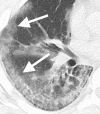
Images in a 29-year-old man with unknown exposure history who presented with fever and cough ultimately requiring admission to intensive care unit. (a) Axial thin-section unenhanced CT scan shows diffuse bilateral confluent and patchy ground-glass (white arrows) and consolidative (black arrows) pulmonary opacities. (b) Axial unenhanced image shows that the disease in the right middle and lower lobes has a striking peripheral distribution (arrows).
Figure 1b:
Images in a 29-year-old man with unknown exposure history who presented with fever and cough ultimately requiring admission to intensive care unit. (a) Axial thin-section unenhanced CT scan shows diffuse bilateral confluent and patchy ground-glass (white arrows) and consolidative (black arrows) pulmonary opacities. (b) Axial unenhanced image shows that the disease in the right middle and lower lobes has a striking peripheral distribution (arrows).
Figure 2:
Image in a 36-year-old man with history of recent travel to Wuhan who presented with fever, fatigue, and myalgias. Coronal thin-section unenhanced CT image shows ground-glass opacities with a rounded morphology in both upper lobes (arrows).
Figure 3:
Image in a 66-year-old woman with history of recent travel to Wuhan who presented with fever and productive cough. Axial thin-section collimated unenhanced CT image shows a crazy-paving pattern, as manifested by right lower lobe ground-glass opacification and interlobular septal thickening (arrow) with intralobular lines.
Figure 4:
Image in a 69-year-old man with history of recent travel to Wuhan who presented with fever. Axial thin-section unenhanced CT scan shows ground-glass opacities in the lower lobes with a pronounced peripheral distribution (arrows).
Figure 5a:
Images in a 43-year-old woman with a history of travel to Wuhan who presented with fever. (a) Axial thin-section unenhanced CT image obtained January 18, 2020, shows normal lung. (b) Follow-up CT image obtained January 21, 2020, shows a new solitary, rounded, peripheral ground-glass lesion in the right lower lobe (arrow).
Figure 5b:
Images in a 43-year-old woman with a history of travel to Wuhan who presented with fever. (a) Axial thin-section unenhanced CT image obtained January 18, 2020, shows normal lung. (b) Follow-up CT image obtained January 21, 2020, shows a new solitary, rounded, peripheral ground-glass lesion in the right lower lobe (arrow).
Similar articles
- Chest CT Findings in 2019 Novel Coronavirus (2019-nCoV) Infections from Wuhan, China: Key Points for the Radiologist.
Kanne JP. Kanne JP. Radiology. 2020 Apr;295(1):16-17. doi: 10.1148/radiol.2020200241. Epub 2020 Feb 4. Radiology. 2020. PMID: 32017662 Free PMC article. No abstract available. - Imaging and clinical features of patients with 2019 novel coronavirus SARS-CoV-2.
Xu X, Yu C, Qu J, Zhang L, Jiang S, Huang D, Chen B, Zhang Z, Guan W, Ling Z, Jiang R, Hu T, Ding Y, Lin L, Gan Q, Luo L, Tang X, Liu J. Xu X, et al. Eur J Nucl Med Mol Imaging. 2020 May;47(5):1275-1280. doi: 10.1007/s00259-020-04735-9. Epub 2020 Feb 28. Eur J Nucl Med Mol Imaging. 2020. PMID: 32107577 Free PMC article. - CT Manifestations of Two Cases of 2019 Novel Coronavirus (2019-nCoV) Pneumonia.
Fang Y, Zhang H, Xu Y, Xie J, Pang P, Ji W. Fang Y, et al. Radiology. 2020 Apr;295(1):208-209. doi: 10.1148/radiol.2020200280. Epub 2020 Feb 7. Radiology. 2020. PMID: 32031481 Free PMC article. No abstract available. - [Diagnostic imaging findings in COVID-19].
Plesner LL, Dyrberg E, Hansen IV, Abild A, Andersen MB. Plesner LL, et al. Ugeskr Laeger. 2020 Apr 6;182(15):V03200191. Ugeskr Laeger. 2020. PMID: 32286216 Review. Danish. - Diagnostic value and key features of computed tomography in Coronavirus Disease 2019.
Li B, Li X, Wang Y, Han Y, Wang Y, Wang C, Zhang G, Jin J, Jia H, Fan F, Ma W, Liu H, Zhou Y. Li B, et al. Emerg Microbes Infect. 2020 Dec;9(1):787-793. doi: 10.1080/22221751.2020.1750307. Emerg Microbes Infect. 2020. PMID: 32241244 Free PMC article. Review.
Cited by
- Predictors of Poor Outcomes in Chronic Obstructive Pulmonary Disease (COPD) Patients Admitted to the Emergency Department With COVID-19: A Prospective Study.
Osoydan Satici M, Satıcı C, İslam MM, Altunok İ, Başlılar Ş, Emir SN, Aksel G, Eroğlu SE. Osoydan Satici M, et al. Cureus. 2024 Oct 9;16(10):e71154. doi: 10.7759/cureus.71154. eCollection 2024 Oct. Cureus. 2024. PMID: 39525229 Free PMC article. - Advanced federated ensemble internet of learning approach for cloud based medical healthcare monitoring system.
Khan R, Taj S, Ma X, Noor A, Zhu H, Khan J, Khan ZU, Khan SU. Khan R, et al. Sci Rep. 2024 Oct 30;14(1):26068. doi: 10.1038/s41598-024-77196-x. Sci Rep. 2024. PMID: 39478132 Free PMC article. - Lung field-based severity score (LFSS): a feasible tool to identify COVID-19 patients at high risk of progressing to critical disease.
Jiang X, Hu J, Jiang Q, Zhou T, Yao F, Sun Y, Liu Q, Zhou C, Shi K, Lin X, Li J, Li Y, Jin Q, Tu W, Zhou X, Wang Y, Xin X, Liu S, Fan L. Jiang X, et al. J Thorac Dis. 2024 Sep 30;16(9):5591-5603. doi: 10.21037/jtd-24-544. Epub 2024 Sep 6. J Thorac Dis. 2024. PMID: 39444869 Free PMC article. - Role of interleukin 6 polymorphism and inflammatory markers in outcome of pediatric Covid- 19 patients.
AbdelAziz RA, Abd-Allah ST, Moness HM, Anwar AM, Mohamed ZH. AbdelAziz RA, et al. BMC Pediatr. 2024 Oct 1;24(1):625. doi: 10.1186/s12887-024-05071-9. BMC Pediatr. 2024. PMID: 39354444 Free PMC article. - Development and validation of a deep learning-based framework for automated lung CT segmentation and acute respiratory distress syndrome prediction: a multicenter cohort study.
Zhou Y, Mei S, Wang J, Xu Q, Zhang Z, Qin S, Feng J, Li C, Xing S, Wang W, Zhang X, Li F, Zhou Q, He Z, Gao Y. Zhou Y, et al. EClinicalMedicine. 2024 Jul 26;75:102772. doi: 10.1016/j.eclinm.2024.102772. eCollection 2024 Sep. EClinicalMedicine. 2024. PMID: 39170939 Free PMC article.
References
- Novel Coronavirus – China. World Health Organization. https://www.who.int/csr/don/12-january-2020-novel-coronavirus-china/en/. Published January 12, 2020.
- Novel Coronavirus (2019-nCoV). World Health Organization. https://www.who.int/emergencies/diseases/novel-Coronavirus-2019. Published January 7, 2020.
- Summary of probable SARS cases with onset of illness from 1 November 2002 to 31 July 2003. World Health Organization. https://www.who.int/csr/sars/country/table2004_04_21/en/. Published April 21, 2004.
- Middle East respiratory syndrome Coronavirus (MERS-CoV). World Health Organization. https://www.who.int/emergencies/mers-cov/en/.
- Situation report - 8. World Health Organization. https://www.who.int/docs/default-source/Coronaviruse/situation-reports/2.... Published January 28, 2020.
MeSH terms
LinkOut - more resources
Full Text Sources
Other Literature Sources
Medical


