A one-billion-year-old multicellular chlorophyte - PubMed (original) (raw)
A one-billion-year-old multicellular chlorophyte
Qing Tang et al. Nat Ecol Evol. 2020 Apr.
Abstract
Chlorophytes (representing a clade within the Viridiplantae and a sister group of the Streptophyta) probably dominated marine export bioproductivity and played a key role in facilitating ecosystem complexity before the Mesozoic diversification of phototrophic eukaryotes such as diatoms, coccolithophorans and dinoflagellates. Molecular clock and biomarker data indicate that chlorophytes diverged in the Mesoproterozoic or early Neoproterozoic, followed by their subsequent phylogenetic diversification, multicellular evolution and ecological expansion in the late Neoproterozoic and Palaeozoic. This model, however, has not been rigorously tested with palaeontological data because of the scarcity of Proterozoic chlorophyte fossils. Here we report abundant millimetre-sized, multicellular and morphologically differentiated macrofossils from rocks approximately 1,000 million years ago. These fossils are described as Proterocladus antiquus new species and are interpreted as benthic siphonocladalean chlorophytes, suggesting that chlorophytes acquired macroscopic size, multicellularity and cellular differentiation nearly a billion years ago, much earlier than previously thought.
Figures
Extended Data Fig. 1. Geological map and stratigraphic column of Proterozoic successions in southern Liaoning Province, North China.
Question mark in stratigraphic column denotes poor age constraint on the Dalinzi Formation, which could be either Neoproterozoic or Cambrian in age. Stars in geological map and stratigraphic column mark sample locality (near Shileicun, 39°35.6566’N, 121°35.8379’E) and sample horizon, respectively. Ca = Cambrian, Pa = Paleoproterozoic, Fm = Formation, CLZ = Changlingzi, NGL = Nanguanling, GJZ = Ganjingzi, YCZ = Yingchengzi, SSLT = Shisanlitai, MJT = Majiatun, CJT = Cuijiatun, XMC = Xingmincun, GT = Getun. Radiometric ages (924 ± 5 Ma and 947.8 ± 7.4 Ma) of diabase sills emplaced in the Cuijiatun and Qiaotou formations are from ref. and ref. ; detrital zircon ages (<924 ± 25 Ma and <1056 ± 22 Ma) are from ref. . See ref. for a compilation of ratiometric ages from Neoproterozoic successions in North China. Geological map drawn by authors based on ref. with permission, and stratocolumn drawn by authors.
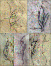
Extended Data Fig. 2. Proterocladus antiquus new species on bedding surface, showing lateral branches.
a–f, VPIGM-4763, VPIGM-4764, VPIGM-4765, VPIGM-4766, VPIGM-4767, and VPIGM-4768, respectively. All photos taken by authors.
Extended Data Fig. 3. P. antiquus preserved on bedding surface, showing multiple orders of lateral branches (a) and aggregates of thalli (b–d).
a–d, VPIGM-4769, VPIGM-4770, VPIGM-4771, and VPIGM-4772, respectively. All photos taken by authors.
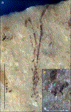
Extended Data Fig. 4. Thallus of P. antiquus with a sub-discoid holdfast preserved on bedding surface.
b is a close-up view of labeled black frame in a. VPIGM-4773. All photos taken by authors.
Extended Data Fig. 5. Heteromorphic cells (a–l) and reproductive cells (m–o) of P. antiquus.
a–l, Filaments with globose (yellow arrowheads), clavate (black arrowheads), doliform (cyan arrowheads), and cyathiform (blue arrowhead) heteromorphic cells. b, k are magnifications of labeled frames in a and j, respectively. VPIGM-4774, VPIGM-4775, VPIGM-4776, VPIGM-4777, VPIGM-4778, VPIGM-4779, VPIGM-4780, VPIGM-4781, VPIGM-4782, and VPIGM-4783, respectively. m–o, Inferred reproductive cells with minute lateral pores (blue arrows), possibly representing openings through which reproductive gametes or zoospores were released. VPIGM-4784, VPIGM-4785, and VPIGM-4786, respectively. Specimens in a and j were photographed on bedding surface, and all other specimens were extracted from the rock matrix using HF acid maceration technique. All scale bars equal 100 μm unless otherwise specified. All photos taken by authors.
Extended Data Fig. 6. Cell branching pattern and apical extensions in extracted specimens of P. antiquus.
a–c, Fragmented filaments with unilateral (a, c) and alternate branches (two lower lateral branches in b). VPIGM-4787, VPIGM-4788, and VPIGM-4789, respectively. d–e, Branching filaments with an inflated apical cell subtending a narrower apical extension (purple arrowhead in d) and an apical cell with septum and constriction (black arrows in e), which is interpreted to have developed from an apical extension. VPIGM-4790 and VPIGM-4791, respectively. All scale bars equal 100 μm. All photos taken by authors.
Extended Data Fig. 7. Branching thallus of P. antiquus with a cell (in black frame) that has a distinct constriction at base (blue arrowhead).
The branching pattern is superficially similar to H-shaped branching in the early vascular plant _Zosterophyllum_73. b is a magnification of white box in a, showing the basal constriction of the cell that initially may represent an apical extension that subsequently develops septa and branches at maturation. VPIGM-4792. All photos taken by authors.
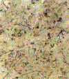
Extended Data Fig. 8. Dense population of fragmented P. antiquus specimens preserved on bedding surface.
VPIGM-4793. All photos taken by authors.
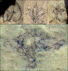
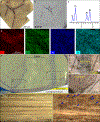
Extended Data Fig. 9. Taphonomy of P. antiquus preserved in the Nanfen mudstone.
a–b, A partially exposed specimen. b is a backscattered electron scanning electron microscopy (BSEM) photograph of the same specimen in a. VPIGM-4794. c, Energy dispersive X-ray spectroscopy (EDS) point analysis at the blue spot in b, showing the presence of carbon in the fossil specimen. d, EDS elemental maps of labeled box in b, showing the enrichment in C and deficiency in O, Al, and Si in fossil relative to matrix. e–g, Nanfen mudstone fractured obliquely relative to bedding plane, showing darker-colored fossil layers and lighter-colored background layers. f and g are magnifications of labeled boxes in e. h–i, Polished slab cut perpendicular to bedding surface, showing darker-colored fossil layers and lighter-colored background layers. i is a close-up view of labeled box in h with blue arrowheads denoting fragmented fossils in a fossil layer. All photos taken by authors.
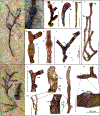
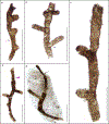
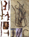
Figure 1 |. Gross morphology of Proterocladus antiquus new species from the Nanfen Formation.
a, Morphological reconstruction and terminology. b–h, Slender thalli preserved on bedding surfaces. c, e, and g are magnifications of white frames in b, d, and f, respectively, showing cyathiform heteromorphic cells (in c and g) and apical extension (in e). VPIGM-4749, VPIGM-4750, VPIGM-4751, and VPIGM-4752, respectively. i–l, Branching thalli extracted from rock matrix using HF acid maceration technique. A close-up view is provided for the holdfast in the black frame in l. VPIGM-4753, VPIGM-4754, VPIGM-4755, and VPIGM-4799 (paratype), respectively. Blue arrowheads in a, c, g, and i: cyathiform heteromorphic cell; purple arrowheads in a and e: apical extension; black arrowheads in a and j: clavate cell; yellow arrowheads in a, j, and k: globose heteromorphic cell; black arrows in a, j, and l: septum and constriction. Scale bars equal 200 μm unless otherwise specified. All photos taken by authors.
Figure 2 |. Cellular structures of Proterocladus antiquus new species.
a–f, Fragmentary specimens extracted from rock matrix using HF acid maceration technique. VPIGM-4756, VPIGM-4757, VPIGM-4758, VPIGM-4759, VPIGM-4760, and VPIGM-4761, respectively. g–k, Well-preserved thallus on bedding surface. VPIGM-4762 (holotype). h–k are magnifications of labeled frames in g. Black and blue arrows denote robust septa and lateral pore, respectively. Cyan, blue, and purple arrowheads denote morphologically differentiated doliform hetermorphic cells, cyathiform hetermorphic cells, and narrow apical extensions, respectively. Scale bars equal 100 μm unless otherwise specified. All photos taken by authors.
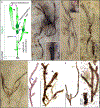
Figure 3 |. Extant Siphonocladales of the genera Cladophora and Rhizoclonium for comparison with Proterocladus.
a, General morphology of a branching thallus of Cladophora, showing elongate cells and unique lateral branching system. Compare with Fig. 1b, d. b, Doliform akinetes (cyan arrowheads) of Rhizoclonium under stressed conditions in contrast to the cylindrical vegetative cells (white arrowhead). Compare with Fig. 2f. c, Reproductive cells of Rhizoclonium with lateral pore (white arrows) after the liberation of gametes or zoospores. Compare with Fig. 2e. d, Lateral branches (blue arrowheads) of Cladophora arising from mother cell subjacent to septa and remaining cytoplasmic contact with mother cell. Compare with Fig. 2a. a adapted from ref. under a Creative Commons License, b from ref. with permission, c from ref. under a Creative Commons License, and d from ref. with permission.
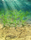
Figure 4 |
An artist’s reconstruction of Proterocladus antiquus. Artwork by Dinghua Yang.
Similar articles
- Tonian carbonaceous compressions indicate that Horodyskia is one of the oldest multicellular and coenocytic macro-organisms.
Li G, Chen L, Pang K, Tang Q, Wu C, Yuan X, Zhou C, Xiao S. Li G, et al. Commun Biol. 2023 Apr 12;6(1):399. doi: 10.1038/s42003-023-04740-2. Commun Biol. 2023. PMID: 37046079 Free PMC article. - Morphological and ecological complexity in early eukaryotic ecosystems.
Javaux EJ, Knoll AH, Walter MR. Javaux EJ, et al. Nature. 2001 Jul 5;412(6842):66-9. doi: 10.1038/35083562. Nature. 2001. PMID: 11452306 - Phosphatized multicellular algae in the Neoproterozoic Doushantuo Formation, China, and the early evolution of florideophyte red algae.
Xiao S, Knoll AH, Yuan X, Pueschel CM. Xiao S, et al. Am J Bot. 2004 Feb;91(2):214-27. doi: 10.3732/ajb.91.2.214. Am J Bot. 2004. PMID: 21653378 - The evolution of modern eukaryotic phytoplankton.
Falkowski PG, Katz ME, Knoll AH, Quigg A, Raven JA, Schofield O, Taylor FJ. Falkowski PG, et al. Science. 2004 Jul 16;305(5682):354-60. doi: 10.1126/science.1095964. Science. 2004. PMID: 15256663 Review. - A bottom-up perspective on ecosystem change in Mesozoic oceans.
Knoll AH, Follows MJ. Knoll AH, et al. Proc Biol Sci. 2016 Oct 26;283(1841):20161755. doi: 10.1098/rspb.2016.1755. Proc Biol Sci. 2016. PMID: 27798303 Free PMC article. Review.
Cited by
- Phylotranscriptomic insights into a Mesoproterozoic-Neoproterozoic origin and early radiation of green seaweeds (Ulvophyceae).
Hou Z, Ma X, Shi X, Li X, Yang L, Xiao S, De Clerck O, Leliaert F, Zhong B. Hou Z, et al. Nat Commun. 2022 Mar 22;13(1):1610. doi: 10.1038/s41467-022-29282-9. Nat Commun. 2022. PMID: 35318329 Free PMC article. - Tonian carbonaceous compressions indicate that Horodyskia is one of the oldest multicellular and coenocytic macro-organisms.
Li G, Chen L, Pang K, Tang Q, Wu C, Yuan X, Zhou C, Xiao S. Li G, et al. Commun Biol. 2023 Apr 12;6(1):399. doi: 10.1038/s42003-023-04740-2. Commun Biol. 2023. PMID: 37046079 Free PMC article. - Organic carbon cycling and black shale deposition: an Earth System Science perspective.
Jin Z, Wang X, Wang H, Ye Y, Zhang S. Jin Z, et al. Natl Sci Rev. 2023 Sep 15;10(11):nwad243. doi: 10.1093/nsr/nwad243. eCollection 2023 Nov. Natl Sci Rev. 2023. PMID: 37900193 Free PMC article. - Fossil-calibrated molecular clock data enable reconstruction of steps leading to differentiated multicellularity and anisogamy in the Volvocine algae.
Lindsey CR, Knoll AH, Herron MD, Rosenzweig F. Lindsey CR, et al. BMC Biol. 2024 Apr 10;22(1):79. doi: 10.1186/s12915-024-01878-1. BMC Biol. 2024. PMID: 38600528 Free PMC article. - Chronic Ionizing Radiation of Plants: An Evolutionary Factor from Direct Damage to Non-Target Effects.
Duarte GT, Volkova PY, Fiengo Perez F, Horemans N. Duarte GT, et al. Plants (Basel). 2023 Mar 4;12(5):1178. doi: 10.3390/plants12051178. Plants (Basel). 2023. PMID: 36904038 Free PMC article. Review.
References
- Falkowski PG et al. The evolution of modern eukaryotic phytoplankton. Science 305, 354–360 (2004). - PubMed
- Keeling PJ The Number, Speed, and Impact of Plastid Endosymbioses in Eukaryotic Evolution. Annu. Rev. Plant Biol 64, 583–607 (2013). - PubMed
- Brocks JJ et al. The rise of algae in Cryogenian oceans and the emergence of animals. Nature 548, 578–581 (2017). - PubMed
- Knoll AH, Summons RE, Waldbauer JR & Zumberge JE in Evolution of Primary Producers in the Sea (eds Falkowski Paul G.& Knoll Andrew H.) 133–163 (Academic Press, 2007).
Publication types
MeSH terms
LinkOut - more resources
Full Text Sources

