Content-Free Awareness: EEG-fcMRI Correlates of Consciousness as Such in an Expert Meditator - PubMed (original) (raw)
Content-Free Awareness: EEG-fcMRI Correlates of Consciousness as Such in an Expert Meditator
Ulf Winter et al. Front Psychol. 2020.
Abstract
The minimal neural correlate of the conscious state, regardless of the neural activity correlated with the ever-changing contents of experience, has still not been identified. Different attempts have been made, mainly by comparing the normal waking state to seemingly unconscious states, such as deep sleep or general anesthesia. A more direct approach would be the neuroscientific investigation of conscious states that are experienced as free of any specific phenomenal content. Here we present serendipitous data on content-free awareness (CFA) during an EEG-fMRI assessment reported by an extraordinarily qualified meditator with over 50,000 h of practice. We focused on two specific cortical networks related to external and internal awareness, i.e., the dorsal attention network (DAN) and the default mode network (DMN), to explore the neural correlates of this experience. The combination of high-resolution EEG and ultrafast fMRI enabled us to analyze the dynamic aspects of fMRI connectivity informed by EEG power analysis. The neural correlates of CFA were characterized by a sharp decrease in alpha power and an increase in theta power as well as increases in functional connectivity in the DAN and decreases in the posterior DMN. We interpret these findings as correlates of a top-down-initiated attentional state excluding external sensory stimuli and internal mentation from conscious experience. We conclude that the investigation of states of CFA could provide valuable input for new methodological and conceptual approaches in the search for the minimal neural correlate of consciousness.
Keywords: EEG-fMRI; consciousness as such; content-free awareness (CFA); default-mode network (DMN); disconnected consciousness; dorsal attention network (DAN); meditation; neural correlate of consciousness (NCC).
Copyright © 2020 Winter, LeVan, Borghardt, Akin, Wittmann, Leyens and Schmidt.
Figures
FIGURE 1
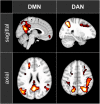
The maps of the two ICA components representing DMN and DAN that have been used in the functional connectivity analysis. The maps are shown in axial and sagittal views superimposed on a standard MNI152 T1 2 mm resolution brain template. The following areas are visible: DMN: posterior cingulate cortex (with the adjacent regions of the precuneus), left and right inferior parietal lobule and medial prefrontal cortex; DAN: left and right intraparietal sulcus, left and right frontal eye field.
FIGURE 2
ECG and breathing measures. The color bars represent the median values of the 5-min epochs of rest (blue), content-related meditation (yellow) and content-free awareness (pink) in each case. From left to right: heart rate (HR), heart-rate variability (HRV, RMSSD = root mean square of successive differences), breathing rate (BR), estimated oxygen consumption (OCe).
FIGURE 3
Topographies of EEG spectral power. Upper row rest condition, lower row difference between content-free awareness (CFA) and rest. Increases in power are displayed in red, decreases in blue. Black dots denote the electrodes of the extended 10–20 system.
FIGURE 4
EEG spectral power during rest, content-related meditation and content-free awareness. For each frequency band, the electrodes with the most significant differences for the main contrast (content-free awareness vs. rest) are shown (theta band: F6, F7, and Pz; alpha band: FCz, P7, and P8). The significance values for the different contrasts are given in Table 2. The color bars represent the median values of spectral power (μV2) of the 5-min epochs of rest (blue), content-related meditation (yellow) and content-free awareness (pink) in each case.
FIGURE 5
fMRI connectivity during rest, content-related meditation and content-free awareness. For each network, only the pairs of nodes with the most significant differences for the main contrast (content-free awareness vs. rest) are shown. A complete list of all significant differences is given in Table 3. The color bars represent the median values of Fisher-transformed correlation coefficients of the 5-min epochs of rest (blue), content-related meditation (yellow) and content-free awareness (pink) in each case. DMN = default mode network, DAN = dorsal attention network, PAC = primary auditory cortex, PCC = posterior cingulate cortex, IPL = inferior parietal lobule, IPS = intraparietal sulcus, FEF = frontal eye field, IFG = inferior frontal gyrus, L = left, R = right.
FIGURE 6
Correlations between the time courses of fMRI connectivity and EEG power. Shown are all Pearson’s correlation coefficients of time courses with significant differences for the main contrast (content-free awareness vs. rest). Blue cells display negative correlations red cells display positive ones. Significant correlations (p < 0.05, FDR-corrected) are printed in bold.
Similar articles
- The Human Default Consciousness and Its Disruption: Insights From an EEG Study of Buddhist Jhāna Meditation.
Dennison P. Dennison P. Front Hum Neurosci. 2019 Jun 12;13:178. doi: 10.3389/fnhum.2019.00178. eCollection 2019. Front Hum Neurosci. 2019. PMID: 31249516 Free PMC article. - Neural correlates of consciousness in patients who have emerged from a minimally conscious state: a cross-sectional multimodal imaging study.
Di Perri C, Bahri MA, Amico E, Thibaut A, Heine L, Antonopoulos G, Charland-Verville V, Wannez S, Gomez F, Hustinx R, Tshibanda L, Demertzi A, Soddu A, Laureys S. Di Perri C, et al. Lancet Neurol. 2016 Jul;15(8):830-842. doi: 10.1016/S1474-4422(16)00111-3. Epub 2016 Apr 27. Lancet Neurol. 2016. PMID: 27131917 - Temporal Dynamics of the Default Mode Network Characterize Meditation-Induced Alterations in Consciousness.
Panda R, Bharath RD, Upadhyay N, Mangalore S, Chennu S, Rao SL. Panda R, et al. Front Hum Neurosci. 2016 Jul 22;10:372. doi: 10.3389/fnhum.2016.00372. eCollection 2016. Front Hum Neurosci. 2016. PMID: 27499738 Free PMC article. - Consciousness supporting networks.
Demertzi A, Soddu A, Laureys S. Demertzi A, et al. Curr Opin Neurobiol. 2013 Apr;23(2):239-44. doi: 10.1016/j.conb.2012.12.003. Epub 2012 Dec 27. Curr Opin Neurobiol. 2013. PMID: 23273731 Review. - Pain and consciousness.
Garcia-Larrea L, Bastuji H. Garcia-Larrea L, et al. Prog Neuropsychopharmacol Biol Psychiatry. 2018 Dec 20;87(Pt B):193-199. doi: 10.1016/j.pnpbp.2017.10.007. Epub 2017 Oct 12. Prog Neuropsychopharmacol Biol Psychiatry. 2018. PMID: 29031510 Review.
Cited by
- Reflections on Inner and Outer Silence and Consciousness Without Contents According to the Sphere Model of Consciousness.
Paoletti P, Ben-Soussan TD. Paoletti P, et al. Front Psychol. 2020 Aug 12;11:1807. doi: 10.3389/fpsyg.2020.01807. eCollection 2020. Front Psychol. 2020. PMID: 32903475 Free PMC article. - Implicit-explicit gradient of nondual awareness or consciousness as such.
Josipovic Z. Josipovic Z. Neurosci Conscious. 2021 Oct 8;2021(2):niab031. doi: 10.1093/nc/niab031. eCollection 2021. Neurosci Conscious. 2021. PMID: 34646576 Free PMC article. - 15 Years MR-encephalography.
Hennig J, Kiviniemi V, Riemenschneider B, Barghoorn A, Akin B, Wang F, LeVan P. Hennig J, et al. MAGMA. 2021 Feb;34(1):85-108. doi: 10.1007/s10334-020-00891-z. Epub 2020 Oct 20. MAGMA. 2021. PMID: 33079327 Free PMC article. Review. - Beyond the veil of duality-topographic reorganization model of meditation.
Cooper AC, Ventura B, Northoff G. Cooper AC, et al. Neurosci Conscious. 2022 Oct 11;2022(1):niac013. doi: 10.1093/nc/niac013. eCollection 2022. Neurosci Conscious. 2022. PMID: 36237370 Free PMC article. Review. - An updated classification of meditation methods using principles of taxonomy and systematics.
Nash JD, Newberg AB. Nash JD, et al. Front Psychol. 2023 Feb 8;13:1062535. doi: 10.3389/fpsyg.2022.1062535. eCollection 2022. Front Psychol. 2023. PMID: 36846482 Free PMC article.
References
LinkOut - more resources
Full Text Sources
Miscellaneous


