Moderate exercise has beneficial effects on mouse ischemic stroke by enhancing the functions of circulating endothelial progenitor cell-derived exosomes - PubMed (original) (raw)
Moderate exercise has beneficial effects on mouse ischemic stroke by enhancing the functions of circulating endothelial progenitor cell-derived exosomes
Jinju Wang et al. Exp Neurol. 2020 Aug.
Abstract
Exosomes (EXs) are emerging as novel players in the beneficial effects induced by exercise on vascular diseases. We have recently revealed that moderate exercise enhances the function of circulating endothelial progenitor cell-derived EXs (cEPC-EXs) on protecting endothelial cells against hypoxia injury. However, the relationship between the changes of cEPC-EXs and the effects of exercise on ischemic stroke (IS) is unknown. Here, we investigated whether exercise-regulated EPC-EXs contribute to the beneficial effects of exercise on IS. C57BL/6 mice received moderate treadmill exercise (10 m/min) for 4-wks and then were subjected to middle cerebral artery occlusion (MCAO) stroke. The neurologic deficit score (NDS), infarct volume, microvessel density, cell apoptosis, angiogenesis/neurogenesis, sensorimotor functions were determined on day 2 (acute stage) and/or day 28 (chronic stage) post-stroke. The miR-126 and EPC-EX levels were analyzed by RT-PCR or nanoparticle tracking analysis combined with microbeads and used for correlation analyses. The function of EPC-EXs from exercised mice was detected in a hypoxia neuron model. Cell apoptosis, axon growth ability and gene expressions (cas-3 and Akt) were measured. Our data showed that: i) On day 2, exercised mice had decreased NDS and infarct volume, reduced cell apoptosis rate and cleaved cas-3 level, and a higher microvessel density than those in control (no-exercise) mice. The levels of EPC-EXs in plasma and brain tissue were raised and positively correlated in exercised mice. Meanwhile, the miR-126 level in cEPC-EXs and in ischemic tissue were upregulated in exercised mice. The EPC-EXs and their carried miR-126 levels negatively correlated with the infarct volume and cell apoptosis, whereas positively correlated with microvessel density. In addition, cEPC-EXs from exercised mice elicited protective effects on neurons against hypoxia-induced apoptosis and compromised axon growth ability which were blocked by miR-126 and PI3k inhibitors in vitro. ii) On day 28, exercised mice had less infarct volume, higher microvessel density, angiogenesis/neurogenesis and better sensorimotor functions. The levels of BDNF, p-TrkB/TrkB and p-Akt/Akt were upregulated in the brain of exercised mice. These recovery indexes correlated with the levels of cEPC-EXs and their miR-126. In conclusion, our data suggest that moderate exercise intervention has protective effects on the brain against MCAO-induced ischemic injury in both acute and chronic stages which might via the release of miR-126 enriched EPC-EXs.
Keywords: EPC-EXs; Exercise; Ischemic stroke; miR-126.
Copyright © 2020 The Authors. Published by Elsevier Inc. All rights reserved.
Conflict of interest statement
Declaration of Competing Interest None.
Figures
Fig. 1.
Exercised mice had reduced NDS and infarct volume, decreased cell apoptosis as well as a higher microvessel density on day 2 post-MCAO surgery. A, NDS; B, representative TTC images and summarized data showing the infarct volume of exercised and control mice; C, representative Tunel staining images and summarized data showing cell apoptosis in the peri-infarct area of exercised and control mice; Green: Tunel labeling; Blue: DAPI staining; scale bar in left panels: 100 um; scale bar in right panels (enlarged images of the box): 25 um; D, cleaved caspase-3 expression in ipsilateral brain of exercised and control mice; E, representative images and summarized data showing the microvessel density in the contralateral and ipsilateral brain; scale bar: 50 um. * p < .05, vs. control; + p < .05, vs. ipsilateral. Data are expressed as mean ± SEM. N = 11/group. (For interpretation of the references to colour in this figure legend, the reader is referred to the web version of this article.)
Fig. 2.
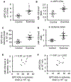
Analyses of the levels of EPC-EXs in the circulation and the brain, as well as the miR-126 expression on day 2 post-MCAO surgery. A-B, the level of cEPC-EXs and their carried miR-126 level in exercised and control mice; * p < .05, vs. control. C-D, EPC-EXs and miR-126 levels in the ischemic brain; * p < .05, vs. control; _E_-F, Pearson’s correlation analyses of the number of EPC-EXs with miR-126 in the ischemic brain, and the level of cEPC-EXs and EPC-EXs in the ischemic brain. Data are expressed as mean ± SEM. N = 11/group.
Fig. 3.
Correlation analyses of the levels of cEPC-EXs and their carried miR-126 with MCAO-induced brain injury on day 2 post-MCAO surgery. A-F, Pearson’s correlation analysis of the level of cEPC-EXs and their carried miR-126 level with NDS, infarct volume and Tunel+ cells in exercised mice; Data is expressed as mean ± SEM. N = 11/group.
Fig. 4.
Exercised mice had alleviated infarct volume, increased microvessel density, angiogenesis and neurogenesis, improved sensorimotor functions as well as upregulated the protein expressions of BDNF/TrkB/Akt signaling pathway proteins in the ischemic brain of exercised mice day 28 post-MCAO surgery. A, representative CV images and summarized data showing the infarct volume of exercised and control mice; Red dash line indicate the infarct area in each brain slide; * p < .05, vs. control. B, microvessel density; C, representative immunofluorescence images and statistical data showing the angiogenesis and neurogenesis. Red: CD31 or NeuN; Green: BrdU; Double positive cells were labeled by arrows. Scale bar: 50 um. * p < .05, vs. control. D, corner test and adhesive remove test analyses. E, the protein levels of BDNF, p-TrkB/TrkB and p-Akt/Akt. * p < .05, vs. day −1, + p < .05, vs. day 0, # p < .05, vs. day 14. Data are expressed as mean ± SEM. N = 11/group. (For interpretation of the references to colour in this figure legend, the reader is referred to the web version of this article.)
Fig. 5.
Correlation analyses of the levels of cEPC-EXs and their carried miR-126 with the infarct volume, angiogenesis, neurogenesis and sensorimotor function of exercised mice on day 28 post-MCAO surgery. A-J, Pearson’s correlation analysis of the level of cEPC-EXs and their carried miR-126 level with the infarct volume, the number of the number of BrdU + CD31 + cells and BrdU + NeuN + cells, and left turns/10 trials and adhesive remove time in exercised mice. Data is expressed as mean ± SEM. N = 11/group.
Fig. 6.
cEPC-EX of exercised mice elevated the miR-126 level and improved the survival ability of H/R-injured N2a cells. A, miR-126 level in different co-culture groups; B, cell viability as revealed by MTT assay; C-D, representative flow cytometry plot and summarized data showing the apoptotic rate of N2a cells; E, cleaved cas-3 level in N2a cells. * p < .05, vs. veh, + p < .05, vs. cEPC-EX, # p < .05, vs. cEPC-EXE. Data are expressed as mean ± SEM. N = 5/group.
Fig. 7.
cEPC-EX of exercised mice restored the neurite length, promoted BDNF secretion and activated the PI3k/Akt signal pathway of N2a cells. A, representative phase-contrast images and summarized data showing the neurite length of N2a cells in different co-culture groups; B, BDNF level in the culture medium of N2a cells in different co-culture groups; C, representative western blot bands and summarized data showing p-AKt/Akt expression of N2a cells in different co-culture groups. * p < .05, vs. veh, + p < .05, vs. cEPC-EX, # p < .05, vs. cEPC-EXE. Data are expressed as mean ± SEM. N = 5/group.
Similar articles
- The miR-210 Primed Endothelial Progenitor Cell Exosomes Alleviate Acute Ischemic Brain Injury.
Wang J, Chen S, Sawant H, Chen Y, Bihl JC. Wang J, et al. Curr Stem Cell Res Ther. 2024;19(8):1164-1174. doi: 10.2174/011574888X266357230923113642. Curr Stem Cell Res Ther. 2024. PMID: 37957914 Free PMC article. - MiR-17-5p Mediates the Effects of ACE2-Enriched Endothelial Progenitor Cell-Derived Exosomes on Ameliorating Cerebral Ischemic Injury in Aged Mice.
Pan Q, Wang Y, Liu J, Jin X, Xiang Z, Li S, Shi Y, Chen Y, Zhong W, Ma X. Pan Q, et al. Mol Neurobiol. 2023 Jun;60(6):3534-3552. doi: 10.1007/s12035-023-03280-4. Epub 2023 Mar 9. Mol Neurobiol. 2023. PMID: 36892728 - Exercise improves cardiac fibrosis by stimulating the release of endothelial progenitor cell-derived exosomes and upregulating miR-126 expression.
Fu G, Wang Z, Hu S. Fu G, et al. Front Cardiovasc Med. 2024 May 9;11:1323329. doi: 10.3389/fcvm.2024.1323329. eCollection 2024. Front Cardiovasc Med. 2024. PMID: 38798919 Free PMC article. Review. - Conventional and Nonconventional Sources of Exosomes-Isolation Methods and Influence on Their Downstream Biomedical Application.
Janouskova O, Herma R, Semeradtova A, Poustka D, Liegertova M, Hana Auer M, Maly J. Janouskova O, et al. Front Mol Biosci. 2022 May 2;9:846650. doi: 10.3389/fmolb.2022.846650. eCollection 2022. Front Mol Biosci. 2022. PMID: 35586196 Free PMC article. Review.
Cited by
- Exosomes as a Promising Therapeutic Strategy for Peripheral Nerve Injury.
Yu T, Xu Y, Ahmad MA, Javed R, Hagiwara H, Tian X. Yu T, et al. Curr Neuropharmacol. 2021;19(12):2141-2151. doi: 10.2174/1570159X19666210203161559. Curr Neuropharmacol. 2021. PMID: 33535957 Free PMC article. Review. - Exercise-Intervened Endothelial Progenitor Cell Exosomes Protect N2a Cells by Improving Mitochondrial Function.
Chen S, Sigdel S, Sawant H, Bihl J, Wang J. Chen S, et al. Int J Mol Sci. 2024 Jan 17;25(2):1148. doi: 10.3390/ijms25021148. Int J Mol Sci. 2024. PMID: 38256220 Free PMC article. - The Mechanism of miR-21-5p/TSP-1-Mediating Exercise on the Function of Endothelial Progenitor Cells in Aged Rats.
Chen X, Xie K, Sun X, Zhang C, He H. Chen X, et al. Int J Environ Res Public Health. 2023 Jan 10;20(2):1255. doi: 10.3390/ijerph20021255. Int J Environ Res Public Health. 2023. PMID: 36674009 Free PMC article. - Exercise mimetics: harnessing the therapeutic effects of physical activity.
Gubert C, Hannan AJ. Gubert C, et al. Nat Rev Drug Discov. 2021 Nov;20(11):862-879. doi: 10.1038/s41573-021-00217-1. Epub 2021 Jun 8. Nat Rev Drug Discov. 2021. PMID: 34103713 Review. - Microglia dynamic response and phenotype heterogeneity in neural regeneration following hypoxic-ischemic brain injury.
Quan H, Zhang R. Quan H, et al. Front Immunol. 2023 Nov 29;14:1320271. doi: 10.3389/fimmu.2023.1320271. eCollection 2023. Front Immunol. 2023. PMID: 38094292 Free PMC article. Review.
References
- Arrick DM, Yang S, Li C, Cananzi S, Mayhan WG, 2014. Vigorous exercise training improves reactivity of cerebral arterioles and reduces brain injury following transient focal ischemia. Microcirculation. 21 (6), 516–523. - PubMed
Publication types
MeSH terms
Substances
LinkOut - more resources
Full Text Sources
Medical


