New Frontiers in Molecular Imaging with Superparamagnetic Iron Oxide Nanoparticles (SPIONs): Efficacy, Toxicity, and Future Applications - PubMed (original) (raw)
Review
New Frontiers in Molecular Imaging with Superparamagnetic Iron Oxide Nanoparticles (SPIONs): Efficacy, Toxicity, and Future Applications
Viviana Frantellizzi et al. Nucl Med Mol Imaging. 2020 Apr.
Abstract
Supermagnetic Iron Oxide Nanoparticles (SPIONs) are nanoparticles that have an iron oxide core and a functionalized shell. SPIONs have recently raised much interest in the scientific community, given their exciting potential diagnostic and theragnostic applications. The possibility to modify their surface and the characteristics of their core make SPIONs a specific contrast agent for magnetic resonance imaging but also an intriguing family of tracer for nuclear medicine. An example is 68Ga-radiolabeled bombesin-conjugated to superparamagnetic nanoparticles coated with trimethyl chitosan that is selective for the gastrin-releasing peptide receptors. These receptors are expressed by several human cancer cells such as breast and prostate neoplasia. Since the coating does not interfere with the properties of the molecules bounded to the shell, it has been proposed to link SPIONs with antibodies. SPIONs can be used also to monitor the biodistribution of mesenchymal stromal cells and take place in various applications. The aim of this review of literature is to analyze the diagnostic aspect of SPIONs in magnetic resonance imaging and in nuclear medicine, with a particular focus on sentinel lymph node applications. Moreover, it is taken into account the possible toxicity and the effects on human physiology to determine the SPIONs' safety.
Keywords: 68Ga-radiolabeled bombesin; Iron oxide nanoparticles; Molecular imaging; Review; SPION.
© Korean Society of Nuclear Medicine 2020.
Conflict of interest statement
Conflict of InterestViviana Frantellizzi, Miriam Conte, Mariano Pontico, Arianna Pani, Roberto Pani, and Giuseppe De Vincentis declare that they have no conflict of interest.
Figures
Fig. 1
The figure shows a supermagnetic iron oxide nanoparticle with a core (radius between 5 to15 nm) and the radius of the whole core with shell and water coat (from 20 to 150 nm)
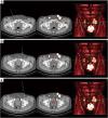
Fig. 2
Uptake of 99mTechnetium-nanocolloid in normal sized right superficial (A), left superficial (B), and left deep (C) inguinal LNs (arrows) seen in SPECT-CT (single-photon emission computed tomography-computed tomography) image
Fig. 3
On the left a representation of 99mTc–Fe3O4–HEDP MNPs, on the right a planar image that shows the uptake of the radiopharmaceutical in liver and bladder in a healthy mouse [52]
Fig. 4
On the left, an image before injection and in the middle after DOTA–BN–TMC–MNPs injection through the tail vein under the 3 T magnetic field. The uptake of NPs is circled in yellow. In the image on the right, it is shown a PET/CT image of a nude mice with a T-47D BC tumor in the right leg: in yellow the uptake by the liver and tumor after the injection of 3.7 MBq 68Ga–DOTA–BN–TMC–MNPs after 120 min [54]
Similar articles
- Superparamagnetic Iron Oxide Nanoparticles-Current and Prospective Medical Applications.
Dulińska-Litewka J, Łazarczyk A, Hałubiec P, Szafrański O, Karnas K, Karewicz A. Dulińska-Litewka J, et al. Materials (Basel). 2019 Feb 19;12(4):617. doi: 10.3390/ma12040617. Materials (Basel). 2019. PMID: 30791358 Free PMC article. Review. - Superparamagnetic Iron-Oxide Nanoparticles Synthesized via Green Chemistry for the Potential Treatment of Breast Cancer.
Tyagi N, Gupta P, Khan Z, Neupane YR, Mangla B, Mehra N, Ralli T, Alhalmi A, Ali A, Al Kamaly O, Saleh A, Nasr FA, Kohli K. Tyagi N, et al. Molecules. 2023 Mar 3;28(5):2343. doi: 10.3390/molecules28052343. Molecules. 2023. PMID: 36903587 Free PMC article. - LHRH-conjugated magnetic iron oxide nanoparticles for detection of breast cancer metastases.
Leuschner C, Kumar CS, Hansel W, Soboyejo W, Zhou J, Hormes J. Leuschner C, et al. Breast Cancer Res Treat. 2006 Sep;99(2):163-76. doi: 10.1007/s10549-006-9199-7. Epub 2006 Jun 3. Breast Cancer Res Treat. 2006. PMID: 16752077 - Potential use of superparamagnetic iron oxide nanoparticles for in vitro and in vivo bioimaging of human myoblasts.
Wierzbinski KR, Szymanski T, Rozwadowska N, Rybka JD, Zimna A, Zalewski T, Nowicka-Bauer K, Malcher A, Nowaczyk M, Krupinski M, Fiedorowicz M, Bogorodzki P, Grieb P, Giersig M, Kurpisz MK. Wierzbinski KR, et al. Sci Rep. 2018 Feb 27;8(1):3682. doi: 10.1038/s41598-018-22018-0. Sci Rep. 2018. PMID: 29487326 Free PMC article. - Superparamagnetic Iron Oxide Nanoparticles: Cytotoxicity, Metabolism, and Cellular Behavior in Biomedicine Applications.
Wei H, Hu Y, Wang J, Gao X, Qian X, Tang M. Wei H, et al. Int J Nanomedicine. 2021 Aug 31;16:6097-6113. doi: 10.2147/IJN.S321984. eCollection 2021. Int J Nanomedicine. 2021. PMID: 34511908 Free PMC article. Review.
Cited by
- In Vivo Distribution of Poly(ethylene glycol) Functionalized Iron Oxide Nanoclusters: An Ultrastructural Study.
Suciu M, Mirescu C, Crăciunescu I, Macavei SG, Leoștean C, Ştefan R, Olar LE, Tripon SC, Ciorîță A, Barbu-Tudoran L. Suciu M, et al. Nanomaterials (Basel). 2021 Aug 25;11(9):2184. doi: 10.3390/nano11092184. Nanomaterials (Basel). 2021. PMID: 34578500 Free PMC article. - How molecular imaging will enable robotic precision surgery : The role of artificial intelligence, augmented reality, and navigation.
Wendler T, van Leeuwen FWB, Navab N, van Oosterom MN. Wendler T, et al. Eur J Nucl Med Mol Imaging. 2021 Dec;48(13):4201-4224. doi: 10.1007/s00259-021-05445-6. Epub 2021 Jun 29. Eur J Nucl Med Mol Imaging. 2021. PMID: 34185136 Free PMC article. Review. - Magnetic Solid Nanoparticles and Their Counterparts: Recent Advances towards Cancer Theranostics.
Cerqueira M, Belmonte-Reche E, Gallo J, Baltazar F, Bañobre-López M. Cerqueira M, et al. Pharmaceutics. 2022 Feb 25;14(3):506. doi: 10.3390/pharmaceutics14030506. Pharmaceutics. 2022. PMID: 35335882 Free PMC article. Review. - Recent nanotheranostics applications for cancer therapy and diagnosis: A review.
Aminolroayaei F, Shahbazi-Gahrouei D, Shahbazi-Gahrouei S, Rasouli N. Aminolroayaei F, et al. IET Nanobiotechnol. 2021 May;15(3):247-256. doi: 10.1049/nbt2.12021. Epub 2021 Feb 14. IET Nanobiotechnol. 2021. PMID: 34694670 Free PMC article. Review. - Current status of Cancer Nanotheranostics: Emerging strategies for cancer management.
Chavda VP, Khadela A, Shah Y, Postwala H, Balar P, Vora L. Chavda VP, et al. Nanotheranostics. 2023 May 1;7(4):368-379. doi: 10.7150/ntno.82263. eCollection 2023. Nanotheranostics. 2023. PMID: 37151802 Free PMC article. Review.
References
- Lassenberger A, Scheberl A, Stadlbauer A, Stiglbauer A, Helbich T, Reimhult E. Individually stabilized, Superparamagnetic nanoparticles with controlled shell and size leading to exceptional stealth properties and high relaxivities. ACS Appl Mater Interfaces. 2017;9:3343–3353. doi: 10.1021/acsami.6b12932. - DOI - PMC - PubMed
Publication types
LinkOut - more resources
Full Text Sources


