COVID-19-Associated Kidney Injury: A Case Series of Kidney Biopsy Findings - PubMed (original) (raw)
. 2020 Sep;31(9):1948-1958.
doi: 10.1681/ASN.2020050699. Epub 2020 Jul 13.
Collaborators, Affiliations
- PMID: 32660970
- PMCID: PMC7461689
- DOI: 10.1681/ASN.2020050699
COVID-19-Associated Kidney Injury: A Case Series of Kidney Biopsy Findings
Purva Sharma et al. J Am Soc Nephrol. 2020 Sep.
Abstract
Background: Reports show that AKI is a common complication of severe coronavirus disease 2019 (COVID-19) in hospitalized patients. Studies have also observed proteinuria and microscopic hematuria in such patients. Although a recent autopsy series of patients who died with severe COVID-19 in China found acute tubular necrosis in the kidney, a few patient reports have also described collapsing glomerulopathy in COVID-19.
Methods: We evaluated biopsied kidney samples from ten patients at our institution who had COVID-19 and clinical features of AKI, including proteinuria with or without hematuria. We documented clinical features, pathologic findings, and outcomes.
Results: Our analysis included ten patients who underwent kidney biopsy (mean age: 65 years); five patients were black, three were Hispanic, and two were white. All patients had proteinuria. Eight patients had severe AKI, necessitating RRT. All biopsy samples showed varying degrees of acute tubular necrosis, and one patient had associated widespread myoglobin casts. In addition, two patients had findings of thrombotic microangiopathy, one had pauci-immune crescentic GN, and another had global as well as segmental glomerulosclerosis with features of healed collapsing glomerulopathy. Interestingly, although the patients had confirmed severe acute respiratory syndrome coronavirus 2 (SARS-CoV-2) infection by RT-PCR, immunohistochemical staining of kidney biopsy samples for SARS-CoV-2 was negative in all ten patients. Also, ultrastructural examination by electron microscopy showed no evidence of viral particles in the biopsy samples.
Conclusions: The most common finding in our kidney biopsy samples from ten hospitalized patients with AKI and COVID-19 was acute tubular necrosis. There was no evidence of SARS-CoV-2 in the biopsied kidney tissue.
Keywords: AKI; ATN; COVID-19; SARS-CoV-2; acute kidney injury; kidney biopsy; kidney pathology.
Copyright © 2020 by the American Society of Nephrology.
Figures
Graphical abstract
Figure 1.
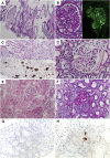
A variety of kidney histopathological findings seen in our patients with COVID-19 and AKI. (A) ATN is often manifested by accumulation of cellular debris in lumens of distal tubules (periodic acid–Schiff [PAS]: ×200). (B) Segmental glomerulosclerosis with features of healing collapse and protein reabsorption granules in podocytes (left, PAS; right, FITC IgG immunofluorescence stain: ×400). (C) Red-brown casts in tubules in the patient with rhabdomyolysis, staining positively for myoglobin stain (upper, hematoxylin and eosin [H&E]; lower, myoglobin immunohistochemistry stain: ×200). (D) Diffuse and early nodular diabetic glomerulosclerosis (H&E, ×400). (E) Diffuse cortical necrosis in a patient with severe TMA (H&E, ×200). (F) Cellular crescent in a glomerulus and surrounding acute tubular injury with flattening of the tubular epithelium in a patient with ANCA disease (PAS, ×200). (G) Representative section of negative immunohistochemistry staining for SARS-CoV-2 nucleocapsid protein after antigen retrieval (×200). (H) Lung tissue as positive control for immunohistochemistry staining for SARS-CoV-2 (×200).
Figure 2.
Ultrastructural examination confirmed the kidney pathological diagnosis in some patients but showed no convincing evidence of viral presence in any of our cases. (A) Electron micrograph showing acute TMA in a patient with gemcitabine exposure; endocapillary space often contains aggregates of crosslinked fibrin (white arrowheads) and fragmented red blood cells (white arrows). Original magnification, ×5,000. (B) Ultrastructural detail of podocyte cytoplasm with clathrin-coated vesicles (black arrows). Original magnification, ×40,000. (C) Multivesicular bodies (black arrowhead) in epithelial cell cytoplasm have been often confused with viral particles. Original magnification, ×50,000.
Similar articles
- Kidney Biopsy Findings in Patients with COVID-19.
Kudose S, Batal I, Santoriello D, Xu K, Barasch J, Peleg Y, Canetta P, Ratner LE, Marasa M, Gharavi AG, Stokes MB, Markowitz GS, D'Agati VD. Kudose S, et al. J Am Soc Nephrol. 2020 Sep;31(9):1959-1968. doi: 10.1681/ASN.2020060802. Epub 2020 Jul 17. J Am Soc Nephrol. 2020. PMID: 32680910 Free PMC article. - Postmortem Kidney Pathology Findings in Patients with COVID-19.
Santoriello D, Khairallah P, Bomback AS, Xu K, Kudose S, Batal I, Barasch J, Radhakrishnan J, D'Agati V, Markowitz G. Santoriello D, et al. J Am Soc Nephrol. 2020 Sep;31(9):2158-2167. doi: 10.1681/ASN.2020050744. Epub 2020 Jul 29. J Am Soc Nephrol. 2020. PMID: 32727719 Free PMC article. - Histopathologic and Ultrastructural Findings in Postmortem Kidney Biopsy Material in 12 Patients with AKI and COVID-19.
Golmai P, Larsen CP, DeVita MV, Wahl SJ, Weins A, Rennke HG, Bijol V, Rosenstock JL. Golmai P, et al. J Am Soc Nephrol. 2020 Sep;31(9):1944-1947. doi: 10.1681/ASN.2020050683. Epub 2020 Jul 16. J Am Soc Nephrol. 2020. PMID: 32675304 Free PMC article. No abstract available. - Renal changes and acute kidney injury in covid-19: a systematic review.
Nogueira SÁR, Oliveira SCS, Carvalho AFM, Neves JMC, Silva LSVD, Silva Junior GBD, Nobre MEP. Nogueira SÁR, et al. Rev Assoc Med Bras (1992). 2020 Sep 21;66Suppl 2(Suppl 2):112-117. doi: 10.1590/1806-9282.66.S2.112. eCollection 2020. Rev Assoc Med Bras (1992). 2020. PMID: 32965368 - Management of acute kidney injury in patients with COVID-19.
Ronco C, Reis T, Husain-Syed F. Ronco C, et al. Lancet Respir Med. 2020 Jul;8(7):738-742. doi: 10.1016/S2213-2600(20)30229-0. Epub 2020 May 14. Lancet Respir Med. 2020. PMID: 32416769 Free PMC article. Review.
Cited by
- Pathology of COVID-19-associated acute kidney injury.
Sharma P, Ng JH, Bijol V, Jhaveri KD, Wanchoo R. Sharma P, et al. Clin Kidney J. 2021 Jan 24;14(Suppl 1):i30-i39. doi: 10.1093/ckj/sfab003. eCollection 2021 Mar. Clin Kidney J. 2021. PMID: 33796284 Free PMC article. Review. - Kidney transplantation and COVID-19 renal and patient prognosis.
Toapanta N, Torres IB, Sellarés J, Chamoun B, Serón D, Moreso F. Toapanta N, et al. Clin Kidney J. 2021 Mar 26;14(Suppl 1):i21-i29. doi: 10.1093/ckj/sfab030. eCollection 2021 Mar. Clin Kidney J. 2021. PMID: 33815780 Free PMC article. Review. - Long-term interplay between COVID-19 and chronic kidney disease.
Schiffl H, Lang SM. Schiffl H, et al. Int Urol Nephrol. 2023 Aug;55(8):1977-1984. doi: 10.1007/s11255-023-03528-x. Epub 2023 Feb 24. Int Urol Nephrol. 2023. PMID: 36828919 Free PMC article. Review. - Liposome encapsulated clodronate mediated elimination of pathogenic macrophages and microglia: A promising pharmacological regime to defuse cytokine storm in COVID-19.
Ravichandran S, Manickam N, Kandasamy M. Ravichandran S, et al. Med Drug Discov. 2022 Sep;15:100136. doi: 10.1016/j.medidd.2022.100136. Epub 2022 Jun 13. Med Drug Discov. 2022. PMID: 35721801 Free PMC article. Review. - Vascular dysfunction in hemorrhagic viral fevers: opportunities for organotypic modeling.
Zarate-Sanchez E, George SC, Moya ML, Robertson C. Zarate-Sanchez E, et al. Biofabrication. 2024 Jun 5;16(3):032008. doi: 10.1088/1758-5090/ad4c0b. Biofabrication. 2024. PMID: 38749416 Free PMC article. Review.
References
- Johns Hopkins University of Medicine Coronavirus Resource Center: COVID-19 Map, 2020. Available at: https://coronavirus.jhu.edu/map.html. Accessed May 15, 2020
MeSH terms
LinkOut - more resources
Full Text Sources
Other Literature Sources
Miscellaneous


